

 PDF
PDF ePub
ePub Citation
 Print
Print


Abstract
Objectives
We wanted to evaluate the effectiveness and safety of anterior interbody fusion (AIF) using cage and plate fixation for treating distractive flexion injury of the cervical spine according to the radiological and clinical outcomes.
Summary of the Literature Review
AIF of the cervical spine using autoiliac bone and plate fixation is known as an effective method for treating not only degenerative disease, but also trauma as well. However, the problem lies in the complications that occur at the donor site. To avoid these complications, the fusion method using a cage is becoming more frequently used, but there are not many reports on using a cage and plate for treating trauma in the cervical spine.
Materials and Methods
We retrospectively analyzed 47 patients with distractive flexion injury of the cervical spine and who underwent anterior decompression and interbody fusion with a autoiliac bone graft and plate fixation (Group I, 32 patients) or who underwent anterior decompression and interbody fusion with cage and plate fixation (Group II, 15 patients). We statistically analyzed the changes of the segmental lordosis, the fused segmental body height, the fusion rate on plain radiography and the neurologic recovery with using an ASIA scoring system.
Results
All the cases were fused by 12.6±2.5weeks after operation. The changes of segmental lordosis shows no statistical difference between the two groups (p=0.69). The anterior and posterior vertebral heights of the fused segments of Group I were more decreased than those of Group II, and there was a statistical difference between the two groups (p=0.03, 0.04). The initial and last follow up neurologic statuses were not statistically difference between the two groups (p=0.11)
Conclusions
For the treatment of fracture-dislocation injury in the cervical spine, AIF using a PEEK cage filled with autoiliac bone and plate fixation is an effective method with the least possibility of complications at the donor site, and at the same time, this surgical method shows equally satisfactory results, both radiologically and clinically, as fusion with using a tricortical autoiliac bone graft.
REFERENCES
1). Allen BL Jr, Ferguson RL, Lehmann TR, O'Brien RP. A mechanistic classification of closed, indirect fractures and dislocations of the lower cervical spine. Spine. 1982; 7(1):1–27.

2). Song KJ, Lee KB, Kim SR. Availability of anterior cervical plating according to the severity of injury in distractive flexion injury in lower cervical spine. J Korean Orthop Assoc. 2005; 40:195–202.
3). Park HJ, Shim YJ. Treatment of distractive flexion injury in lower cervical spine using anterior cervical fusion. J Korean Soc Spine Surg. 2007; 14:221–228.
4). Cybulsky GR, Douglas RA, Meyer PR Jr, et al. Complications in three column cervical spine injuries requiring anterior-posterior stabilization. Spine. 1992; 17:253–256.
5). Henriques T, Olerud C, Bergman A, Jonsson H Jr. Distractive flexion injuries of the subaxial cervical spine treat with anterior plate alone. J Spinal Disord Tech. 2004; 17:1–7.
6). Ulrich C, Worsdorfer O, Claes L, Magerl F. Comparative study of the stability of anterior and posterior cervical spine fixation procedures. Arch Orthp Trauma Surg. 1987; 106:226–231.
7). Robinson R, Smith G. Anterlateral cervical disc removal and interbody fusion for cervical disc herniation. Bull Johns Hopkins Hosp. 1955; 96:223–224.
8). Cloward R. The anterior approach for removal of ruptured cervical discs. J Neurosurg. 1958; 15:602–617.
9). Wilson DH, Campbell DD. Anterior cervical discectomy without bone graft. Report 71 cases. J Neurosurg. 1977; 47:551–555.
10). Silber JS, Anderson DG, Daffner SD, et al. Donor site morbidity after anterior iliac crest bone harvest for single-level anterior cervical discectomy and fusion. Spine. 2003; 28:134–139.
11). Arringon ED, Smith VJ, Chambers HG, et al. Complication of iliac crest bone graft harvesting. Clin Orthop. 1996; 329:300–309.
12). Cho DY, Lee WY, Sheu PC. Treatment of multilevel cervical fusion with cages. Surg Neurol. 2004; 62:378–385. discussion 385-386.
13). Rawlinson JN. Morbidity after anterior cervical decompression and fusion: The influence of the donor site on recovery, and the results of a trial of Surgibone compared to autologous bone. Acta Neurochir. 1994; 131:106–118.
14). Summer BN, Eisenstein SM. Donor site pain from the ilium. A complication of lumbar spine fusion. J Bone Joint Surg Br. 1998; 71:677–680.
15). Malloy KM, Hilibrand AS. Autograft versus allograft in degenerative cervical disease. Clin Orthop Relat Res. 2002; 394:27–38.
16). Zdeblick TA, Ducker TB. The use of freeze-dried allograft bone for anterior cervical fusions. Spine. 1991; 16:726–729.
17). Deutsch H, Haid R, Rodts G Jr, Mummaneni PV. The dicision-making process: allograft versus autograft. Neurosurgery. 2007; 60:98–102.
18). Bagby GW. Arthrodesis by the distraction-compression method using a stainless steel implant. Orthopaedics. 1988; 11:931–944.
19). Brantigan JW, Steffee AD. A carbon fiber implant to aid interbody lumbar fusion. Two-year clinical results in the first 26 patients. Spine. 1993; 18:2106–2107.
20). Kuslich SD, Ulstrom CL, Griffith SL, Ahern JW, Dow-dle JD. The Bagby and Kuslich method of lumbar interbody fusion. History, techniques, and 2-year followup results of a United States prospective, multicenter trial. Spine. 1998; 23:1267–1279.
21). Ray CD. Threaded titanium cages for lumbar interbody fusion. Spine. 1997; 22:667–679.
22). Yu-Cheng Chou, Der-Cherng Chen, Wanhua Annie Hsieh, et al. Efficacy of anterior cervical fusion: Comparison of titanium cages, polyetheretherketone (PEEK) cages and autogenous bone grafts. J Clin Neurosci. 2008; 15:1240–1245.
23). Park HJ, Shim YJ, Yang JH. Anterior decompression and fusion in the treatment of single-level cervical disc herniation: Plate vs Cage. J Korean Soc Spine Surg. 2008; 15:140–148.
24). Cho DY, Liau WR, Lee WY, et al. Preliminary experience using a polyetheretherketone (PEEK) cage in the treatment of cervical disc disease. Neurosurgery. 2002; 51:1343–1349. discussion 1349-1350.
25). Erich Kast, Sharam Derakhshani, Matthias Bothmann, Joachim Oberle. Subsidence after anterior cervical interbody fusion: A randomized prospective clinical trial. Neurosurg Rev. 2008.
26). Kemmesies D, Meier U. Experience with five different intervertebral disc spacers for cervical spondylodesis. Zentralbl Neurochir. 2005; 66:24–33.
27). G. Matge. Cervical cage fusion with 5 different implants: 250 cases. Acta Neurochir. 2002; 144:539–550.
28). Kettler A, Wilke HJ, Claes L. Effects of neck movements on stability and subsidence in cervical interbody fusion: an in vitro study. J Neurosurg. 2001; 94:97–107.
29). Demircan MN, Kutlay AM, Colak A, et al. Multilevel cervical fusion without plates, screws or autogenous iliac crest bone graft. J Clin Neurosci. 2007; 14:723–728.
30). Kulkarni AG, Hee HT, Wong HK. Solis cage (PEEK) for anterior cervical fusion: preliminary radiological results with emphasis on fusion and subsidence. Spine J. 2007; 2:205–209.
31). Delepine F, Jund S, Schlatterer B, de Peretti F. Experience with poly ether ether ketone (PEEK) cages and locking plate for anterior cervical fusion in the treatment of spine trauma without cord injury. Rev Chir Orthop Reparatrice Appar Mot. 2007; 93:789–797.
Figures and Tables%
Fig. 1.
A 52-year old male with C6 nerve root injury due to unilateral facet dislocation. (A) Preoperative lateral roentgenogram shows anterior displacement of C5 on C6 body. (B) T2 weighted sagittal MR image shows a C5-6 disc protrusion. (C) Lateral radiograph, immediately after surgery, shows anterior cervical fusion with cervical spine locking plate and tricortical autoiliac bone. (D, E) Lateral roentgenogram of flexion/extension views that show no motion and solid fusion.
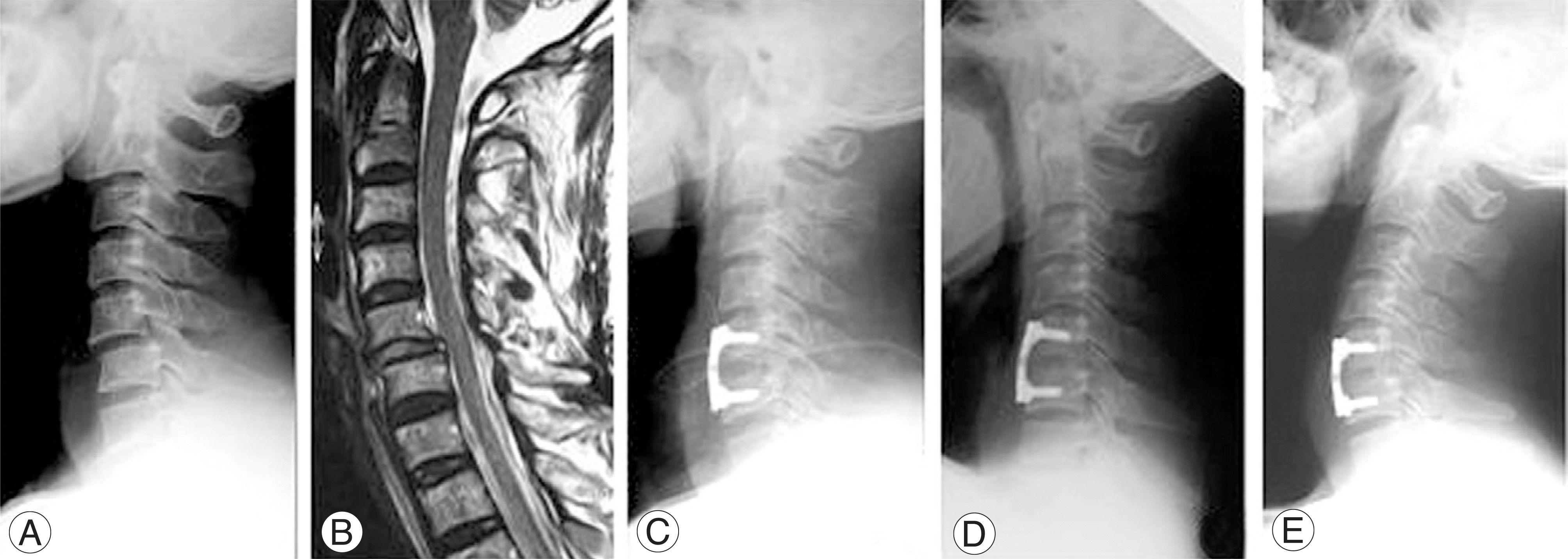
Fig. 2.
A 43-year old male with C5 nerve root injury due to bilateral facet dislocation. (A) Preoperative lateral roentgenogram shows anterior displacement of C5 on C6 body. (B) T2 weighted sagittal MR image shows a C5-6 disc protrusion. (C) Lateral radiograph, immediately after surgery, shows anterior cervical fusion with cervical spine locking plate and Solis PEEK cage that packed with cancellous iliac bone. (D, E) Lateral roentgenogram of flexion/extension views that show no motion and solid fusion.
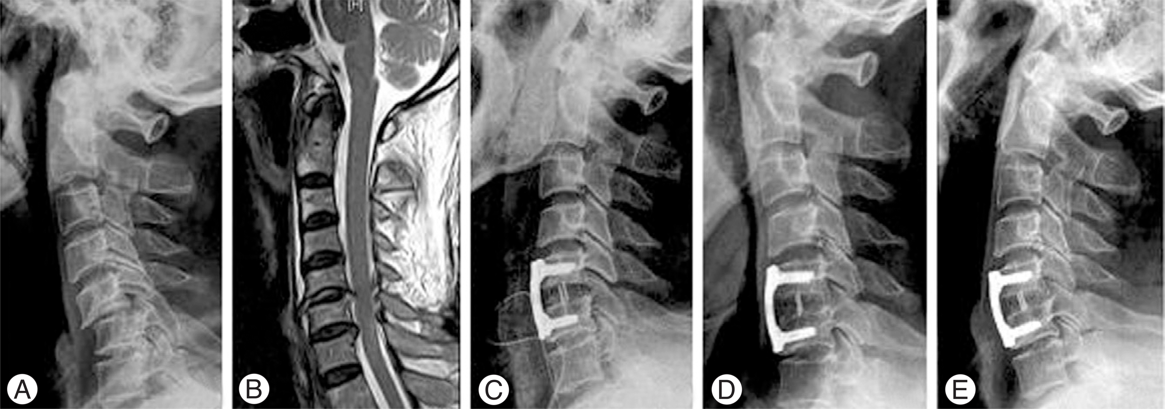
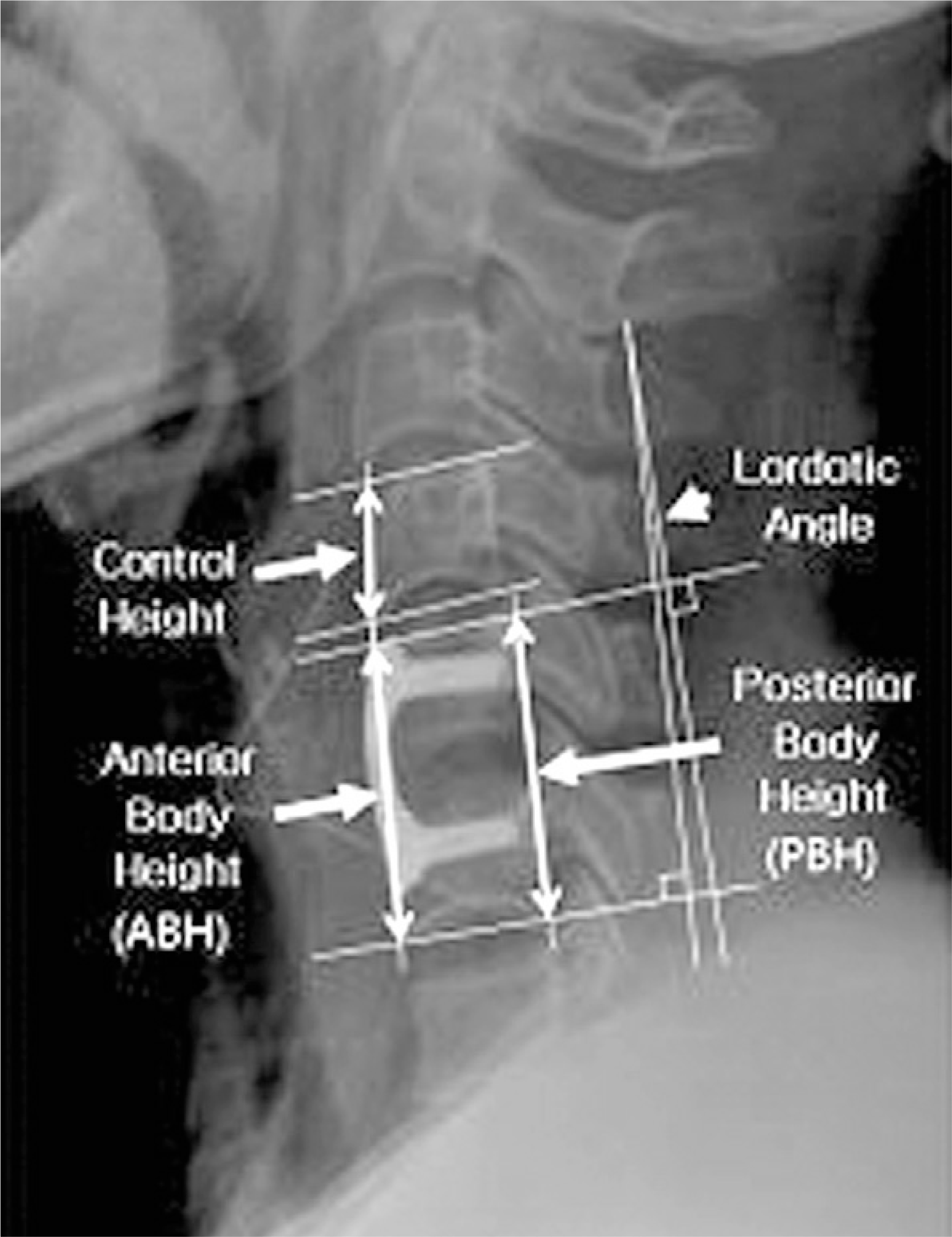
Table 1.
Demography of patients
Table 2.
Change of local lordosis and body height between two groups
| Autobone group (n=32) | Cage group (n=15) | P-value | ||
|---|---|---|---|---|
| Lordotic angle (。) | Postoperation. | 5.1±5.6 | 5.8±4.4 | |
| Last F/U | 4.7±4.8 | 5.3±4.1 | ||
| Change of angle (▲) | 0.4±4.8 | 0.5±3.8 | 0.69 | |
| Ant. body height (ABH*,%) | Postoperation. | 3.0±0.9 | 2.4±0.2 | |
| Last F/U | 2.7±0.8 | 2.3±0.2 | ||
| Change of Ht.(▲)† | -9.2±7.7 | -3.4±3.8 | 0.03 | |
| Post. body height. (PBH*,%) | Postoperation | 2.9±0.9 | 2.3±0.2 | |
| Last F/U | 2.7±0.7 | 2.2±0.1 | ||
| Change of Ht.(▲)† | -5.3±6.2 | -3.4±2.7 | 0.04 |
Table 3.
Change of local lordosis and body height by injury level (Group II)
 XML Download