

 PDF
PDF ePub
ePub Citation
 Print
Print


Abstract
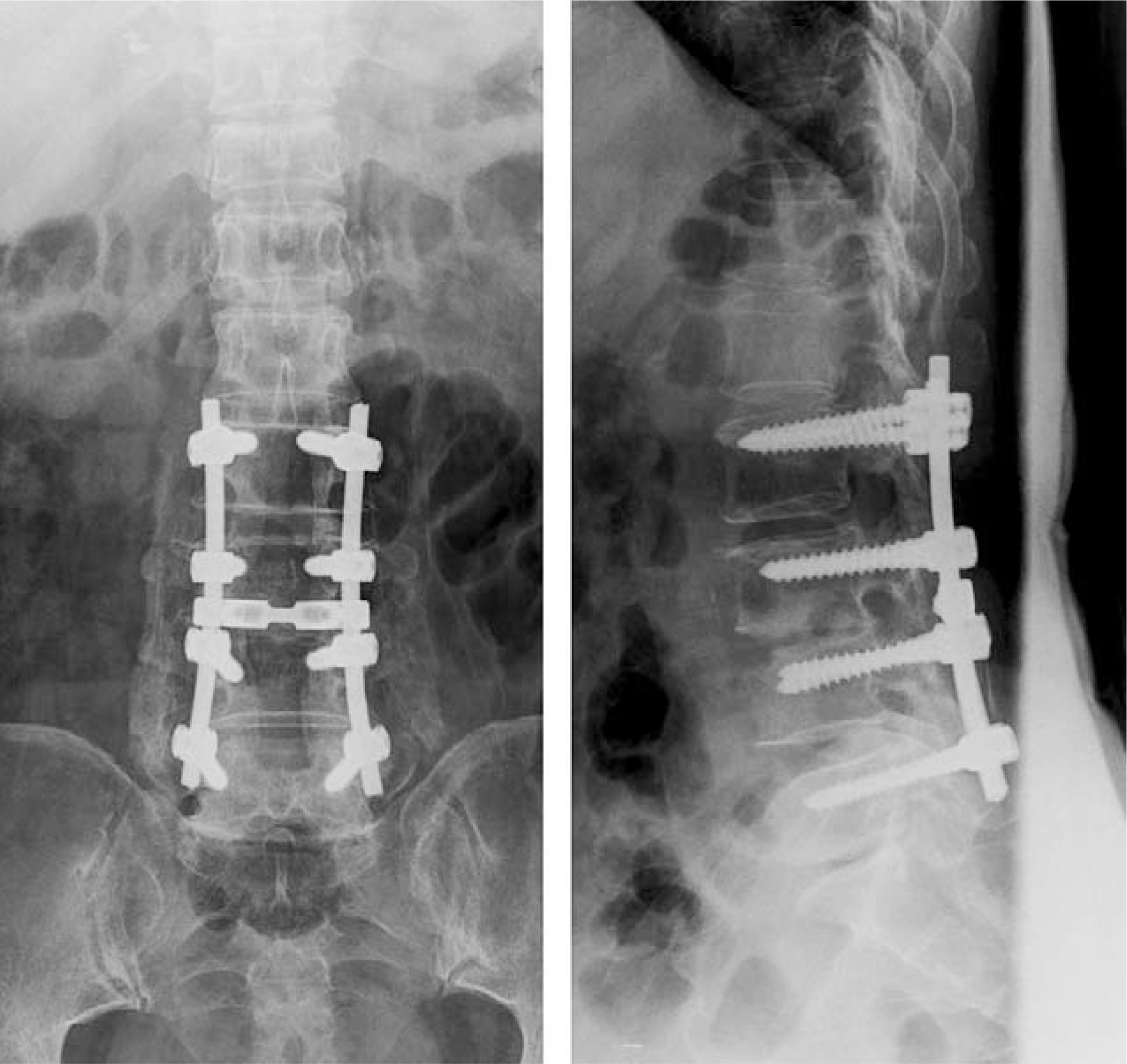
Epidural abscess is a rare disease that can cause severe neurological complications or death if it is not recognized and treated early. Authors report a case of panspinal epidural abscess, which is diagnosed by MRI and treated with surgical drainage and antibiotics.
REFERENCES
1). Bluman EM, Palumbo MA, Lucas PR. Spinal epidural abscess in adults. J Am Acad Orthop Surg. 2004; 12:155–163.

2). Baker AS, Ojeman RG, Swarz MN, Richardson EP Jr. Spinal epidural abscess. N Engl J Med. 1975; 293:463–468.
3). Kim H, Oh SH, Choi IS, et al. .:. Acute panspinal epidural abscess. J Korean Neurosurg Soc. 1999; 28:392–397.
4). Rigamonti D, Leim L, Sampath P, et al. .:. Spinal epidural abscess: Contemporary trends in etiology, evaluation and management. Surg Neurol. 1988; 52:189–197.
5). Simpson RK Jr, Azordegan PA, Sirbasku DM, Weath-ers SW, Lidsky MD, Baskin DS. Rapid onset of quadri-plegia from a panspinal epidural abscess. Spine. 1991; 16:1002–1005.
6). Reihsaus F, Waldbaur H, Seeling W. Spinal epidural abscess: A meta-analysis of 915 patients. Neurosurg Rev. 2000; 23:175–205.
7). Danner RL, Hartmann BJ. Update of spinal epidural abscess: 35 cases and review of the literature. Rev Infect Dis. 1987; 9:265–274.
8). Lang IM, Hughes JPR, St Clair Forbes W, MaKenna F. MR imaging appearances of cervical epidural abscess. Clinical Radiology. 1995; 50:446–471.
9). Choi WT, Choi BY, Lee JW, Moon MS. Pyogenic spinal epidural abscess - A case report -. J Korean Orthop Assoc. 2002; 37:319–323.
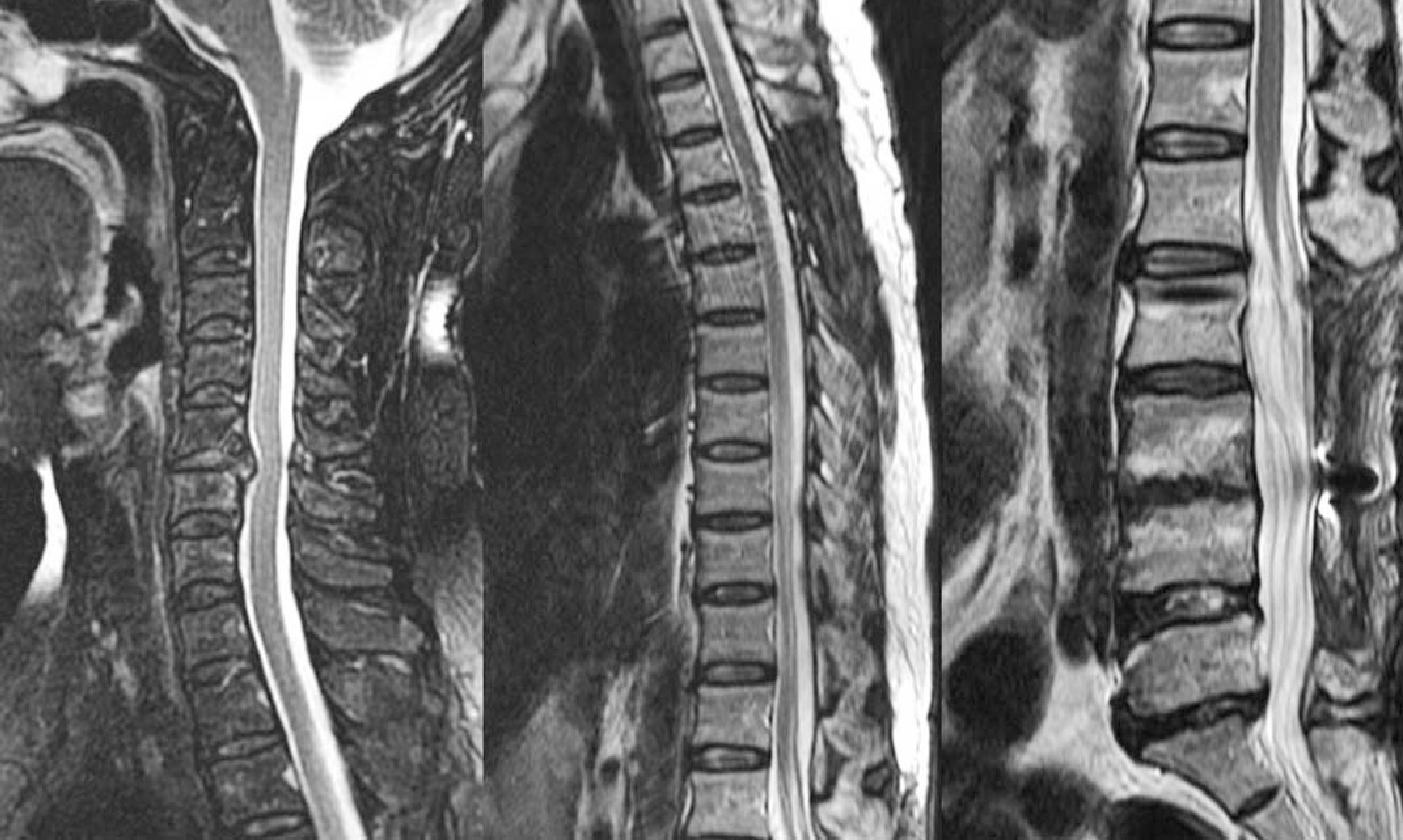
Fig. 1.
Peripheral enhanced epidural fluid collection can be seen in lumbar epidural space on T2 weighted sagittal image and gadolinium-enhanced sagittal image.

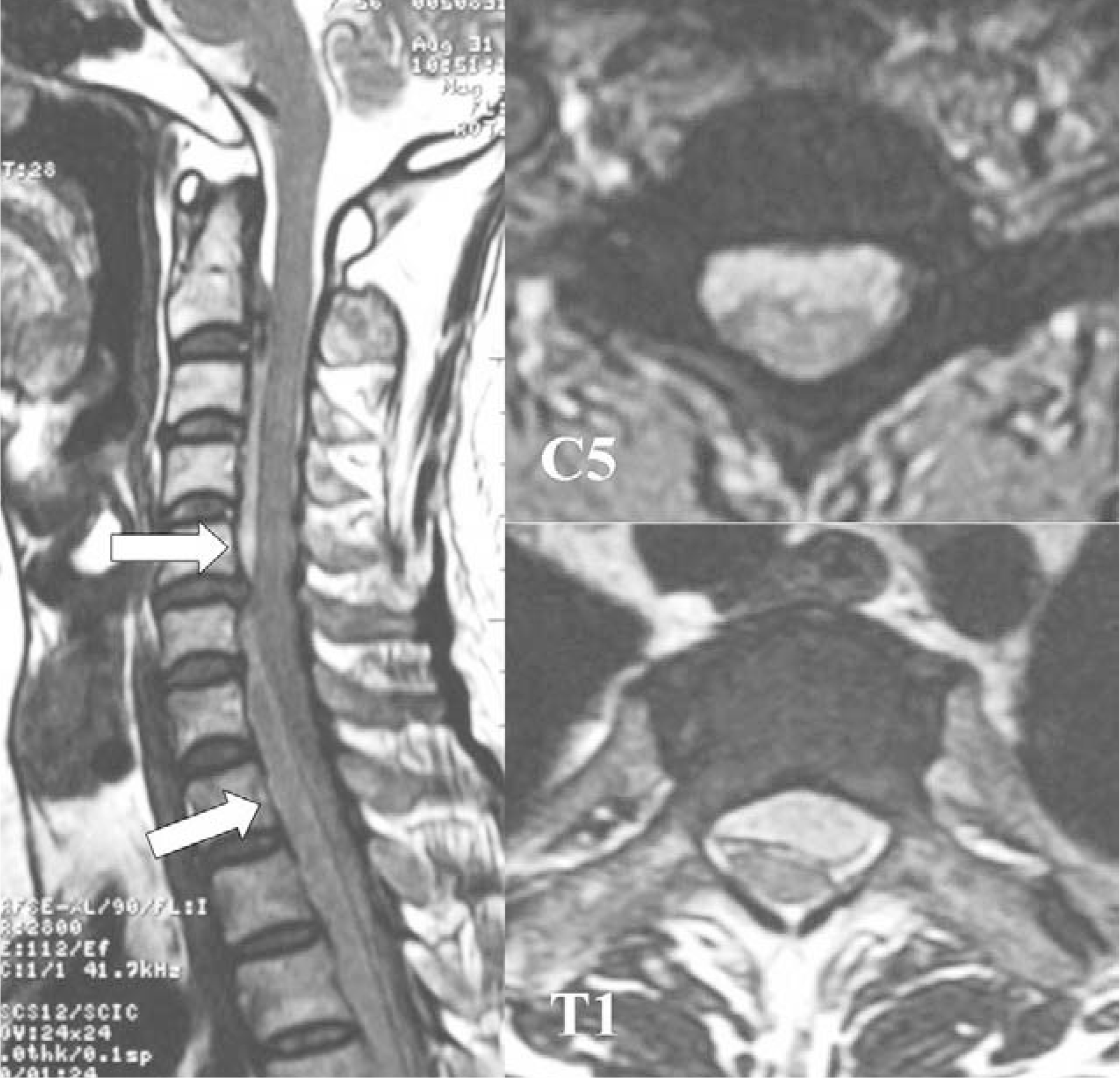
Fig. 2.
Intermediated signal epidural abscess in cervical epidural space, which compresses the spinal cord posteriorly can be seen on T2 weighted images.
 XML Download