Journal List > Infect Chemother > v.49(1) > 1035490

Heidari, Hasanpour, Ebrahim-Saraie, and Motamedifar: High Incidence of Virulence Factors Among Clinical Enterococcus faecalis Isolates in Southwestern Iran

Abstract

Background

Over the past two decades, enterococci have emerged as an important agent responsible for hospital acquired infection. Several virulence factors contribute to the adherence, colonization, evasion of the host immune response, and pathogenicity and severity of the infection. Enterococcus faecalis is the most common and virulent species causing infections in hospitalized patients. The aim of the present study was to examine the prevalence of genes encoding virulence factors and antimicrobial resistance patterns of E. faecalis strains isolated from hospitalized patients in Shiraz, south west of Iran.

Materials and Methods

A total of 51 E. faecalis isolates from the urine, blood, pleural fluid, peritoneal fluid, eye discharge, endotracheal tube (ETT) and transjugular intrahepatic portosystemic shunt (TIPS) specimens of patients were identified by phenotypic and genotypic methods. Antimicrobial sensitivity tests and detection of virulence factors were performed using standard methods.

Results

The efa and asa1 were the most frequently detected gene (100%) among the isolates, followed by esp (94.1%), ace (90.2%), gelE (80.4%), cylA (64.7%), and hyl (51%). More than half of the isolates (52.9%) were high level gentamicin resistant (HLGR). Vancomycin resistance was observed among 23 (45.1%) isolates. The lowest antimicrobial activity was related to erythromycin (3.9%), tetracycline (5.9%) and ciprofloxacin (9.8%). No isolate was found resistant to fosfomycin and linezolid.

Conclusion

Our data indicated a high incidence of virulence factors among E. faecalis strains isolated from clinical samples. Colonization of drug resistant virulent isolates in hospital environment may lead to life threatening infection in hospitalized patients. Therefore, infection control procedures should be performed.

Introduction

Enterococcus is a Gram-positive and common commensal bacterium of human gastrointestinal and genitourinary tracts [1]. Over the past two decades, enterococci have emerged as an important agent responsible for hospital acquired infection, particularly bacteremia, sepsis, endocarditis, urinary tract infection (UTI), and abdominal and pelvic infections [2]. Certain conditions, such as prolonged hospitalization, immunodeficiency and antibiotic therapy, causes a high risk for acquiring of enterococcal infections in patients [34].
Several virulence factors including enterococcal surface protein (Esp), hyaluronidase (Hyl), gelatinase (GelE), aggregation substance (AS) proteins (Asa1), collagen-binding protein (Ace), and cytolysin (CylA) contribute to the adherence, colonization, evasion of the host immune response, extracellular production of enzymes, pathogenicity, and severity of the infection [56]. Moreover, treatment of enterococcal infections has been clinically challenging due to increasing resistance to a broad range of antimicrobial agents, including β‑lactams, macrolides, fluroquinolones, glycopeptides, and aminoglycosides [47].
Enterococci possess efficient genetic exchange systems. The genes encoding virulence determinants can be transferred to resistant strains via these systems [48]. Emergence of antimicrobial resistant virulent enterococci is a serious problem in the treatment and control of nosocomial infections [9].
Several studies have shown that Enterococcus faecalis is an important and virulent species causing various infections [461011]. There are few data regarding the enterococcal virulence determinants in hospitalized patients in south-west of Iran. Therefore, the aim of the present study was to examine the prevalence of genes encoding virulence factors and antimicrobial resistance patterns of E. faecalis strains isolated from hospitalized patients in Shiraz, Iran.

Materials and Methods

1. Bacterial isolates

A total of 51 E. faecalis isolates were collected from specimens of patients hospitalized in Nemazee hospital (the main hospital affiliated to Shiraz University of Medical Sciences with 1,000 beds and 50,000 inpatients per year) during September 2015 to March 2016. They were obtained from the urine (n = 38), blood (n = 4), pleural fluid (n = 4), peritoneal fluid (n = 2), eye discharge (n = 1), endotracheal tube (ETT) (n = 1), and transjugular intrahepatic portosystemic shunt (TIPS) (n = 1). Only one isolate per patient was included. Identification of enterococcal isolates was performed based on conventional microbiological tests (Gram staining, catalase test, growth on Brain Heart Infusion agar (Conda, Madrid, Spain) with 6.5% NaCl, and bile-esculin test) [1213]. The ddlE gene was targeted by polymerase chain reaction (PCR) using species specific primers (ddlE. faecalis F-5'-ATCAAGTACAGTTAGTCT-3' and R-5'-ACGATTCAAAGCTAACTG-3') for molecular confirmation of E. faecalis [414]. The PCR protocol consisted of a pre-denaturation step at 95 °C for 5 min, followed by 30 cycles of 60s at 95°C, 45s at 45°C and 50s at 72°C. A final extension step was performed at 72°C for 5 min.

2. Antimicrobial susceptibility testing

Antimicrobial sensitivity tests were performed by disc diffusion method on the Mueller- Hinton Agar (Merck Co., Darmstadt, Germany) based on Clinical and Laboratory Standards Institute (CLSI) guideline [15]. The tested antibiotics (Mast Group Ltd., UK.) were penicillin (10 units), ampicillin (10 µg), ciprofloxacin (5 µg), erythromycin (15 µg), tetracycline (30 µg), nitrofurantoin (300 µg), rifampin (5 µg), fosfomycin (200 µg), quinupristin- dalfopristin (15 µg), linezolid (5 µg), vancomycin, (30 µg) and teicoplanin (30 µg). High-level gentamicin resistant (HLGR) isolates were also detected by disc diffusion method (with 120 µg gentamicin disks) on the Mueller- Hinton Agar (Merck Co., Darmstadt, Germany) [15]. E. faecalis ATCC 29212 was used as the reference strain for antibiotic susceptibility testing.

3. DNA extraction and detection of virulence genes

Genomic DNA was extracted from fresh colonies as described previously [4]. PCR was done for detection of the genes encoding virulence factors (esp, cylA, hyl, gelE, asa1, ace, efa) [48]. The products were separated by electrophoresis in 1.5% agarose gels with 1 X TAE (Tris/Acetate/EDTA) buffer, stained with KBC power load dye (CinnaGen Co. Tehran, Iran) and visualized under ultraviolet illumination.

4. Statistical analysis

The distribution of virulence genes among HLGR and non-HLGR isolates was calculated by Chi-square and Fisher's exact test for each gene. In addition, differences in the incidence of virulence genes with respect to the origin of specimen were also analyzed by Chi-square test. A P-value of ≤0.05 was considered as statistically significant.

Results

1. Antimicrobial resistance pattern

During 6 months of the study, a total of 51 E. faecalis strains were collected randomly with the possibility of true infections. No isolate was found to be resistant to fosfomycin and linezolid. Vancomycin resistance was observed among 23 (45.1%) isolates (Table 1). More than half of the isolates (52.9%) were HLGR (Table 2).
Table 1

Antimicrobial resistance rates among Enterococcus faecalis isolates.
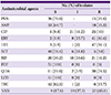
ic-49-51-i001
Antimicrobial agents No. (%) of isolates
S I R
PEN 36 (70.6) - 15 (29.4)
AMP 33 (64.7) - 18 (35.3)
CIP 5 (9.8) 21 (41.2) 25 (49)
ERY 2 (3.9) 8 (15.7) 41 (80.4)
TET 3 (5.9) 1 (2) 47 (92.1)
NIT 40 (78.4) 6 (11.8) 5 (9.8)
RIF 20 (39.2) 10 (19.6) 21 (41.2)
FOF 51 (100) 0 (0) 0 (0)
QDA 11 (21.6) 2 (3.9) 38 (74.5)
LZD 51 (100) 0 (0) 0 (0)
TEC 42 (82.3) 1 (2) 8 (15.7)
VAN 9 (17.6) 19 (37.3) 23 (45.1)
S, susceptible; I, intermediate; R, resistant; PEN, penicillin; AMP, ampicillin; CIP, ciprofloxacin; ERY, erythromycin; TET, tetracycline; NIT, nitrofurantoin; RIF, rifampin; FOF, fosfomycin; QDA, quinupristin- dalfopristin; LZD, linezolid; TEC, teicoplanin; VAN, vancomycin.
Table 2

The distribution of virulence genes and high level resistance to gentamicin among Enterococcus faecalis isolates.

ic-49-51-i002
Clinical samples (N) Virulence genes HLGR / non-HLGR
Urine (38) efa, asa1, esp, ace, cylA, hyl HLGR
efa, asa1, esp, ace, gelE, cylA non-HLGR
efa, asa1, esp, ace, cylA, hyl non-HLGR
efa, asa1, esp, ace, gelE, cylA, hyl HLGR
efa, asa1, esp, ace, cylA, hyl HLGR
efa, asa1, esp, ace, gelE, cylA, hyl HLGR
efa, asa1, esp, ace, gelE, cylA non-HLGR
efa, asa1, esp, ace, cylA, hyl non-HLGR
efa, asa1,esp, gelE HLGR
efa, asa1, esp, ace, gelE, hyl non-HLGR
efa, asa1, esp, ace, gelE, hyl HLGR
efa, asa1, esp, ace, gelE, hyl non-HLGR
efa, asa1, esp, ace, gelE, cylA HLGR
efa, asa1, esp, ace, gelE, cylA, hyl HLGR
efa, asa1, esp, ace, gelE, cylA non-HLGR
efa, asa1,esp, gelE non-HLGR
efa, asa1, esp, ace, gelE, cylA, hyl non-HLGR
efa, asa1, esp, ace, cylA HLGR
efa, asa1, esp, ace, gelE, cylA HLGR
efa, asa1, esp, ace, gelE, hyl non-HLGR
efa, asa1, esp, gelE, cylA, hyl HLGR
efa, asa1, ace, gelE, cylA non-HLGR
efa, asa1, esp, ace, gelE, cylA, hyl non-HLGR
efa, asa1, ace, gelE, hyl HLGR
efa, asa1, esp, ace, cylA HLGR
efa, asa1, esp, gelE HLGR
efa, asa1, esp, ace, gelE, cylA HLGR
efa, asa1, esp, ace, cylA, hyl non-HLGR
efa, asa1, esp, ace, gelE, cylA HLGR
efa, asa1, esp, ace, gelE HLGR
efa, asa1, esp, ace, gelE, cylA, hyl HLGR
efa, asa1, esp, ace, gelE, cylA non-HLGR
efa, asa1, esp, ace, cylA, hyl non-HLGR
efa, asa1, esp, ace, gelE, cylA HLGR
efa, asa1, esp, ace, gelE, cylA, hyl non-HLGR
Blood (4) efa, asa1, esp, ace, gelE non-HLGR
efa, asa1, esp, ace, gelE, cylA, hyl non-HLGR
efa, asa1, esp, ace, gelE, cylA non-HLGR
efa, asa1, ace, gelE HLGR
Pleural (4) efa, asa1, esp, ace, gelE HLGR
efa, asa1, esp, ace, gelE, cylA, hyl HLGR
efa, asa1, esp, ace, gelE, cylA, hyl non-HLGR
efa, asa1, esp non-HLGR
Peritoneal (2) efa, asa1, esp, ace, gelE, cylA HLGR
efa, asa1, esp, ace, gelE, hyl non-HLGR
Eye (1) efa, asa1, esp, ace, gelE, hyl HLGR
ETT (1) efa, asa1, esp, ace, hyl non-HLGR
TIPS (1) efa, asa1, esp, ace, gelE, cylA HLGR
ETT, Endotracheal tube; TIPS, Transjugular intrahepatic portosystemic shunt; HLGR, High level gentamicin resistant.

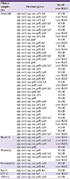
2. Distribution of virulence genes

The efa and asa1 were the most frequently detected gene (100%) among isolates, followed by esp (94.1%), ace (90.2%), gelE (80.4%), cylA (64.7%), and hyl (51%) (Table 2). The frequency of genes encoding virulence factors among HLGR and non-HLGR isolates is shown in Table 3. There was no significant difference between the distribution of virulence genes among HLGR and non-HLGR isolates (P > 0.05).
Table 3

Distribution of genes encoding virulence factors among HLGR and non-HLGR isolates.

ic-49-51-i003
Virulence gene
hyl N(%) cylA N(%) gelE N(%) ace N(%) esp N(%)
HLGR 27(52.9) 11 (40.7) 18 (66.6) 23 (85.2) 24 (88.8) 25 (92.6)
Non-HLGR 24(47.1) 15 (62.5) 15 (52.5) 18 (75) 22 (91.6) 23 (95.8)
Total (51) 26 (51) 33 (64.7) 41 (80.4) 46 (90.2) 48 (94.1)
HLGR, High level gentamicin resistant.

Discussion

Enterococci, particularly E. faecalis, are common pathogens that can cause severe nosocomial infections [7]. In our study, E. faecalis isolates were obtained from various clinical specimens. Similar to other reports from Iran, noticeable resistance to several antimicrobial agents among E. faecalis strains was observed [416]. Nearly half of the isolates were resistant to vancomycin (45.1%). Similarly, high prevalence of vancomycin-resistant enterococci has been previously reported from southwestern Iran [1317]. All of isolates were susceptible to fosfomycin and linezolid (Table 1). Our findings indicate that these agents are efficient choices against enterococcal infections.
More than 52% of the strains were HLGR. Resistance to high concentrations of gentamicin among E. faecalis isolates has been reported in previous studies [89] Presence of genes encoding aminoglycoside-modifying enzymes and transmission among enterococcal strains lead to high levels resistance to gentamicin [18]. It seems that gentamicin has become an ineffective antibiotic against hospital-acquired enterococcal infections.
In addition to antimicrobial resistance, the presence of virulence factors is another concern about enterococci that may contribute to the severity of enterococcal infections [8]. In the present study, the distribution of virulence genes among E. faecalis strains was determined.
All of the isolates harbored efa and asa1 genes (Table 2). In various studies, similar frequencies of the efa have been reported [489]. In a study conducted by Gaglio et al., the efa gene was detected among 25% of the strains isolated from dairy products [19]. It appears that the efa gene is always present in clinical E. faecalis strains. In the current study, the prevalence of asa1 was more than that observed in previous studies [48192021]. AS encoded by asa1 causes clumping of E. faecalis strains and mediates efficient DNA exchange between donor and recipient bacteria [522]. The presence of asa1 gene among all of our isolates can facilitate exchange of resistance and virulence associated genes in hospital setting.
The esp was the second most detected gene among the isolates (94.1%). The prevalence of esp was also 94.7% in strains isolated from UTI. High frequency of esp among UTI causative E. faecalis isolates is consistent with other studies [623]. The enterococcal surface protein is encoded by the esp gene facilities colonization and persistence of E. faecalis in acute urinary tract infections [524].
We found that the prevalence of ace, gelE, cylA and hyl genes was 90.2%, 80.4%, 64.7%, and 51%. Our results indicated that E. faecalis isolates carried more virulence genes in comparison to various reports [49181925262728]. These differences probably reflect variability in the types of samples or geographic regions.
There was no significant difference between the distribution of virulence factors among HLGR and non-HLGR isolates, although cylA and gelE were more prevalent in HLGR strains (Table 3). This result is in contrast with that of a previous study which has reported that the hyl gene is significantly the most frequent virulence gene in HLGR isolates [4].
In the current study, the antimicrobial resistant isolate obtained from TIPS had 6 virulence genes. Colonization of same isolates in hospital environment may lead to life threatening infection in hospitalized patients. Therefore, to prevent colonization of such drug resistant virulent isolates, infection control procedures should be performed.
To the best of our knowledge, this study is the first investigation regarding enterococcal virulence determinants in Shiraz, the south west of Iran. Moreover, high virulence strains were identified. Different profiles of virulence factors can be important for understanding the enterococcal pathogenicity and their effects on the host.
There were some limitations related to the present study. First, small number of isolates were investigated. Second, to identification of the source of infection and take preventive measures in hospital setting molecular analysis was required.
In conclusion, a high incidence of genes encoding virulence factors among E. faecalis strains isolated from clinical samples was observed. Our results indicated that all of the isolates carried efa and asa1 genes. The esp was the second most prevalent one. Gene encoding Hyl (hyl) was the least frequent one that presented in more than half of the isolates. To prevent colonization of such virulent strains in hospital environment, infection control procedures are recommended to be performed.

Acknowledgment

This study was supported by Shiraz University of Medical Sciences grant No. 94-9225.

Notes

Conflict of Interest No conflicts of interest.

References

1. Fisher K, Phillips C. The ecology, epidemiology and virulence of Enterococcus . Microbiology. 2009; 155:1749–1757.
2. Guerrero-Ramos E, Molina-González D, Blanco-Morán S, Igrejas G, Poeta P, Alonso-Calleja C, Capita R. Prevalence, antimicrobial resistance, and genotypic characterization of vancomycin-resistant enterococci in meat preparations. J Food Prot. 2016; 79:748–756.
crossref
3. Emaneini M, Hosseinkhani F, Jabalameli F, Nasiri MJ, Dadashi M, Pouriran R, Beigverdi R. Prevalence of vancomycin-resistant Enterococcus in Iran: a systematic review and meta-analysis. J Clin Microbiol Infect Dis. 2016; 35:1387–1392.
crossref
4. Heidari H, Emaneini M, Dabiri H, Jabalameli F. Virulence factors, antimicrobial resistance pattern and molecular analysis of Enterococcal strains isolated from burn patients. Microb Pathog. 2016; 90:93–97.
crossref
5. Barbosa-Ribeiro M, De-Jesus-Soares A, Zaia AA, Ferraz CC, Almeida JF, Gomes BP. Antimicrobial susceptibility and characterization of virulence genes of Enterococcus faecalis isolates from teeth with failure of the endodontic treatment. J Endod. 2016; 42:1022–1028.
crossref
6. Strateva T, Atanasova D, Savov E, Petrova G, Mitov I. Incidence of virulence determinants in clinical Enterococcus faecalis and Enterococcus faecium isolates collected in Bulgaria. Braz J Infect Dis. 2016; 20:127–133.
crossref
7. Singh KV, La Rosa SL, Somarajan SR, Roh JH, Murray BE. The fibronectin-binding protein EfbA contributes to pathogenesis and protects against infective endocarditis caused by Enterococcus faecalis . Infect Immun. 2015; 83:4487–4494.
crossref
8. Choi JM, Woo GJ. Molecular characterization of high-level gentamicin-resistant Enterococcus faecalis from chicken meat in Korea. Int J Food Microbiol. 2013; 165:1–6.
crossref
9. Niu H, Yu H, Hu T, Tian G, Zhang L, Guo X, Hu H, Wang Z. The prevalence of aminoglycoside-modifying enzyme and virulence genes among enterococci with high-level aminoglycoside resistance in Inner Mongolia, China. Braz J Microbiol. 2016; 47:691–696.
crossref
10. Ceci M, Delpech G, Sparo M, Mezzina V, Sánchez Bruni S, Baldaccini B. Clinical and microbiological features of bacteremia caused by Enterococcus faecalis . J Infect Dev Ctries. 2015; 9:1195–1203.
crossref
11. Al-Talib H, Zuraina N, Kamarudin B, Yean CY. Genotypic variations of virulent genes in Enterococcus faecium and Enterococcus faecalis isolated from three hospitals in Malaysia. Adv Clin Exp Med. 2015; 24:121–127.
crossref
12. Honarm H, Falah Ghavidel M, Nikokar I, Rahbar Taromsari M. Evaluation of a PCR assay to detect Enterococcus faecalis in blood and determine glycopeptidesresistance genes: van A and van B. Iran J Med Sci. 2012; 37:194–199.
13. Kaveh M, Bazargani A, Ramzi M, Sedigh Ebrahim-Saraie H, Heidari H. Colonization rate and risk factors of vancomycin resistant enterococci among patients received hematopoietic stem cell transplantation in Shiraz, Southern Iran. Int J Organ Transplant Med. 2016; 7:197–205.
14. Dutka-Malen S, Evers S, Courvalin P. Detection of glycopeptide resistance genotypes and identification to the species level of clinically relevant enterococci by PCR. J Clin Microbiol. 1995; 33:1434.
crossref
15. Clinical and Labratory Standard Institute (CLSI). Performance standards for antimicrobial susceptibility testing. 25th informational supplement. Wayne, PA: CLSI;2015. p. M100-S23.
16. Mohammadi F, Ghafourian S, Mohebi R, Taherikalani M, Pakzad I, Valadbeigi H, Hatami V, Sadeghifard N. Enterococcus faecalis as multidrug resistance strains in clinical isolates in Imam Reza hospital in Kermanshah, Iran. Br J Biomed Sci. 2015; 72:182–184.
crossref
17. Karimzadeh I, Mirzaee M, Sadeghimanesh N, Sagheb MM. Antimicrobial resistance pattern of Gram-positive bacteria during three consecutive years at the nephrology ward of a tertiary referral hospital in Shiraz, Southwest Iran. J Res Pharm Pract. 2016; 5:238–247.
crossref
18. Filipová M, Bujdákova H, Drahovská H, Lisková A, Hanzen J. Occurrence of aminoglycoside-modifying-enzyme genes aac(6′)-aph(2"), aph(3′), ant(4′) and ant(6) in clinical isolates of Enterococcus faecalis resistant to high-level of gentamicin and amikacin. Folia Microbiol (Praha). 2006; 51:57–61.
crossref
19. Gaglio R, Couto N, Marques C, de Fatima Silva Lopes M, Moschetti G, Pomba C, Settanni L. Evaluation of antimicrobial resistance and virulence of enterococci from equipment surfaces, raw materials, and traditional cheeses. Int J Food Microbiol. 2016; 236:107–114.
crossref
20. Gulhan T, Boynukara B, Ciftci A, Sogut MU, Findik A. Characterization of Enterococcus faecalis isolates originating from different sources for their virulence factors and genes, antibiotic resistance patterns, genotypes and biofilm production. Iran J Vet Res. 2015; 16:261–266.
21. Hällgren A, Claesson C, Saeedi B, Monstein HJ, Hanberger H, Nilsson LE. Molecular detection of aggregation substance, enterococcal surface protein, and cytolysin genes and in vitro adhesion to urinary catheters of Enterococcus faecalis and E. faecium of clinical origin. Int J Med Microbiol. 2009; 299:323–332.
crossref
22. Arias CA, Murray BE. The rise of the Enterococcus: beyond vancomycin resistance. Nat Rev Microbiol. 2012; 10:266–278.
crossref
23. Kafil HS, Mobarez AM. Spread of enterococcal surface protein in antibiotic resistant Enterococcus faecium and Enterococcus faecalis isolates from urinary tract infections. Open Microbiol J. 2015; 9:14–17.
crossref
24. Ferguson DM, Talavera GN, Hernández LA, Weisberg SB, Ambrose RF, Jay JA. Virulence genes among Enterococcus faecalis and Enterococcus faecium isolated from coastal beaches and human and nonhuman sources in Southern California and Puerto Rico. J Pathog. 2016; 2016:3437214.
25. Molale LG, Bezuidenhout CC. Antibiotic resistance, efflux pump genes and virulence determinants in Enterococcus spp. from surface water systems. Environ Sci Pollut Res Int. 2016; 23:21501–21510.
crossref
26. Guerrero-Ramos E, Cordero J, Molina-González D, Poeta P, Igrejas G, Alonso-Calleja C, Capita R. Antimicrobial resistance and virulence genes in enterococci from wild game meat in Spain. Food Microbiol. 2016; 53:156–164.
crossref
27. Yang JX, Li T, Ning YZ, Shao DH, Liu J, Wang SQ, Liang GW. Molecular characterization of resistance, virulence and clonality in vancomycin-resistant Enterococcus faecium and Enterococcus faecalis: a hospital-based study in Beijing, China. Infect Genet Evol. 2015; 33:253–260.
crossref
28. Arbabi L, Boustanshenas M, Rahbar M, Owlia P, Adabi M, Koohi SR, Afshar M, Fathizadeh S, Majidpour A, Talebi-Taher M. Antibiotic susceptibility pattern and virulence genes in Enterococcus spp. isolated from clinical samples of Milad hospital of Tehran, Iran. Arch Clin Infect Dis. 2016; 11:e36260.
TOOLS
Similar articles