

 PDF
PDF ePub
ePub Citation
 Print
Print


Introduction
Cytomegalovirus (CMV) is a major pathogen causing considerable morbidity and mortality by infecting the host. It is also responsible for causing graft failure or loss due to episodes of rejection, in absence of the anti-CMV prevention strategies in solid organ transplantation (SOT) recipients [123456789]. The harmful effects of CMV infection in SOT recipients are categorized into two main types, the direct and indirect effects. CMV infection can directly cause symptomatic diseases (such as tissue-invasive end-organ damage, and complications like pneumonia and colitis), as well as mononucleosis-like syndrome and asymptomatic viremia [471011]. In addition, CMV infection indirectly results in various general or transplant-specific ill-effects on both the short and long-term graft outcome mediated via immunomodulation [35671112131415161718192021]. The lytic or cytopathic replication of CMV in SOT recipients may be due to the reactivation of life-long latent CMV infection, de novo primary CMV infection by transmission from donor, reinfection, or superinfection [129112223].
Several clinical risk factors responsible for increasing the incidence or severity of CMV infection after SOT have been elucidated. These include the CMV IgG serostatus of the donor (D) and recipient (R) (which indicates the probability of transmission and pre-existing CMV-specific immunity; from high to low risk: D+/R− > D+/R+ > D−/R+ > D−/R−), type of organ transplant (from high to low risk: lung, intestine > heart, liver > kidney), immunological sensitization of recipients with a high degree of human leukocyte antigen (HLA) mismatch, maintenance of immunosuppressive (IS) regimens [by using T lymphocyte-depleting antibodies like thymoglobulin® or anti-thymocyte globulin (ATG®)], coinfection with human herpes virus (HHV)-6 or HHV-7, and the existence of specific genetic polymorphisms regulating innate immunity (such as Toll-like receptors 2 and 4) [12711242526272829].
In SOT recipients at high risk for CMV replication post-transplantation, CMV infection and disease can be treated using multiple strategies (depending on the clinical situations and risk categories), which are classically divided into universal prophylaxis and preemptive treatment for CMV viremia [410112829]. Some methods can help in measuring the extent of CMV replication in the peripheral blood samples of SOT recipients after transplantation and then direct to the beginning and interruption time of preemptive management [4101129]. These prevention strategies could be used successfully; however, depending on the clinical situation, they are associated with their respective pros and cons [4101129]. In case of universal prophylaxis, severe CMV viremia and tissue-invasive diseases (especially, the late-onset CMV disease) were observed after discontinuation of the prevention strategies for some time [4101129]. In addition, the CMV quantitative nucleic acid testing (QNAT; using real-time polymerase chain reaction [PCR]) and phosphoprotein (pp) 65 antigenemia assays have been used to monitor CMV replication and to guide the initiation of preemptive treatment after SOT; however, these assays lack standardization, despite the release of the standardized International Unit (IU) by the World Health Organization (WHO) to address the discrepancy regarding clinically meaningful cut-off levels for CMV infection [411243031].
These observations have necessitated the development of novel diagnostic and/or prognostic methods for the efficient diagnosis of CMV replication for regulation of prevention strategies after SOT. Immunological monitoring for CMV management in SOT recipients was performed using a novel clinical method that specifically determined an individual’s CMV-specific cell-mediated immunity (CMV-CMI), among other complex immune responses against CMV [3233]. Immune monitoring of CMV has been broadly classified as non-CMV-specific and CMV-specific monitoring [323334]. The non-CMV-specific immune monitoring includes monitoring the intracellular concentration of ATP in the stimulated CD4+ T lymphocytes (ImmuKnowTM assay), soluble CD30, serum complement factors (including C3, C4, and mannose-binding lectin), as well as the QuantiFERON® Monitor assay [34]. The CMV-CMI can be measured using a variety of methods including enzyme-linked immunosorbent assay (ELISA), enzyme-linked immunospot (ELIspot) assay, and flow cytometry. Measurement of the interferon-gamma (IFN-γ) levels, among the levels of various cytokines produced in the activated CMV-specific CD8+ T lymphocytes, after stimulation of peripheral blood mononuclear cells (PBMCs) by specific CMV antigens (Ags) ex vivo, is primarily used for quantifying CMI responses [3234]. In this review, we have focused on CMV-specific immune monitoring in SOT recipients.
CMV-CMI
Although CMV can trigger immune responses from virtually every arm of the host immune system, including innate immunity [from dendritic cells (DC) and natural killer (NK) cells] and adaptive immunity [from the αβ and γδ regulatory T cells], the cell-mediated adaptive immunity is thought to play a pivotal role in controlling CMV replication [35]. Both CD4+ (type I T helper cell, Th1) and CD8+ memory T lymphocytes have been largely implicated in protection against CMV infection [32]. The IFN-γ-producing CMV-specific CD8+ cytotoxic T lymphocytes (CTL) have a crucial role in limiting CMV viremia during the initial acute phase of primary infection, whereas the CD4+ T lymphocyte subset is responsible for establishing long-term immune control for CMV infection. Therefore, the CMV-CMI response plays a crucial role during the development of primary CMV infection and disease, as well as in the recurrent episodes in SOT recipients [32].
In general, CMV infection and replication elicits an increase in IFN-γ release during the CMV-CTL response. This response often results in the production of a diverse variety of CMV-associated antigenic proteins such as tegument phosphoproteins pp50 and pp65, glycoprotein B (gB), and immediate early (IE)-1, 2. Finally, as a result of the CMV-specific CD8+ memory T lymphocyte response, the CMV-associated antigens pp50, pp65, pp150, gB, IE-1, and IE-2 stimulate the secretion of IFN-γ, whereas the specific HLA class I alleles act as restriction determinants of the immune response [36–40]. The enumeration and ex vivo assessment of the functionality of CMV-specific CD4+ and/or CD8+ T lymphocytes could help in predicting the actual risk of developing CMV disease in SOT recipients.
The basic principle and importance of immune monitoring against CMV in SOT recipients
The shortcomings in the measurement of CMV viral load (VL) using QNAT assay and the WHO standardized IU, and the disadvantages associated with the current prevention strategies, elicited the need for a novel biomarker and a laboratory technique to accurately predict or diagnose CMV replication with higher level of sensitivity, consistency, and standardization. This advancement would ultimately enable efficient control of CMV and improve long-term outcome in SOT recipients.
The extent of CMV-CMI response in SOT recipients shall be important in inhibiting immunological evasion and preventing lytic replication of latent CMV. It could assist in making crucial decisions related to the modification of current prevention strategies, and tailoring them more accurately for the respective recipient. If the recipient displays poor CMV-CMI response, it indicates an increased risk for CMV replication. Thus, the clinicians could choose between long-term primary prevention treatment, secondary prophylaxis after discontinuation of the primary universal prophylaxis, or preemptive treatment to prevent the recurrence of CMV replication and late-onset CMV disease [4142]. In contrast, if the recipient displays robust CMV-CMI response, it indicates a decreased risk of CMV infection after SOT. In such cases, antiviral treatment for prevention of CMV infection can be discontinued with increased confidence [3342]. The novel prevention strategies against CMV involving the measurements of CMV-CMI and plasma CMV VL, could be potentially used for real-time monitoring and tailoring treatment in clinical settings [323443]. The clinical benefit of monitoring CMV-CMI does not lie in measuring the magnitude of the response at a certain time point, but its lies in understanding whether the response increases, decreases, or remains constant over time. The measurement of CMV-CMI, at varying time intervals before and after SOT, is the best indicator of potential immunity against CMV disease [32344344]. Tables 1 and 2 summarize the immunological hallmarks and the ongoing clinical trials (using ELISA, ELISpot, and flow cytometry) for CMV prediction in SOT recipients, respectively. Figure 1 shows a schematic representation of the potential clinical tools used for CMV-specific immune monitoring.
Table 1
Immunological hallmarks of the commercially available assays for CMV-specific immune monitoring
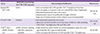
| Assay | Method for measuring the CMV-CMI response | Immunological hallmarks | References |
|---|---|---|---|
| QuantiFERON®-CMV | ELISA | • Does not analyze the CMV-specific CD4+ T lymphocyte function | [44, 48, 54, 55] |
| • Dose not apply to recipients with not-covered HLA class I haplotypes | |||
| • Restricted to particular class I HLA types | |||
| • Measurement cannot be performed at single-cell level | |||
| • High rate of indeterminate results (which cannot be interpreted) | |||
| T-Track® CMV | ELISpot | • Not restricted to particular HLA types | [32, 34, 62, 63] |
| • Measures the functionality of a broad array of effector cells including CD4+/CD8+ T lymphocytes, NK, and NKT cells | |||
| iTAg™ Class I pMHC Tetramers | pMHC tetramer staining with flow cytometry standard | • High sensitivity (since results are strictly dependent upon the coverage of specific HLA types in individuals) | [34, 85, 87] |
| • Does not assess the function of CMV-specific CD8+ T lymphocytes |
Table 2
List of prospective multi-center clinical studies (either ongoing or completed but unreported) using CMV-specific immune monitoring assays in SOT recipients [courtesy: clinicaltrials.gov (July 12, 2017)]
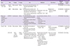
SOT, solid organ transplantation; R, recipient; N/A, not available; CMV, cytomegalovirus; Tx, transplantation; IE, immediate-early protein; pp, phosphoprotein; D, donor; Px, prophylaxis against CMV; CMI, cell-mediated immunity, Cx, complication; ATG, anti-thymocyte globulin; ELISpot, Enzyme-linked immunospot; RCT, randomized controlled trial
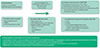
Figure 1
Schematic representation of immune monitoring for cytomegalovirus-specific cell-mediated immune response
aIt needs the immunodominant synthetic peptides. These generally comprise of long synthetic peptides (13–22 amino acids) for CD4+ T lymphocyte stimulation, and short synthetic peptides (8–10 amino acids) for CD8+ T lymphocyte stimulation [97].
w/, with; w/o, without; PBMC, peripheral blood mononuclear cell; BAL, bronchoalveolar lavage; CMV, cytomegalovirus; DC, dendritic cell; IE, immediate-early; pp, phosphoprotein; gB, glycoprotein B; CMV-CMI, cytomegalovirus-specific cell-mediated immune response; IFN-γ, interferon-gamma; TNF-α, tumor necrosis factor alpha; CCL, chemokine (C-C motif) ligand; CXCL, C-X-C motif chemokine; ICS, intracellular staining; ELISA, enzyme-linked immunosorbent assay; ELISpot, enzyme-linked immunosorbent spot assay; pMHC, peptide-major histocompatibility complex; CyTOF, cytometry by time of flight; HLA, human leukocyte antigen
The QuantiFERON®-CMV assay
1. Basic principle and protocol
The basic principle and procedure for performing the QuantiFERON®-CMV assay are identical to the QuantiFERON®-TB Gold In-Tube test, except for the Ags used for in vitro stimulation of the CD8+ T lymphocytes. This assay belongs to a class of diagnostic tests called interferon-gamma release assays (IGRAs) [4546]. The QuantiFERON®-CMV assay was used for the first time by Walker et al. in 2006 [47]. This original assay measured the CMI response by quantitating the IFN-γ levels released after in vitro stimulation of the CMV-specific CD8+ memory T lymphocytes with 21 CMV CD8+ T lymphocyte epitopes (used as Ags) from the human CMV (HCMV) proteins (including pp50, pp65, gB, IE-1, and IE-2) that were specific and restricted for various class I HLA-A and HLA-B alleles. The IFN-γ levels of ≥ 0.2 IU/mL were considered to be positive. Walker et al. confirmed that each HCMV peptide epitope could induce IFN-γ secretion that was sufficiently measurable using ELISA. They demonstrated that 10 HCMV seropositive healthy volunteers displayed high IFN-γ levels measured by QuantiFERON®-CMV and ELISPOT assays using ELISA and all the 21 HCMV Ags [47].
Presently, the commercially available standardized QuantiFERON®-CMV assay kit (Cellestis Ltd, Qiagen Inc., Melbourne, Australia) uses three specific collection tubes containing phytohemagglutinin [PHA (a mitogen), as the positive control], CMV Ags (CMV tube), and only heparin (no Ag, labelled “nil”, used as the negative control). This assay has been approved and is commercially used in the European Union (EU); however, it has not been approved by the Food and Drug Administration (FDA). The CMV Ag tube contains a mixture of the 22 CMV CD8+ T lymphocyte-specific synthetic peptide epitopes, composed of 8 to 13 amino acids and derived from the 6 uniquely immunodominant HCMV proteins (namely pp28, pp50, pp65, gB, IE-1 and IE-2) that are specific and restricted for various class I HLA (A, B, and C) alleles. This CMV Ag tube consists of an epitope peptide (amino acid sequence TRATKMQVI), derived from the HCMV pp65 protein restricted through CwB (A30/B13), in addition to those present in the original assay performed by Walker et al. [4547]. These 22 epitopes cover 20 HLA class I haplotypes, accounting for > 98% of the human population. About 1 mL of the whole peripheral blood drawn from the recipients is directly added into the three collection tubes. After shaking and incubating for 16–24 h or overnight at 37 °C, the IFN-γ levels in supernatants harvested from each tube are measured using ELISA [45].
The interpretation of test is performed after subtracting the IFN-γ level of the nil tube from the IFN-γ levels of the HCMV Ag or PHA tubes. The result is reported as “reactive” or “positive” if the IFN-γ levels for CMV Ags are ≥0.2 IU/mL (irrespective of the level for PHA). The results are reported as “non-reactive” if the IFN-γ levels are <0.2 IU/mL for CMV Ags and ≥0.5 IU/mL for PHA. When IFN-γ levels are <0.2 IU/mL for CMV Ags and <0.5 IU/mL for PHA, the result is reported as “indeterminate”. These cut-off values were defined according to the study by Walker et al. [47].
2. Application of the QuantiFERON®-CMV assay for SOT recipients
Initially, when Walker et al. analyzed the 21 CMV Ags in peripheral blood from 25 SOT recipients at various time intervals post-transplant using the QuantiFERON®-CMV assay, all the CMV-seronegative recipients showed nearly undetectable IFN-γ levels (<0.1 IU/mL) and all the CMV-seropositive recipients showed high IFN-γ levels (19 ± 22.5 IU/mL, positive test). The IFN-γ levels stimulated by the CMV Ags correlated well with those stimulated by PHA [47].
Several prospective observational studies using the commercially available QuantiFERON®-CMV assay have demonstrated the potential of this assay for measuring the CD8+ T lymphocyte responses, in order to predict clinically relevant events related to CMV lytic replication [4849]. These events may include either the risk of initial CMV reactivation (for example, after discontinuation of primary prophylaxis) or recurrence of CMV viremia after the initial treatment.
1) Pre-transplant risk-stratification in CMV-seropositive recipients
The baseline assessment of risk of CMV infection conventionally relies on the pre-transplant CMV IgG serostatus, under the assumption that CMV-seropositive recipients (R+) have pre-existing CMV-specific immunity. Nevertheless, Cantisan et al. recently observed that about one-third of the R+ transplant candidates actually lacked a proper CMV-CMI response (evaluated using the QuantiFERON®-CMV assay) [50]. Interestingly, the recipients with a non-reactive test result were more likely to develop post-transplant CMV replication than those with a reactive test result before SOT. The authors concluded that this strategy may eventually contribute towards reclassification of the current CMV risk stratification, and the R+ recipients with non-reactive test results pre-transplant should be regarded as high-risk recipients [50].
2) Predicting the occurrence of late-onset CMV disease after discontinuation of primary prophylaxis in high-risk D+/R− recipients
The major disadvantage of universal anti-CMV prophylaxis (for 3–6 months), which is the most effective strategy for D+/R− recipients with highest risk of post-transplant CMV infection and disease, is the development of CMV lytic replication after discontinuation of prophylaxis (called the late-onset CMV disease) [1032]. Although longer prophylactic duration of 6 months can reduce the frequency of late-onset CMV disease, it may also lead to a delay in the spontaneous development of CMV-specific memory and effector T lymphocyte-mediated immune responses [3251].
A few studies for assessing the efficiency of the QuantiFERON®-CMV assay for the prediction of late-onset CMV-disease have been reported. Kumar et al. monitored 108 recipients with high risk for CMV disease using the QuantiFERON®-CMV assay, before transplantation and after 1-month intervals for 3 months after SOT. These were D+/R− or D+/R+ recipients who were previously administrated the T lymphocyte-depleting antibodies (ATG®) or those who underwent lung transplantation. All enrolled recipients had been receiving the anti-CMV prophylaxis for 3 months. After the completion of anti-CMV prophylaxis, the recipients who tested positive for the QuantiFERON®-CMV assay had a significantly lower incidence of late-onset CMV disease (cut-off IFN-γ levels ≥0.2 IU/mL, for positive test: 3.3% [1/30] versus for negative test: 21.8% [17/78], P = 0.044; cut-off IFN-γ levels ≥0.1 IU/mL, for positive test: 5.3% [2/38] versus for negative test: 22.9% [16/70], P = 0.038). However, in a subgroup analysis using 35 (32.4%) D+/R− recipients, the frequency of late-onset CMV disease was not significantly different between the recipients who tested positive and those who tested negative (10% versus 40%, P = 0.12). These results suggested that immune monitoring for CMV-CMI response using the QuantiFERON®-CMV assay may be helpful in predicting the occurrence of late-onset CMV disease in selected SOT recipients at a high risk for CMV replication [44].
These data were validated using a multicenter study with 124 D+/R− recipients, in which the QuantiFERON®-CMV assay was monitored after 1 and 2 months of completing the 3-month anti-CMV prophylaxis. Recipients with a reactive result (IFN-γ level ≥0.1 IU/mL) at any time point showed a significantly lower cumulative incidence of the late-onset CMV disease 12-month post-transplantation compared to those with a non-reactive result (including negative or indeterminate results [6.4%, 2/31 versus 26.8%, 25/93; P = 0.02]). This study revealed that the QuantiFERON®-CMV assay showed good positive and modest negative predictive values of 0.93 (95% confidence interval, CI = 0.78−0.99) and 0.27 (95% CI = 0.18−0.37), respectively, for predicting the development of late-onset CMV disease in D+/R− SOT recipients. A drawback of this study was that the number of indeterminate results (9.7%), which were difficult to interpret, was considerably high [48].
Based on these findings, we can use the QuantiFERON®-CMV assay for predicting the development of late-onset CMV disease after the completion of primary prophylaxis in the D+/R− recipients, every month for a year after transplantation. For positive test results, which indicated good CMV-CMI response, regular monitoring using the QuantiFERON®-CMV assay may be sufficient. However, in case of negative test results at the end of primary prophylaxis or during any follow-up time point, we should consider continuing the primary prophylaxis with regular monitoring [32].
3) Predicting spontaneous clearance of asymptomatic low-level CMV viremia without anti-CMV prevention
It is understood that the CMV-seropositive recipients at low or intermediate risk for developing post-transplant CMV infection or disease can spontaneously suppress several episodes of asymptomatic temporal low-level CMV viremia, which result from the reactivation of latent CMV infection or superinfection in the D+/R+ recipients, even if they did not receive the anti-CMV prevention [52]. Lisboa et al. reported that a high CMV-CMI response during immune monitoring predicted a high frequency of spontaneous clearance of asymptomatic CMV viremia, without preemptive prevention against CMV [49]. They analyzed 37 recipients at intermediate risk for developing CMV infection or disease. These recipients were CMV-seropositive and did not receive the T lymphocyte-depleting antibodies (ATG®) or lung transplants. It was observed that the recipients with a positive QuantiFERON®-CMV test result (IFN-γ level ≥0.2 IU/mL) during the onset of detectable CMV viremia showed significantly higher frequency of spontaneous CMV clearance, compared to those with a negative QuantiFERON®-CMV result (IFN-γ level <0.2 IU/mL) and then progression to anti-CMV treatment (92.3% versus 45.5%, P = 0.004) [49]. This preliminary evidence suggests that the measurement of CMV-CMI response using the QuantiFERON®-CMV assay can be used as biomarker or end-point for predicting the development of CMV lytic replication, similar to the QNAT assay for monitoring the recipients undergoing preemptive treatment. This can be helpful in deciding the initiation or discontinuation of the anti-CMV preemptive treatment, depending on the level of the CMV-CMI response in the recipient at a specific time point [43]. If the recipient shows asymptomatic low-level viremia in the QNAT assay, the QuantiFERON®-CMV assay can be performed at the onset of low-level viremia for 3 weeks at 1-week intervals. The preemptive CMV treatment can be initiated when the QuantiFERON®-CMV results are negative, and can be discontinued when the results are positive. Subsequently, monitoring using the QuantiFERON®-CMV assay should be continued over time. This approach could be applied to another possible option of preemptive treatment [32].
4) A novel biomarker for the prediction of CMV replication in R+ recipients undergoing secondary prophylactic or preemptive treatment: prediction of relapse of CMV disease or viremia
Evaluating the CMV-CMI response can be useful in deciding whether modification of preemptive strategies is necessary or not and can also be helpful in deciding secondary prophylaxis in the R+ recipients with a history of CMV disease treatment, in order to prevent the onset or relapse of CMV replication [32]. To tailor the CMV prevention strategies for individual patients in clinical practice via real-time measurement of the CMV-CMI response, the first interventional study was performed by Kumar et al. They used the QuantiFERON®-CMV assay results to decide upon the early discontinuation of the CMV prevention therapy in SOT recipients. The 27 recipients (44.4% D+/R−, 48.1% D+/R+) who enrolled at the start of anti-CMV treatment (after the first episode of asymptomatic CMV viremia) were included in this study. The anti-CMV treatment was discontinued, if VL was undetectable (<137 IU/mL) at a single time point or at two time points spaced one week apart. They performed the QuantiFERON®-CMV assay after the completion of treatment and obtained the results within 3 days of blood collection. By doing so, they applied this test in real-time practice. According to the results of the QuantiFERON®-CMV assay, the subsequent anti-CMV treatment strategy was divided into two categories: (1) if the test was positive (IFN-γ level ≥0.2 IU/mL), the anti-CMV treatment was discontinued, and (2) if the test was negative, the secondary anti-CMV prophylaxis was initiated for 2 months. Only 1 out of 14 (7.1%) recipients with positive QuantiFERON®-CMV assay results and early discontinuation of the anti-CMV treatment without secondary prophylaxis showed the recurrence of asymptomatic low-level CMV viremia. However, 9 out of 13 (69.2%) recipients who received additional secondary prophylaxis for 2 months showed clinically significant CMV recurrence (IFN-γ level ≥ 500 IU/mL) despite prolonged treatment. The positive QuantiFERON®-CMV assay group displayed significantly lower CMV recurrence rate compared to the negative QuantiFERON®-CMV group (P = 0.001) [53].
ELISpot assay
The ELISpot assay quantifies the number of IFN-γ-producing CMV-specific effector T lymphocytes upon ex vivo stimulation as spot-forming units (SFUs). The number of SFUs in a given number of PBMCs was enumerated using a standard automated imaging scanner. Since different CMV ELISpot assays use different CMV-stimulating proteins, the results showed less clarity and reliability in determining the cut-off value for the number of SFUs that defined adequate CD4+ and CD8+ T lymphocyte response. Thus, clinical applicability of the ELISpot assay in SOT recipients has been limited [54–58]. Abate et al. evaluated the patterns of CMV-specific T lymphocyte immune reconstitution using the ELISpot assay in 117 kidney transplant recipients, before and one year after SOT at regular intervals, and reported that the recipients who did not have CMV viremia had significantly higher SFUs compared to those having viremia (median value = 138 versus 28 SFUs/200,000 PBMCs; P <0.001) [55]. Bestard et al. reported that the observation of low number of SFUs in kidney transplant recipients at the pre-transplant stage after stimulation with the IE-1 peptide pool, predicted the risk of both primary and late-onset CMV infection with good sensitivity (> 80%). The negative predicted values (>90%) and the high area under the curve (AUC > 0.70) in the receiver operating characteristic (ROC) curve, calculated the optimal cut-off value as 7 or 8 SFUs/300,000 PMBCs [59].
An optimized and standardized ELISpot assay using the commercially available Conformité Européenne (CE)-marked T-Track® CMV kit (Lophius Biosciences GmbH, Regensburg, Germany) has been recently developed, which consists of the recombinant urea-formulated (T-activated®) CMV IE-1 and pp65 stimulants [106061]. The principle and protocol of the T-Track® CMV assay are similar to the QuantiFERON®-CMV assay (an IGRA), except for the measurement of the IFN-γ-releasing T lymphocytes in PBMCs using IFN-γ ELISpot and IFN-γ levels in supernatant using ELISA [61]. In contrast to other immunodominant epitopes, unmodified proteins, and peptides used for the in-house ELISpot assays, the urea-formulated proteins are processed and presented via both the exogenous MHC class II and endogenous MHC class I pathways, using cross-presentation by Ag-presenting cells (APC), which mimics the naturally acquired CMV infection [62]. Theoretically, they can activate a broader range of Ag-reactive effector cells, including the CD4+/CD8+ T lymphocytes as well as the bystander NK and NK T cells (NKT) via HLA-independent stimulation [6263]. Banas et al. compared three commercially available assay kits, namely QuantiFERON®-CMV, T-Track® CMV, and a kit containing a mixture of six class I iTAg™ MHC Tetramers, for evaluating the CMV-CMI response in 124 hemodialysis patients [60]. They reported that the positive-detection rate in the CMV-seropositive patients was the highest (90%) using the T-Track® CMV assay, compared to that using the QuantiFERON®-CMV (73%) and iTAg™ MHC Tetramers (77%) assays [60]. Two studies using the T-Track® CMV assay in SOT recipients are currently underway (Table 2). The T-SOPT®.CMV assay is also available as a CE-marked commercial kit (Oxford Immunotec Ltd., Abingdon, UK). However, the T-Track® CMV and T-SOPT®.CMV assays are not currently available for use outside the EU. The applicability of T-SOPT®.CMV test in SOT recipients has not been evaluated yet.
Measurement of cell surface proteins and intracellular inflammatory cytokines using multi-color flow cytometry or mass cytometry
1. Flow cytometry (or flow cytometry standard)
Flow cytometry standard (FCS) is a single-cell-based platform, which allows the separation of subsets of unique immunologic cell populations using fluorescently labeled proteins. Multi-color (polychromatic) multi-channel flow cytometric analysis is a widely used standard method for immune monitoring. Analysis of the phenotypes (called immunophenotyping) and function of specific immune cells is performed using gating strategies for FCS data [64]. Immunophenotyping is typically performed by measuring cluster of differentiation (CD) markers, which are cell surface proteins. Enumeration of CD4+/CD8+ T lymphocytes in HIV-infected individuals using FCS is the most popular immunophenotyping test used routinely in clinical practice in the field of infectious diseases. Functional analysis of immune cells can be conducted using intracellular cytokine staining (ICS) after permeabilizing the cell membrane. The fluorescently labelled proteins in FCS are separated into monoclonal Abs and soluble peptide-major histocompatibility complex (pMHC) multimers (tetra, penta, or dextramers) [65]. A strong interaction between the αβ T cell Ag receptor (TCR), and the fluorescence [phycoerythrin (PE) or allophycocyanin (APC)]- and streptavidin-labeled biotinylated pMHC (class I or II) tetramers in conjunction with Ag epitopes enables immunophenotyping via costaining of the Ag-specific receptor-carrying T lymphocytes [6566].
2. Intracellular cytokine staining (ICS)
Using FCS, ICS detects diverse inflammatory cytokines, such as IFN-γ, tumor necrosis factor alpha (TNF-α), interleukin-2 (IL-2), and IL-6, present in whole blood or PBMCs and produced by the CMV-specific effector memory Th1 cells. For this, the ex vivo CMV-specific stimulation was performed for 48 h using various stimulants, including the CMV-infected dendritic cells, CMV-infected cell lysates, or several CMV-specific immunodominant epitopic peptides (such as pp65, pp72, and IE-1) [326768697071727374757677]. In several studies, the CMV-specific CMI assay using ICS has been demonstrated to be useful in predicting the risk of CMV viremia or disease after kidney, lung, heart, liver, and small bowel transplantation [3244566768697071727375767778798081]. These results also demonstrate the importance of polyfunctionality in the CMV-specific CD4+ and CD8+ T lymphocytes, in order to control CMV replication after SOT [566869707577818283].
Few clinical studies indicating the cut-off value for a homogeneous subpopulation of CMV-specific T lymphocytes have been performed in a large number of SOT recipients. Gerna et al. investigated the CMV-CMI response in 134 SOT recipients using FCS and ICS. They suggested that the presence of CMV-specific CD4+IFN-γ+ and CD8+IFN-γ+ T lymphocytes (≥0.4 cells/mm3) in peripheral whole blood or ≥0.05% of either cell subset at any time point monthly till 6 months and 9, 12 months after SOT was considered as a protective immune response against CMV disease [70]. Mena-Romo et al. evaluated the CMV-CMI response of the CD8+CD69+IFN-γ+ T lymphocytes in 106 CMV-seropositive (R+) SOT recipients using FCS and ICS. The presence of CD8+CD69+IFN-γ+ cells (≥0.25%) in total PBMCs 4-weeks post-SOT was independently associated with the significant reduction of high-grade CMV viremia (≥2,000 IU/mL; odd ratio, OR = 0.31, 95% CI = 0.02 – 0.74, P = 0.021) and served as an indication of preemptive CMV treatment (OR = 0.22, 95% CI = 0.07 – 0.74, P = 0.014) [80]. Thus, they demonstrated clinical utility of the unique CMV-specific CD8+ T lymphocyte subsets and their meaningful cut-off values using a multivariate model including other clinical risk factors [80]. The CMV-CMI assay using FCS and ICS can be used in clinical practice, subject to corroboration of the above results with additional large-scale clinical studies.
3. pMHC tetramer staining
The extent of pMHC-tetramer staining with high specificity could be diminished due to the recipient HLA restriction of a large panel of tetramers [65]. Clinical studies demonstrating the role of pMHC-tetramer staining on CMV replication in SOT are limited. Sund et al. performed a pilot study for monitoring the levels of tetramer-selected CD8+ T lymphocytes (tetraCD8) at 1-month intervals for a year after SOT. The study was performed with 17 kidney transplant recipients and employed the in-house pMHC (class I) tetramers loaded with pp65 peptides [84]. Immediately after SOT, the tetraCD8 counts were significantly lower compared to the pre-transplant baseline. However, one year after SOT, the tetraCD8 levels were not significantly different compared to the baseline levels. Thus, the tetraCD8 counts could not predict the development of CMV viremia after SOT [84]. The commercially developed iTAgTM [Ni2+-nitrilotriacetic acid (NTA)–His-tag Chelate complexes] class I MHC Tetramers (Beckman Coulter, Krefeld, Germany) allow the staining of epitope-specific CD8+ T lymphocytes [85]. Mees et al. evaluated the CMV-specific CD8+ T lymphocytes in 23 kidney transplant recipients using the CMV-specific iTAgTM class I pMHC Tetramers (Beckman Coulter, Germany) with the CMV-Ag specific pMHC-tetramers restricted by five different HLA-A and HLA-B alleles for 6 months after SOT [86]. The CMV-specific tetramers did not play a significant role in predicting CMV replication after SOT, because of small number of CMV viremia or disease [86]. A study by Brooimans et al. suggested that the standardized single-platform iTAgTM class I CMV-specific pMHC Tetramer assays against the TCRs specific for the three different MHC class I CMV peptides were reproducible and useful for enumerating the CMV-specific T lymphocytes [87]. Other CMV-specific pMHC dextramer reagent tubes have been developed in Denmark (Immudex, Copenhagen, Denmark), which are composed of a “Dextramer” tube with CMV-TCR-specific pMHC-dextramers, a “Negative Control” tube, and a “FMO (Fluorescence Minus One)” tube without dextramers. Despite being inconvenient, the HLA-A and HLA-B alleles of the individuals corresponded to the MHC class I haplotypes within these dextramers [88]. The quantity and function of the CMV-specific T lymphocytes have not evaluated using the Immudex MHC Dextramer® in SOT recipients.
4. Mass cytometry or cytometry by time of flight (CyTOF)
Mass cytometry or CyTOF is recently emerging as a novel innovative technology in the field of immunology, although it is associated with some problems [648990]. Its principle is similar to that of matrix-assisted laser desorption/ionization time-of-flight (MALDI-TOF) mass spectrometry, which is commonly used in microbial species identification and measures the vaporized or nebulized ions using TOF mass spectrometry [649091]. The most promising aspect of CyTOF, which uses monoclonal Abs tagged with rare heavy-metal-isotope probes (instead of a fluorochrome), is its unique ability for high-throughput high-content data collection using over 50 high-dimension parameters [64899092]. On the other hand, the fluorescence-based polychromatic flow cytometer currently do not increase more than 18 color-channels [64]. Using several analytical tools or plotting methods [such as CITRUS (cluster identification, characterization, and regression), SPADE (spanning-tree progression of density-normalized events), PCA (principal component analysis), and t-SNE (t-distributed stochastic neighbor embedding)], we can extract the highly multiplexed imaging results from the CyTOF data and understand the current status of immunophenotypes or specific cellular components (such as proliferation or activation markers, intracellular cytokines, and transcription factors) in various immune cells [649093]. These immune cells include the naïve/activated/effector/memory CD4+/CD8+ T lymphocytes, immature/mature B lymphocytes, plasma cells, NK cells, NKT cells, non-classical/classical monocytes, and myeloid/plasmacytoid dendritic cells [92]. In addition, we could obtain a more comprehensive immunological profile for functional markers and cell surface/intracellular proteins, such as the repertories of NK cell receptors [for example, killer cell immunoglobulin-like receptor (KIR)] [9294].
To date, studies involving the use of CyTOF in the field of transplantation biology and CMV replication are in their preliminary stages [90]. Horowitz et al. examined the association between CMV reactivation, and the regulation of NK cells and CD8+ T lymphocytes during lymphocyte reconstitution after allogeneic hematopoietic cell transplantation using mass cytometry [94]. In 15 pediatric liver transplant recipients, using CyTOF, Lau et al. compared the immunological profiles of the seven recipients with operational tolerance defined as stable graft function in the absence of IS, with the immunological profiles of the eight recipients on IS drug [95]. This analysis revealed that the pediatric liver transplant recipients with operational tolerance showed a distinct subset (CD5+CD25+CD38−/loCD45RA−) of the CD4+ T lymphocytes [95]. However, studies involving mass immunological profiling in SOT adult recipients with CMV replication using CyTOF have not been reported.
The problems associated with CyTOF include low sampling efficiency, low acquisition rate, low sensitivity (low staining index), and sorting inability. These problems need to be resolved, since this technique can be used as a promising tool for immune monitoring. Unlike multi-color conventional flow cytometry, high-parameter CyTOF does not suffer from interference (spill over), compensation owing to spectral overlap, or background signals due to autofluorescence [64909296].
Conclusion
The drawbacks associated with the QuantiFERON®-CMV and ELISpot assays hinder their routine application in clinical studies. These drawbacks include: (1) class I HLA restriction in the QuantiFERON®-CMV assay, (2) low sensitivity and poor specificity, (3) high indeterminate rate or unacceptably high rate of uninterpretable results, (4) inability to distinguish between the CMV-CMI response mediated by the CD4+ and CD8+ T lymphocytes, (5) unreliable cut-off levels in various clinical scenario, and (6) measurement of a single cytokine (IFN-γ) [44485455]. However, recently, Kumar et al. suggested that immune monitoring could be used as a promising biomarker for deciding the anti-CMV prevention strategies in SOT recipients [53]. Coupled with the highly standardized and easy-to-use techniques like ELISA, ELISpot, and FCS, these tools for immune monitoring in conjunction with the QNAT assay can be used for administering more efficient therapies for CMV prevention (involving the limited use of antiviral agents). Finally, due to the inhibition of CMV lytic replication, this would result in the improvement of short- and long-term outcomes in SOT recipients.
 XML Download