

 PDF
PDF ePub
ePub Citation
 Print
Print


Introduction
Henoch-Schönlein purpura (HSP) is a systemic vasculitis involving the small blood vessels and has distinct clinical features. The main clinical symptoms are purpura, arthralgia, abdominal pain, and nephritis. Cutaneous involvement is the most common presentation and half of the patients experience diverse gastrointestinal symptoms [1]. The pathogenesis of HSP is still unknown. Several factors such as infections, medications, malignancy, and their combinations were reported as a cause of HSP [2].
Human cytomegalovirus (CMV) is a member of the Herpes virus family and may cause wide variety of diseases depending on a patient’s immune status. CMV can cause local vasculitis by damaging the vascular endothelial cells and may lead to tissue destruction [3]. Moreover, the infection can spread to lungs, gastrointestinal tract and retina.
CMV antibody is commonly detected in 91% of adults in Korea. Once infected, the virus remains in the body and reactivates in immunodeficiency condition. CMV infection has been considered as a causative factor of HSP or can present opportunistic infection in an immunocompromised state. But there have been several reports on CMV infection involving immunocompetent patients [4]. Herein, we report a case of a small cell lung cancer patient affected with CMV in a relatively immune-competent state, and association of CMV with HSP.
Case Report
A 68-year-old male was admitted for multiple, palpable whole body purpura. He complained of concomitant diffused abdominal pain with hematochezia developed 2 days before visiting the hospital. Nine days after the completion of mediastinal radiation, skin rash began to spread from the lower legs and throughout the body and hematochezia occurred 5 day subsequent to skin rash (Fig. 1A and 1B). Patient was diagnosed with limited stage of small cell lung cancer and was initially treated with four cycles of etoposide (120 mg/m2 intravenous, days 1-3) and cisplatin (60 mg/m2 intravenous, day 1) combination chemotherapy followed by mediastinal irradiation on the tumor mass (50 Gy / 25 fx). The patient had achieved a partial response after the completion of treatment based on the findings of chest CT scan and [18F]-FDG PET-CT scan. On admission, he appeared to be ill, and complained of vague pain on entire abdomen; vital sign was stable without fever. On complete blood count, white blood cell (WBC) count was 8,520/mm3 (polymorphonuclear leukocytes 72.7%, lymphocytes 18.0%, and eosinophils 0.7%), the hemoglobin level was 15 g/dl and the platelet count was 179,000 /mm3. On blood chemistry, total protein was 6.9 g/dl and serum albumin was decreased to 3.2 g/dl. Renal function and liver function were normal. Serum cholesterol, lipid profile, and prothrombin time/activated partial thromboplastin time were normal. Direct and indirect coombs’ test was also found to be negative. Urine dipstick test was positive for hematuria (3+) and proteinuria (2+). The 24-hour urine analysis showed urine levels of protein and creatinine as 3,005 g/day and 0.6 g/day respectively. Serum C3 and C4 level was 68 mg/dl and 10.6 mg/dl, respectively (reference range, 76-139 mg/dl, and 12-17 mg/dl, respectively). Rheumatoid factor was 105.5 IU/ml (reference range, 0-20 IU/ml). In addition, the test for anti-neutrophilcytoplasmic Ab (ANCA), anti-myeloperoxidase (Anti-MPO) test, cryoglobulin, and antinuclear antibody (ANA) were negative. Ig G, A, and M were all within the normal range. Serologic test for hepatitis B antigen, parvovirus and HIV virus were all negative. Chest X-ray was unremarkable. Abdominal CT revealed diffuse edematous thickening of the duodenum to proximal jejunum with prominent mucosal enhancement (Fig. 2A).
Figure 1
Skin lesions on the lower extremities: multiple purpura were observed on both legs (A), and abdomen (B).

Figure 2
Abdominal computed tomography revealed diffuse edematous thickening of the duodenum to proximal jejunum with prominent mucosal enhancement (A). Upper gastrointestinal endoscopy showed multiple ulceration of duodenal mucosa (B). Histopathological examination of duodenal ulcers revealed hyperchromatic increased large nuclei of glandular epithelium (C) and cytomegalovirus inclusion bodies (D, arrow).

Skin biopsy revealed leukocytoclastic vasculitis in the dermis and IgA deposition on the endothelial cells and negative CMV immunohistochemical staining. Colonoscopy showed no specific findings. Upper gastrointestinal duodenoscopy showed multiple ulceration of second portion of duodenal mucosa (Fig. 2B). Histopathological examination of the duodenal ulcers revealed hyperchromatic increased large nuclei of glandular epithelial and CMV inclusion bodies (Fig. 2C and 2D). The immunofluorescence staining for IgA showed positive findings. The copy number of CMV Real-time Quantitative polymerase chain reaction (RQ-PCR) and DNA titer was elevated to 1,360 copies/ml. Based on the detection of CMV inclusion body in duodenal ulcer and increased CMV DNA titer, diagnosis of CMV duodenitis was confirmed and administration of intravenous ganciclovir (5 mg/kg every 12 hours, intravenous) was initiated.
The renal biopsy to identify the cause of severe hematuria and proteinuria displayed mesangial cell proliferation of the glomerulus and interstitial fibrosis of tubulointerstitium. The electron microscopy disclosed increased mesangial matrices with a few mesangial deposits and moderate effacement of epithelial cell foot processes. Immunofluorescence staining showed mesangial staining for IgA and C3, which was found to be compatible with HSP. Detection of CMV in kidney biopsies was negative. The ophthalmology inspection revealed no evidences of CMV retinitis. With continuation of ganciclovir, systemic steroid (prednisolone 1 mg/kg/day) and polyclonal intravenous immunoglobulin (100 mg/kg/day, 7 days) were also administered for the treatment of HSP. After 5 days of treatment, relief in the gastrointestinal symptoms and skin rash were observed. After 14 days of ganciclovir treatment, CMV RQ-PCR was not detected. Improvement in multiple duodenal ulcerations was seen on follow up duodenoscopy with negative CMV staining. However, in spite of prolonged steroid treatment for 18 days, massive proteinuria (over 4 g/day) persisted. Therefore, it was decided to add methotrexate (10mg daily) and hydroxychloroquine sulfate (200 mg daily) along with prednisolone (30 mg/day). After 21 days of treatment, patient was discharge with oral steroid treatment, at 30th day of treatment of outpatient clinic, proteinuria decreased to 1.5 g/day and improvement in patient’s condition was observed. However, at 75th day of treatment, patient readmitted with pneumocystis jiroveci pneumonia. In spite of intravenous trimethoprim and sulfamethoxazole (20 mg/kg/day) and steroid treatment (40 mg prednisolone daily), patient died of acute respiratory failure.
Discussion
We report HSP with renal, gastrointestinal manifestation in small cell lung cancer patient, with underlying active CMV infection. Clinical presentation of palpable purpura, diffuse abdominal pain, IgA deposition in skin biopsy specimen, and renal involvement were compatible with diagnostic criteria of HSP. HSP is a systemic vasculitis with IgA-dominant immune deposits affecting small vessels [5]. Many factors such as infection, food, drug, autoimmune connective diseases are reported to function as a trigger factor [6]. Solid tumor is also known as a trigger factor of HSP in about 5% of reported cases [7]. Non-small cell lung cancer is the most common solid malignancy associated with HSP [8]. There are reports on development of HSP after active chemotherapy or radiation, but, pathogenic mechanism is not still clearly defined [9]. One of the possible explanations is that abnormal production of tumor associated antigen and antibody results in immune complex deposition in vessel walls, and this tumor associated antigen decreases clearance of circulating immune complex [1011]. In this case, patient had underlying small cell carcinoma of lung which could be one of the considerations in pathogenesis of HSP. On the basis of the clinical features, it was difficult to distinguish CMV-induced HSP from idiopathic HSP with coincidental CMV infection. As the clinical symptoms developed 9 days after the completion of prolonged mediastinal radiation, small cell lung cancer itself could be the cause to induce immunosuppressive state in patient. But the onset of HSP associated with malignancy is closely associated with the initial diagnosis of malignancy or after first chemotherapy [12]. The patient finished chemotherapy and radiation therapy with partial response at the time of diagnosis of HSP. Considering the disease status of underlying small cell lung cancer, the possibility of malignancy associated HSP was considered to be low. The patient showed symptoms and signs of HSP and CMV duodenitis was diagnosed thereafter. Considering the notation that most of the Asian population is predisposed to latent CMV infection [1314], it is hypothesized that active CMV infection might be a cause of HSP in the present case. Although our patient was in immunocompetent state, there is a possibility that latent CMV might be activated by systemic chemotherapy and mediastinal irradiation, predisposing the development of HSP. It was difficult to distinguish CMV induced HSP from idiopathic HSP of coincidental CMV infection. However, Considering the fact that abdominal pain and hematochezia were improved immediately after antiviral therapy, it is rational that CMV may be a cause of HSP rather than coincidental infection associated with malignancy [15]. The patient demonstrated marked proteinuria. Despite active antiviral and steroid treatment, nephrotic range of proteinuria (4 g/day) persisted. After addition of immunosuppressive agent, protein excretion in urine was gradually reduced. These findings strongly supported the explanation that HSP was not directly caused by CMV infection but CMV disease was a predisposing factor in the pathogenesis of HSP. In general, the frequency of renal involvement is 10-50% in adult HSP patients [16]. HSP nephritis is considered to be the most serious complication. Prognosis in the majority of patients is favorable, while the condition in 5-15% patients may progress to renal failure [17]. The outcome and clinical presentation are more severe in adults [18]. There are many studies regarding the prognostic factors of HSP nephritis. Severe abdominal pain, marked proteinuria have been closely associated with prognosis of HSP [19]. In the present case, old age, and abdominal pain could be considered as unfavorable risk factors. The treatment of HSP nephritis depends on the severity of disease. In cases of accompanying severe nephritis, immunosuppressive therapy is required.
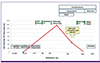
Initially, we did not consider CMV infection, but surveillance study revealed active CMV infection. High CMV titer does not necessarily relates to CMV disease Presence of CMV inclusion body in duodenal biopsy specimen clarified the presence of CMV duodenitis in the patient. In the present case, long period of mediastinal irradiation with concomitant chemotherapy may cause patient’s immunity decrease, leading to reactivation of latent state of CMV. Although CMV duodenitis and HSP were diagnosed independently, authors postulated that CMV duodenitis may be a predisposing factor in the occurrence of HSP. We summarized schematic presentation of the patient’s clinical course (Fig. 3).
Figure 3
Schematic presentation of the patient’s clinical course.
MTX, methotrexate; HCQ, Hydroxychloroquine sulfate; IVGV, intravenous immunoglobulin; CMV, cytomegalovirus; PCR, polymerase chain reaction; CT, computed tomography; F/U, follow-up; CTx, chemotherapy; RTX, radiation therapy.
Although the etiology of HSP is heterogeneous, the possibility of viral infection should be considered as differential diagnosis like in our patient. Different treatment approaches are needed for viral associated vasculitis [20]. Other than systemic steroid and immune suppressant, active antiviral therapy should be considered for the treatment. But in the patients with no organ transplantation, ganciclovir treatment for CMV disease is not still a standard of care. No conclusive statement can be made about the potential benefit of antiviral therapy for severe CMV immunocompetent patient. However, in severe CMV disease, the delayed treatment gives rise to disease progression with ultimate increase in morbidity and mortality, so many studies recommend initiation of antiviral therapy regardless of immune status in severe CMV disease [4]. In our case, because the patient had severe abdominal pain with marked proteinuria, with confirmed CMV duodenitis, we started ganciclovir with consideration of severe CMV disease.
Consequently, although the patient was in a relatively immunocompetent state, CMV infection should be considered [4]. Meticulous screening of CMV as a potential causative factor for HSP can facilitate early diagnosis and initiation of appropriately directed antiviral therapy.
 XML Download