

 PDF
PDF ePub
ePub Citation
 Print
Print


INTRODUCTION
Cryptococcosis is a systemic and local fungal infection caused by Cryptococcus spp. It is generally agreed that most cryptococcal infections are acquired by the inhalation of infectious propagules [1].
Cryptococcosis, which is usually due to Cryptococcus neoformans and Cryptococcus gattii, is considered to be one of the most serious fungal infections in immunocompromised patients [2]. Cryptococcus spp. other than C. neoformans and C. gattii were previously considered to be saprophytes and nonpathogenic to humans; however, opportunistic infections associated with rare Cryptococcus spp., such as Cryptococcus laurentii and Cryptococcus albidus, have increased over the past four decades [1, 3].
Despite changing trends in opportunistic infections, infections caused by non-neoformans and non-gattii Cryptococcus are rare. We recently experienced an immunocompromised patient with refractory acute myeloid leukemia (AML) after allogeneic hematopoietic stem cell transplantation (HSCT) who presented with fungemia and a disseminated cutaneous infection caused by non-neoformans and non-gattii Cryptococcus.
CASE REPORT
A 47-year-old female diagnosed with AML underwent induction and consolidation chemotherapy. Allogeneic HSCT was subsequently performed after achieving complete remission in February 2009. Over the next 3 years, the patient showed no evidence of relapse. Then, the patient developed extramedullary relapse in the breast which was diagnosed by breast tissue biopsy.
Reinduction chemotherapy with a total of 40 Gy of radiation was administered to treat the breast mass. However, the mass increased in size after radiation therapy and a subsequent bone marrow biopsy showed the full-blown hematologic recurrence of AML. Therefore, salvage chemotherapy with fludarabine, cytarabine, and idarubicin was started for progressive AML.
During salvage chemotherapy, the patient took 250 mg of ciprofloxacin twice daily and 100 mg of fluconazole per day for prophylaxis. On day 4 after starting chemotherapy, the patient developed a fever of 38.4°C. It subsided over the course of a day following empirical antibiotic treatment with 4.5 g of piperacillin/tazobactam four times per day.
On day 18, the patient again developed a high fever (38.8°C) and she exhibited multiple erythematous papules on her back, right thigh, and both arms. All other vital signs were stable, with a blood pressure of 120/80 mmHg, pulse rate of 104/min, and a breathing rate of 20/min. At this point, the patient did not have any other symptoms or signs except for an itching sensation, pain, and multiple pinhead- to matchhead-sized erythematous papules and vesicles with erythema on her back, right thigh, and extremities. Laboratory studies revealed a white blood cell count of 30/µL (neutrophils, 0%; lymphocytes, 80%; and monocytes, 20%), absolute neutrophil count (ANC) of 0/mm3, hemoglobin level of 7.5 g/dL, platelet count of 32 ⨯ 10³/mm3, and total bilirubin level of 1.3 mg/dL. Chest and abdominal X-rays did not reveal any specific abnormalities.
Empirical antibiotic therapy was changed to cefepime (2 g, three times per day) and vancomycin (1 g, twice daily) because of the patient’s persistent fever. Blood cultures, fungal cultures, and viral serologic tests were negative at this time.
Though the administered antibiotics had been modified, the patient’s fever and skin lesions worsened. The cutaneous lesions changed to bullae with hemorrhagic patches (Fig. 1A) and edematous plaques (Fig. 1B). Repeated culture studies were performed; then, on day 21 after salvage chemotherapy, one set of blood culture from peripheral vein in four sets of them showed round to oval budding encapsulated yeast cells that were confirmed to be C. laurentii. In repeated blood culture on day 24, two sets of peripheral blood culture in four sets of blood culture examinations showed C. laurentii again. Biochemical identification of the culture was conducted by automated Vitek-II® (bioMérieux, Durham, NC, USA). A skin biopsy was also performed on day 26, and it revealed the presence of fungal hyphae (Fig. 2A and B).
Figure 1
A skin lesion observed on day 18 after salvage chemotherapy. Multiple pinhead- to matchhead-sized papules and vesicles with erythema were observed initially, followed by 1cm-sized bullae with hemorrhagic patches (A) and 2cm-sized erythematous and edematous plaques with central bullous changes (B).
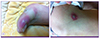
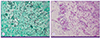
Figure 2
Pathologic findings from a skin biopsy. Fungal hyphae were observed using Grocott’s methenamine silver stain (A, ×400) and Periodic acid-Schiff staining (B, ×400).
The patient was administered conventional amphotericin B (1 mg/kg, daily) beginning on day 27, immediately after the cryptococcosis was documented. The patient’s cutaneous lesions started to improve and her fever subsided after 2 days of treatment. Follow-up blood cultures became negative within 4 days. On day 29 after salvage chemotherapy, the ANC was recovered to 1,500/mm3. Amphotericin B was given for 3 weeks (cumulative dose, 1,284 mg), after which the disseminated cryptococcosis was deemed completely controlled.
DISCUSSION
Cryptococcus spp. are encapsulated, basidiomycetous yeasts that are present in the environment worldwide. Cryptococcus spp. generally occur in soil contaminated with pigeon feces and are often transmitted to humans through inhaled fomites [4, 5]. There have been increasing reports of non-neoformans and non-gattii cryptococcosis over the past four decades [1, 6].
Cryptoccus spp. other than C. neoformans or C. gatii is extremely rare and includes C. laurentii and C. albidus. We identified C. laurentii from presented case by automated Vitek-II® (bioMérieux, USA), and it has high probability to be C. laurentii. Although we tried to perform DNA sequencing with paraffin-embedded skin block, we failed to amplify fungal DNA.
Although non-neoformans and non-gattii cryptococcosis has been reported as occurring worldwide, its natural habitat or clinical characteristics have not been thoroughly established because of a lack of cases [1, 2]. Bloodstream and central nervous system infections are the most common forms of non-neoformans and non-gattii cryptococcosis. In the current case, the patient had a disseminated infection that presented with early skin manifestations. The patient had a refractory hematologic malignancy after allogeneic HSCT and was treated with multi-agent chemotherapy.
Cryptococcus spp. is capable of assuming different morphotypes: yeast, pseudohyphae, and hyphae. The yeast form is the most common cell type observed clinically [7]. The hyphal and pseudohyphal forms are rarely observed in the clinical setting and are considered attenuated in virulence during cryptococcosis in a mammalian host [7, 8]. The regulation of Ace2 and morphogenesis (RAM) pathway and the transcription factor ZNF2 is the master activator of the yeast transformation [7]. Any mutations of RAM pathway in Cryptococcus render cells constitutively in the pseudohyphal form and elevated expression of ZNF2 drives hyphal growth [7, 9].
The existing antifungal options and duration of treatment for cryptococcosis have been established largely for C. neoformans infections. The mainstay of treatment for cryptococcosis is combination therapy with amphotericin B and 5-flucytosine or monotherapy with amphotericin B [1, 10]. According to the case reports published thus far, the initial treatment for a rare Cryptococcus spp. infection is removal of the infection source either through the combined use of amphotericin B and 5-flucytosine or monotherapy with amphotericin B or fluconazole [1, 3]. Monotherapy with fluconazole or itraconazole can be used for patients that have only regional symptoms without a central nervous system or systemic infection [11, 12]. In patients with fungemia, combined treatment with amphotericin B and 5-flucytosine for the first 10–14 days followed by monotherapy with fluconazole or itraconazole for several weeks has been reported [1, 11]. However, there is no validated standard treatment for rare Cryptococcus spp. infections because of the lack of cases. In the presented case, the infection occurred during fluconazole prophylaxis, indicating the possibility of an azole-resistant pathogen. Therefore, we switched to amphotericin B, which showed good efficacy.
Non-neoformans and non-gattii cryptococcosis, especially disseminated infections with a cutaneous manifestation, are rare; However these rare Cryptococcus spp. Infections have increased in recent years. In severely immunocompromised patients with persistent febrile neutropenia, non-neoformans and non-gattii cryptococcosis should be considered, especially when an azole-resistant fungal infection is suspected.
 XML Download