

 PDF
PDF ePub
ePub Citation
 Print
Print


Tuberculosis (TB) is challenging in patients harbouring malignancy as the imaging features of both these entities are not overtly different. Early identification in such patients is imperative as the disease can flare during chemotherapy or radiation. It is well established that whole body 18Fluorodeoxyglucose Positron emission tomography-computed tomography imaging (18F FDG PET-CT) is useful in identifying occult infections apart from its oncological utilities. However visual interpretation alone is not enough. Dual-time-point imaging is an additional technique used during routine 18F-FDG PET/CT imaging to differentiate malignancy from underlying inflammatory or infectious diseases. To avoid false-positive 18F FDG scans in patients with inflammatory or granulomatous disease, the conventional protocol of single time-point (STP) scanning, commonly acquired 60 min after FDG injection may not be enough. Based on the theory that FDG uptake increases over time in mitotically active malignant lesion in contrast to stable or decreasing FDG uptake in benign disorders, dual time-point 18F-FDG PET/CT has an important role [12].
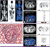
We present a 59 year old immunocompromised male with skeletal lesions proven to be a metastasis from a poorly differentiated adenocarcinoma. A whole body PETCT imaging was requested to look for unknown site of primary malignancy. 296 MBq of FDG was injected intravenously in euglycemic status and an hour later whole body images PET - CT (contrast enhanced CT) from head to mid thigh was acquired. Images revealed a thick walled irregularly marginated cavitatory lesion in left lung upper lobe with surrounding lymphangitic spread (SUV Max 3.3) with mediastinal lymph nodal, right adrenal and skeletal deposits; indicating a primary left lung malignancy with distant metastases. 18F-FDG PET/CT in addition revealed bilateral renal lesions which were FDG avid (Fig. 1A and B) (SUV Max, standard uptake value maximum of 6.2). Dual time-point imaging (Fig. 1C) (performed 2 hours post injection) showed partial clearance of FDG from the renal lesions (SUV max of 4.2) raising a possibility of an infective pathology which is otherwise difficult to differentiate from a primary or secondary deposit. Histological proof was sought, and a co existing extra pulmonary tuberculosis of both kidneys was confirmed (Fig. 1D). Patient was thus treated not only with chemotherapy but anti tuberculosis therapy was also instituted.
Although anatomical imaging modalities such as CT, MRI, and ultrasonography are conventionally used to identify infective and inflammatory processes, 18F-FDG PET/CT imaging provides additional advantages like identifying unsuspected sites of occult infection elsewhere by way of whole body screening (with no additional radiation exposure), high sensitivity, no interpretational difficulties due to metallic hardware implanted in patient, and absence of adverse reactions. FDG being a simple glucose molecule is trapped by malignant and inflammatory cells alike making it a challenging task for interpretation by eye balling alone. Higher the FDG uptake more active is the infection; or more aggressive the malignancy. An additional technique known as Dual-time-point imaging is used to differentiate malignant and inflammatory pathologies which is crucial for diagnosis and optimizing patient management. A semiquantitative index known as standardized uptake value (SUV) is obtained which serves as a measure of glucose uptake. It is seen that inflammatory lesions tend to maintain a stable or reduced SUV over time as the intracellular FDG uptake either remains unaltered or slowly gets washed away; while malignant lesions show a higher retention of FDG in actively dividing cells thus exhibiting a higher SUV at 2 hours delayed imaging. This specialised semiquantitative imaging technique indirectly helps in interpreting whether a lesion is inflammed or malignant in certain confounding situations.
18F-FDG PET/CT and dual time - point maging were crucial in identifying lung primary, metastatic deposits and coexisting extrapulmonary bilateral renal tuberculosis in our patient. Identifying extrapulmonary tuberculosis in a relatively rare site (kidneys) is crucial as management from malignancy differs. This imaging technique can also be used to monitor response to antituberculous therapy. Diagnosis of extrapulmonary TB can be elusive, necessitating a high index of suspicion. To conclude 18F FDG PET/CT is popular in the evaluation and followup of infective etiologies because of its favourable kinetics like relative long half life (110 min), high production yield, transportability to distant centres and most importantly high sensitivity as the degree of FDG uptake is directly related to the severity of infection. And dual time point technique adds an extra edge to enhance the specificity in diagnosing infections.
 XML Download