

 PDF
PDF ePub
ePub Citation
 Print
Print


Abstract
Background
Although erysipelas and cellulitis are common soft tissue infectious diseases, there have been a few studies which investigate clinical characteristics and causative organisms in Korea.
Materials and Methods
We retrospectively reviewed the medical records of patients who had been diagnosed with erysipelas or cellulitis from ten general hospitals between January 2009 and February 2011.
Results
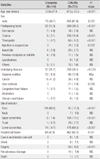
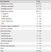
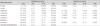
During the study period, a total of 144 patients with erysipelas and 735 with cellulitis were recruited. The mean age of erysipelas patients was 53.6 years, and that of cellulitis patients was 47.5 years. Diabetes mellitus was the most common underlying disease in both groups. The most common site of erysipelas was the face (80.6%) and that of cellulitis was the lower extremity (64.9%). Culture studies have been done in 31.9% (46/144) of patients with erysipelas, and 41.1% (302/735) with cellulites. Causative organisms were identified in 3 patients (2.1%) with erysipelas and 57 (7.8%) with cellulitis. Streptococcus pyogenes was isolated from two patients with erysipelas, and group G streptococcus from one. Staphylococcus aureus (44.0%) was the most common isolate in patients with cellulitis, followed by streptococci (27.1%), Enteobateriaceae (11.9%), and Vibrio species (6.8%). First-generation cephalosporin was the most commonly used antimicrobial agent in both groups.
Figures and Tables
References
1. Swartz MN. Clinical practice. Cellulitis. N Engl J Med. 2004. 350:904–912.
2. Morris AD. Cellulitis and erysipelas. Clin Evid (Online). 2008. pii:1708.
3. Pasternack MS, Swartz MN. Mandell GL, Bennett JE, Dolin R. Cellulitis, necrotizing fascitis, and subcutaneous tissue infections. Mandell, Douglas, and Bennett's Principles and Practice of Infectious Diseases. 2009. 7th ed. Philadelphia: Saunders Elsevier;1289–1312.
4. Baddour LM, Bisno AL. Recurrent cellulitis after saphenous venectomy for coronary bypass surgery. Ann Intern Med. 1982. 97:493–496.

5. Dankert J, Bouma J. Recurrent acute leg cellulitis after hysterectomy with pelvic lymphadenectomy. Br J Obstet Gynaecol. 1987. 94:788–790.
6. Simon MS, Cody RL. Cellulitis after axillary lymph node dissection for carcinoma of the breast. Am J Med. 1992. 93:543–548.
7. Dupuy A, Benchikhi H, Roujeau JC, Bernard P, Vaillant L, Chosidow O, Sassolas B, Guillaume JC, Grob JJ, Bastuji-Garin S. Risk factors for erysipelas of the leg cellulitis casecontrol study. BMJ. 1999. 318:1591–1594.
8. Chartier C, Grosshans E. Erysipelas. Int J Dermatol. 1990. 29:459–467.
9. Chartier C, Grosshans E. Erysipelas: an update. Int J Dermatol. 1996. 35:779–781.
10. Stevens DL, Bisno AL, Chambers HF, Everett ED, Dellinger P, Goldstein EJ, Gorbach SL, Hirschmann JV, Kaplan EL, Montoya JG, Wade JC. Infectious Diseases Society of America. Practice guidelines for the diagnosis and management of skin and soft-tissue infections. Clin Infect Dis. 2005. 41:1373–1406.
11. Perl B, Gottehrer NP, Raveh D, Schlesinger Y, Rudensky B, Yinnon AM. Cost-effectiveness of blood cultures for adult patients with cellulitis. Clin Infect Dis. 1999. 29:1483–1488.
12. Woo PC, Lum PN, Wong SS, Cheng VC, Yuen KY. Cellulitis complicating lymphoedema. Eur J Clin Microbiol Infect Dis. 2000. 19:294–297.
13. Hook EW 3rd, Hooton TM, Horton CA, Coyle MB, Ramsey PG, Turck M. Microbiologic evaluation of cutaneous cellulitis in adults. Arch Intern Med. 1986. 146:295–297.
14. Duvanel T, Auckenthaler R, Rohner P, Harms M, Saurat JH. Quantitative cultures of biopsy specimens from cutaneous cellulitis. Arch Intern Med. 1989. 149:293–296.
15. Kielhofner MA, Brown B, Dall L. Influence of underlying disease process on the utility of cellulitis needle aspirates. Arch Intern Med. 1988. 148:2451–2452.
16. Sachs MK. The optimum use of needle aspiration in the bacteriologic diagnosis of cellulitis in adults. Arch Intern Med. 1990. 150:1907–1912.
17. Bernard P, Bedane C, Mounier M, Denis F, Catanzano G, Bonnetblanc JM. Streptococcal cause of erysipelas and cellulitis in adults. A microbiologic study using a direct immunofluorescence technique. Arch Dermatol. 1989. 125:779–782.
18. Lee JY, Park SD. A study for improvement of the positive culture rate using skin biopsy specimens in patients with cellulitis. Korean J Dermatol. 2007. 45:134–139.
19. Hahn SH, Yang BK, Kim CH, Ahn TW, Suh SK. Cellulitis in young adults. J Korean Orthop Assoc. 1998. 33:614–619.
20. Grayson ML, McDonald M, Gibson K, Athan E, Munckhof WJ, Paull P, Chambers F. Once-daily intravenous cefazolin plus oral probenecid is equivalent to once-daily intravenous ceftriaxone plus oral placebo for the treatment of moderate-to-severe cellulitis in adults. Clin Infect Dis. 2002. 34:1440–1448.
21. Chan JC. Ampicillin/sulbactam versus cefazolin or cefoxitin in the treatment of skin and skin-structure infections of bacterial etiology. Adv Ther. 1995. 12:139–146.
 XML Download