

 PDF
PDF ePub
ePub Citation
 Print
Print


Abstract
Conventional denture impression techniques have limitations for edentulous patients with severe alveolar bone resorption and can cause problems from excessive border extension. Especially when a patient has movable tissue it is difficult to make accurate impression, thus might interrupt stable seating of complete denture. Fabrication of complete denture using closed mouth technique for edentulous patient with severe ridge resorption is thought to provide better stability and retention. In this case, an 86-year-old patient had both edentulous jaws with epulis fissuratum on maxillary anterior ridge and severe mandibular ridge resorption. Thus, tentative vertical dimension was determined by using Centric tray and individual tray attached with gothic arch tracer was fabricated. Complete denture was fabricated using closed mouth technique and the patient was satisfied with better stability and retention of the complete denture. (J Korean Acad Prosthodont 2018;56:120-5)
REFERENCES
1.Pietrokovski J., Massler M. Alveolar ridge resorption following tooth extraction. J Prosthet Dent. 1967. 17:21–7.

2.Tallgren A. The continuing reduction of the residual alveolar ridges in complete denture wearers: a mixed-longitudinal study covering 25 years. J Prosthet Dent. 1972. 27:120–32.
3.Misch CM., Misch CE. Introral autogenous bone grafts for implant dentistry. Misch CE, editor. Contemporary implant dentistry. 2nd ed.St. Louis, MO: Mosby Inc.;1999. p. 90.
4.Zarb GA., Bolender CL., Eckert SE. Prosthodontic treatment for edentulous patients. St. Louis: Mosby;2004.
5.Nagle RJ., Sears VH. Dental prosthetics; complete dentures. Mosby;1958.
6.Klein IE. Complete denture impression technique. J Prosthet Dent. 1955. 5:739–55.
7.Basker RM., Harrison A., Ralph JP. A survey of patients referred to restorative dentistry clinics. Br Dent J. 1988. 164:105–8.
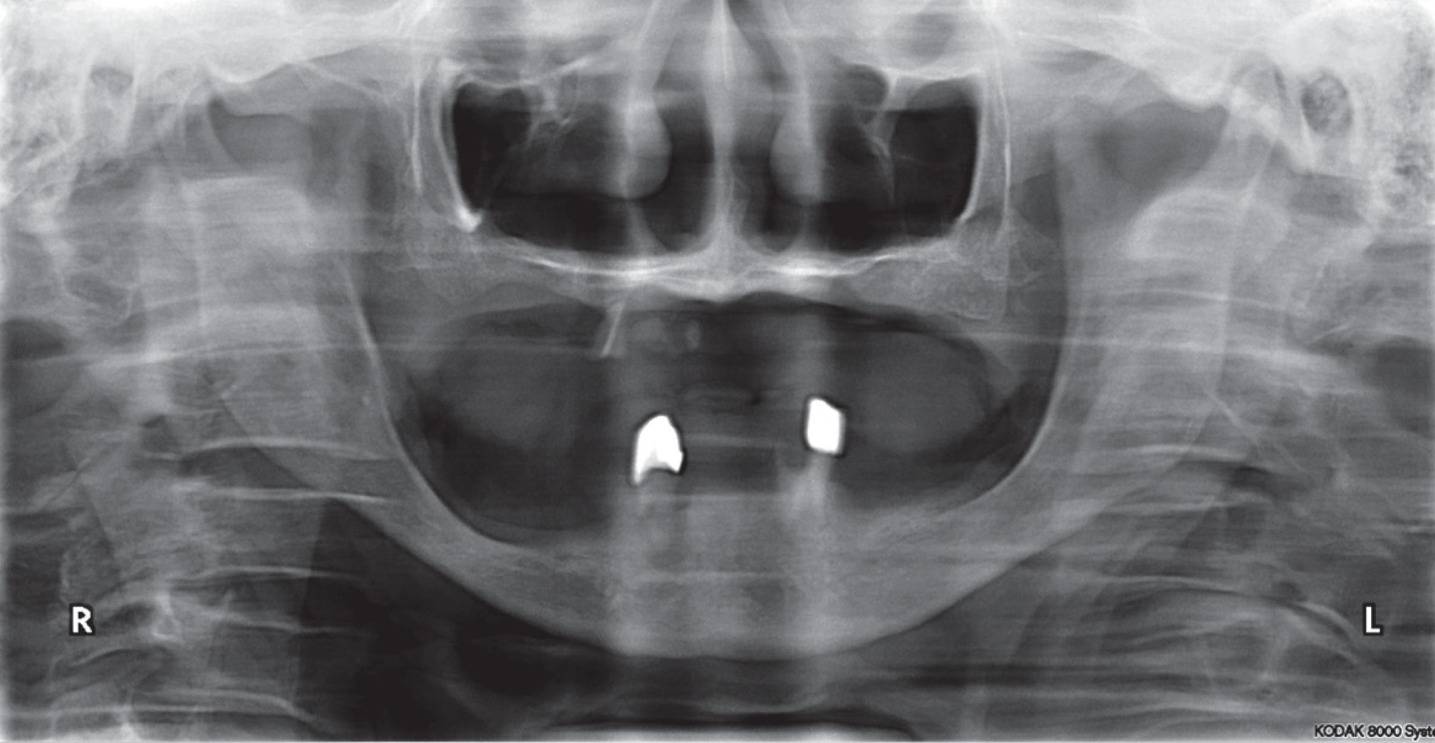
Fig. 2.
Initial intraoral photographs. (A) Frontal view, (B) Maxillary occlusal view, (C) Mandibular occlusal view.

Fig. 3.
Intraoral photographs after extraction, epulis fissuratum removal surgery. (A) Maxillary occlusal view, (B) Mandibular occlusal view.

Fig. 4.
Preliminary vertical dimension taking with Centric tray and mounting in semi-adjustable articulator. (A) Preliminary vertical dimension taking at resting position, (B) Centric tray, (C) Preliminary cast mounting.

Fig. 5.
Fabrication individual tray and Gnathometer M placement. (A) Gnathometer M placement, (B) Bite rim mount application on maxillary individual tray (open tray in anterior region), (C) Bite rim mount application on mandibular individual tray.


 XML Download