

 PDF
PDF ePub
ePub Citation
 Print
Print


Abstract
Cleft lip and palate is congenital deformity in oral and maxillofacial area. Normal soft palate has velopharyngeal closure action by connecting oral cavity and nasal cavity at rest and moving upward at swallowing and specific pronunciation. Cleft palate patients with velopharyngeal insufficiency have difficulty in mastication, swallowing and pronunciation because velopharyngeal closure is incomplete. At this time, a prosthetic device used to cover palate defects is called a palatal obturator. A palatal obturator separates oral cavity and nasal cavity and recovers pronunciation, mastication, swallowing and esthetic function. The purpose of this case study is to report the results because it reaches a satisfactory result in functional and esthetic aspects through functional impression procedures using modeling compound and tissue conditioner for restoration of a cleft palate patient with velopharyngeal insufficiency. (J Korean Acad Prosthodont 2013;51:353-60)
REFERENCES
1.Vojvodic D., Jerolimov V., Celebic A. Prosthetic rehabilitation of a cleft palate patient: a clinical report. J Prosthet Dent. 1996. 76:230–2.

2.Veau V., Borel S. Division palatine; anatomie, chirugie, phone-tique. Paris, Masson et cie;1931.
3.Baik HS., Keem JH., Kim DJ. The prevalence of cleft lip and/or cleft palate in Korean male adult. Korean J Orthod. 2001. 31:63–9.
4.Olinger NA. Cleft palate prosthesis rehabilitation. J Prosthet Dent. 1952. 2:117–35.
5.Brown KE. Peripheral consideration in improving obturator retention. J Prosthet Dent. 1968. 20:176–81.
6.Khan Z. Soft palate obturator prosthesis made with visible light-cured resin. J Prosthet Dent. 1989. 62:671–3.
7.Desjardins RP. Obturator prosthesis design for acquired maxillary defects. J Prosthet Dent. 1978. 39:424–35.
8.Casey DM. Palatopharyngeal anatomy and physiology. J Prosthet Dent. 1983. 49:371–8.
Fig. 1.
Initial photographs. A: Maxillary occlusal view. There is linear defect on anterior alveolus and hard palate, B: Mandibular occlusal view, C: Frontal view. D: There is defect on soft palate and pharyngeal portion, E: Extraoral view, F: Panoramic radiograph.
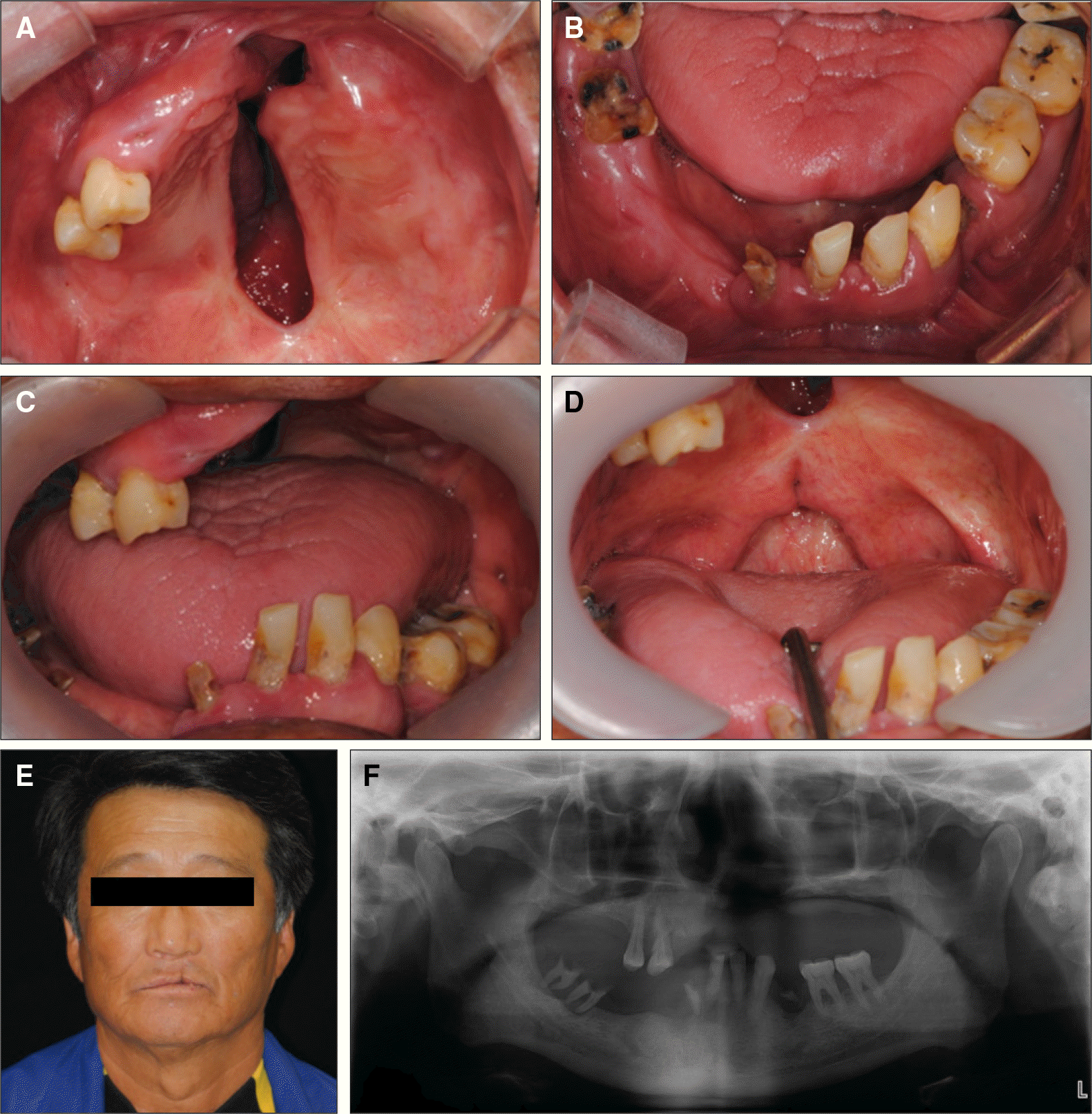
Fig. 2.
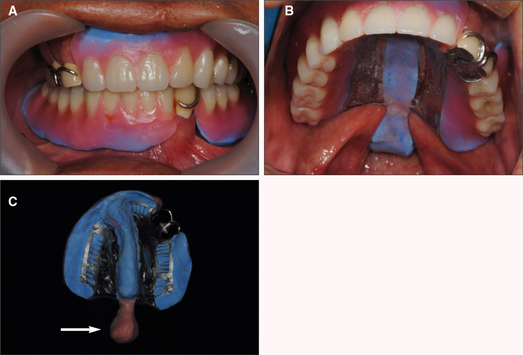
A: Temporary denture, B: Intraoral photograph after the placement of temporary denture and crown.
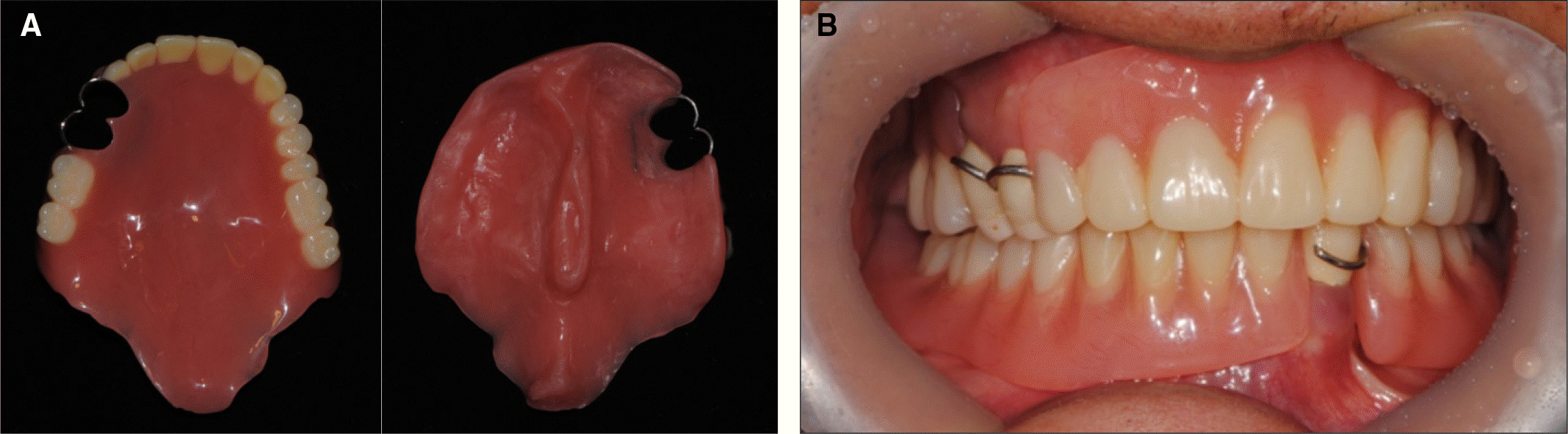
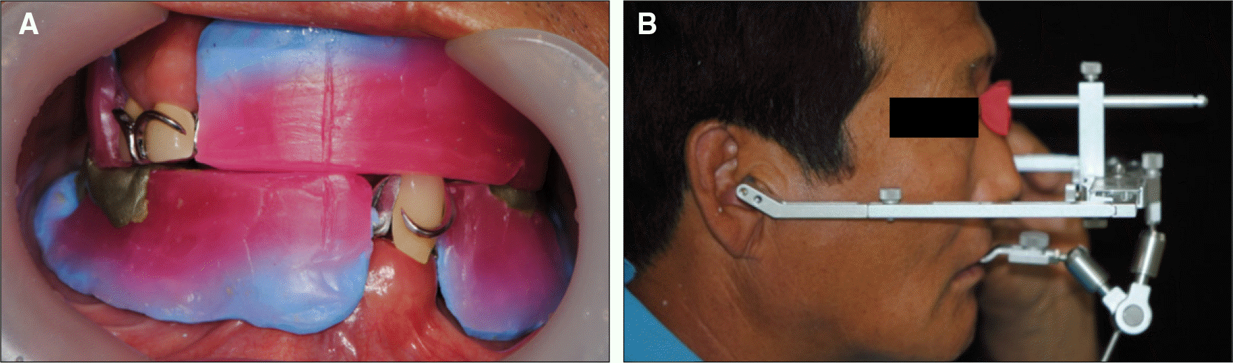
Fig. 3.
Final impression. A: Upper functional impression including velar and pharyngeal portion, B: Lower functional impression.
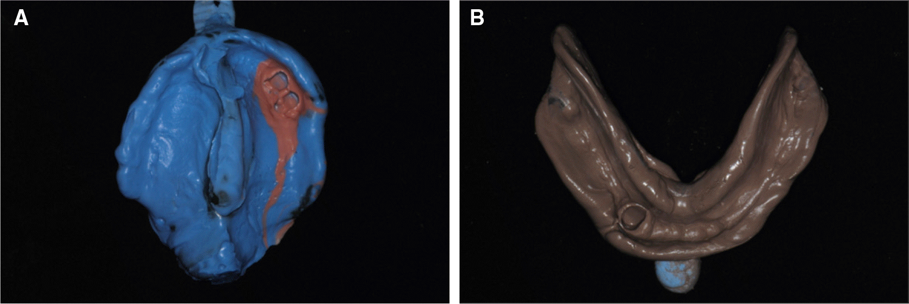
Fig. 4.
Master cast. A: Upper master cast including velar and pharyngeal portion, B: Lower master cast.
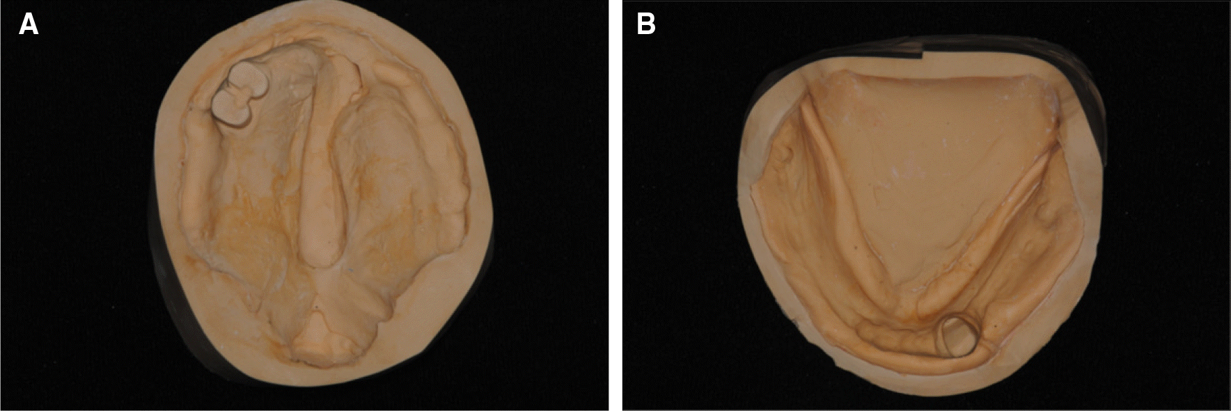
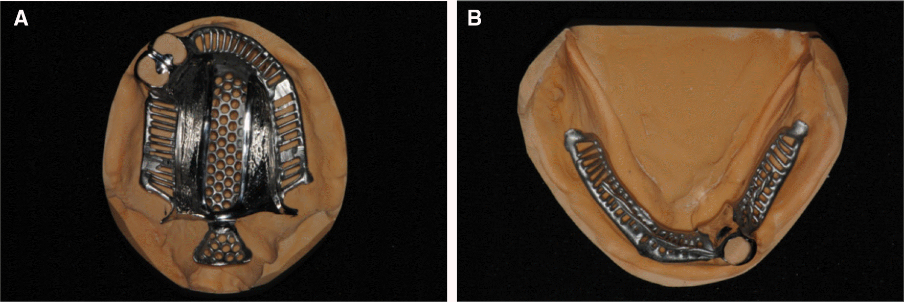
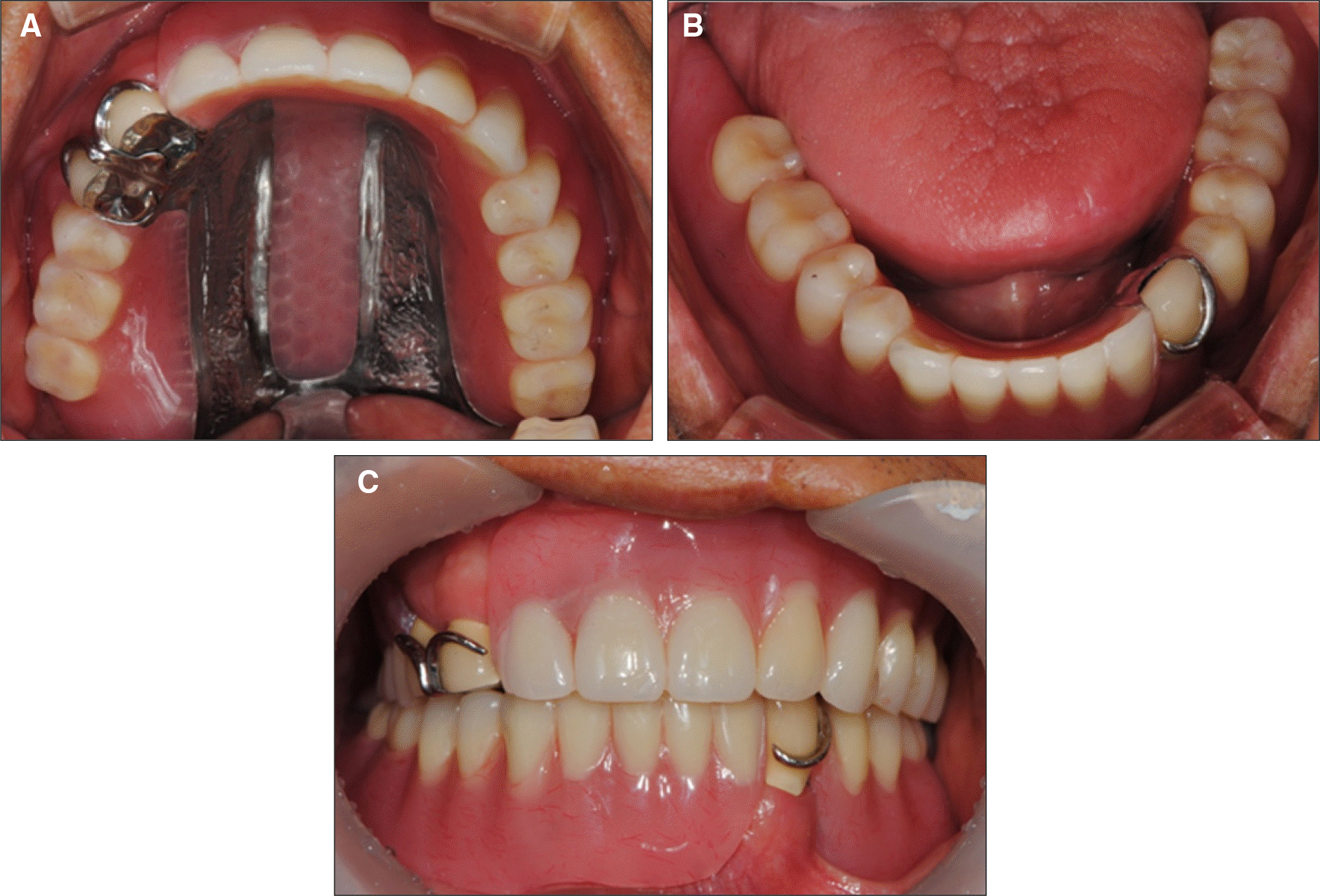
Fig. 5.
Metal framework. A: Upper framework including velar and pharyngeal part, B: Lower framework.
 XML Download