Abstract
Statement of problem
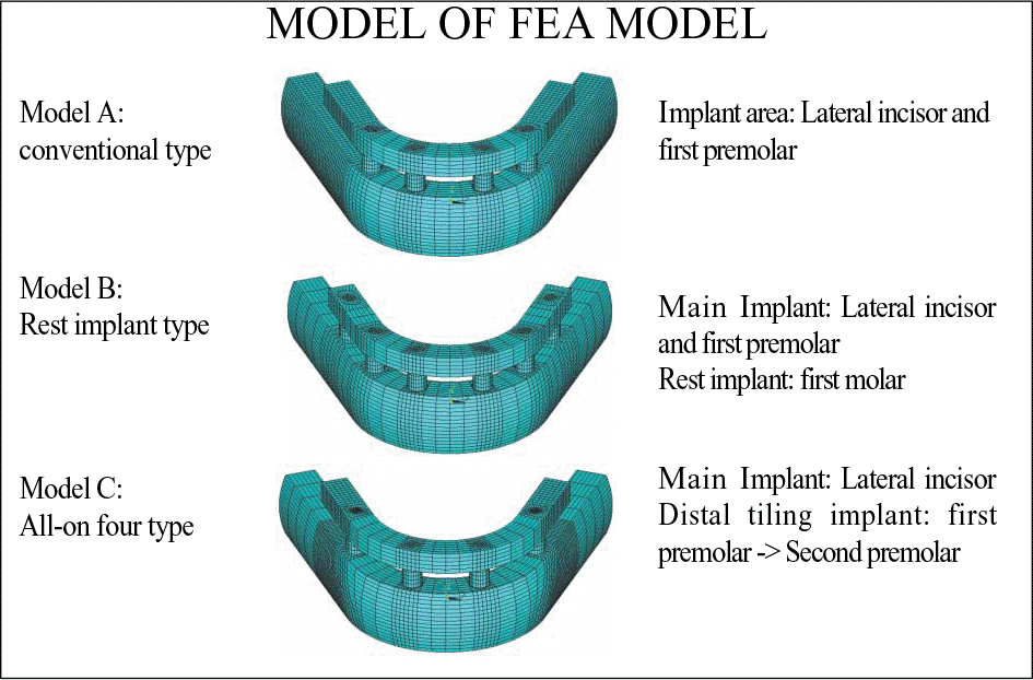
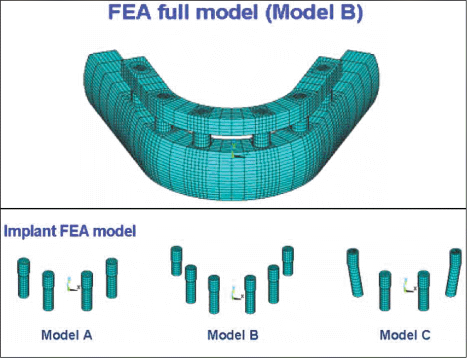
The position and length of cantilever influence on the stress distribution of implants, superstructure and bone. In edentulous mandible, implant-supported cantilever prostheses that based 4 or 6 implants between mental foramens has been attempted. Excessive bite force loaded at cantilever prosthesis causes bone resorption and breakage of superstructure prosthesis around posterior implants. To complement the cantilever length of conventional prosthesis, In 1992, (McCartney) introduced "cantilever - rest - implant" and Malo reported "All-on-Four" in 2003.
Purpose
Analyze and compare the stress distribution of conventional cantilever prostheses with rest implant and Allon-Four TM implant prostheses.
Material and method
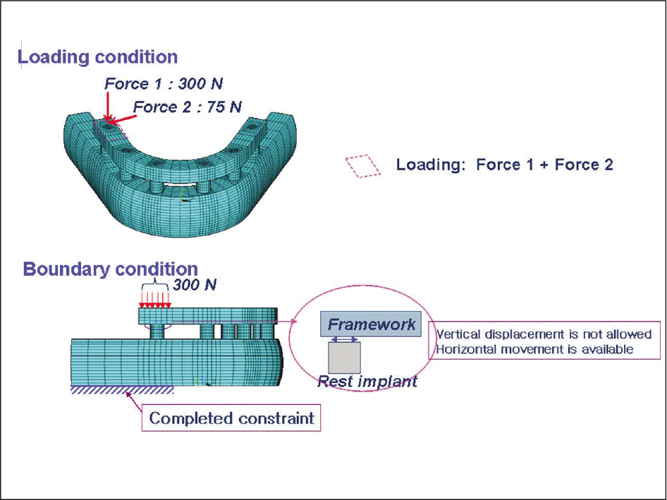
The external loads(300 N vertically, 75 N horizontally) are applied to first molar area. The stress value, stress distribution and aspect of stress dispersion are analyzed by three-dimensional finite element analysis program, ANSYS ver. 10.0.
Results
1. The rest implant and “ All-on-Four” implant system are superior to conventional cantilever prostheses to reduce stress on the bone and the superstructure around implants. 2. The rest implant was of the greatest advantage to stress distribution on bone, implant and superstructure. 3. With same number of implants, distally tilted implants are preferred to conventional cantilever prostheses for reducing the length of cantilever. (J Korean Acad Prosthodont 2009;47:70-81)
REFERENCES
1.Bra ° nemark PI. Osseointegration and its experimental background. J Prosthet Dent. 1983. 50:399–410.
2.Bra ° nemark PI., Adell R., Breine U., Hansson BO., Lindstro ¨ m J., Ohlsson A. Intra-osseous anchorage of dental prostheses. I. Experimental studies. Scand J Plast Reconstr Surg. 1969. 3:81–100.
3.Weinberg LA., Kruger B. Biomechanical considerations when combining tooth-supported and implant-supported prostheses. Oral Surg Oral Med Oral Pathol. 1994. 78:22–7.

4.Holmes DC., Grigsby WR., Goel VK., Keller JC. Comparison of stress transmission in the IMZ implant system with polyoxymethylene or titanium intramobile element: a finite element stress analysis. Int J Oral Maxillofac Implants. 1992. 7:450–8.
5.Rangert B., Jemt T., Jo ¨ rneus L. Forces and moments on Bra ° nemark implants. Int J Oral Maxillofac Implants. 1989. 4:241–7.
6.Richter EJ. Basic biomechanics of dental implants in prosthetic dentistry. J Prosthet Dent. 1989. 61:602–9.

7.Bra ° nemark PI. Osseointegration and its experimental background. J Prosthet Dent. 1983. 50:399–410.
8.Van Rossen IP., Braak LH., de Putter C., de Groot K. Stress-absorbing elements in dental implants. J Prosthet Dent. 1990. 64:198–205.

9.Van Zyl PP., Grundling NL., Jooste CH., Terblanche E. Three-dimensional finite element model of a human mandible incorporating six osseointegrated implants for stress analysis of mandibular cantilever prostheses. Int J Oral Maxillofac Implants. 1995. 10:51–7.
10.Zarb GA., Schmitt A. The longitudinal clinical effectiveness of osseointegrated dental implants: the Toronto study. Part III: Problems and complications encountered. J Prosthet Dent. 1990. 64:185–94.

11.McCartney JW. Cantilever rests: an alternative to the unsupported distal cantilever of osseointegrated implant-supported prostheses for the edentulous mandible. J Prosthet Dent. 1992. 68:817–9.

12.Benzing UR., Gall H., Weber H. Biomechanical aspects of two different implant-prosthetic concepts for edentulous maxillae. Int J Oral Maxillofac Implants. 1995. 10:188–98.
13.Lewinstein I., Banks-Sills L., Eliasi R. Finite element analysis of a new system (IL) for supporting an implant-retained cantilever prosthesis. Int J Oral Maxillofac Implants. 1995. 10:355–66.
14.Malo ´ P., Rangert B., Nobre M. "All-on-Four" immediate-function concept with Bra ° nemark System implants for completely edentulous mandibles: a retrospective clinical study. Clin Implant Dent Relat Res. 2003. 5:2–9.
15.Cho C., Shin SW., Kwon JJ. Three dimensional finite element analysis on the mandibular cantilevered prosthesis supported by implants. J Korean Acad Prosthodont. 2000. 38:724–43.
16.Jang BS., Kim CW., Kim YS. A three dimensional finite element stress analysis of osseointegrated prosthesis according to the location and length of cantilever. J Korean Acad Prosthodont. 1996. 34:501–32.
17.Adell R., Lekholm U., Rockler B., Bra ° nemark PI. A 15-year study of osseointegrated implants in the treatment of the edentulous jaw. Int J Oral Surg. 1981. 10:387–416.

18.De Boever JA., McCall WD Jr., Holden S., Ash MM Jr. FFunctional occlusal forces: an investigation by telemetry. J Prosthet Dent. 1978. 40:326–33.
19.Ericsson I., Lekholm U., Bra ° nemark PI., Lindhe J., Glantz PO., Nyman S. A clinical evaluation of fixed-bridge restorations supported by the combination of teeth and osseointegrated titanium implants. J Clin Periodontol. 1986. 13:307–12.

20.Mathews MF., Breeding LC., Dixon DL., Aquilino SA. The effect of connector design on cement retention in an implant and natural tooth-supported fixed partial denture. J Prosthet Dent. 1991. 65:822–7.

21.Van Steenberghe D. A retrospective multicenter evaluation of the survival rate of osseointegrated fixtures supporting fixed partial prostheses in the treatment of partial edentulism. J Prosthet Dent. 1989. 61:217–23.

22.Borchers L., Reichart P. Three-dimensional stress distribution around a dental implant at different stages of interface development. J Dent Res. 1983. 62:155–9.

23.Clelland NL., Ismail YH., Zaki HS., Pipko D. Three-dimensional finite element stress analysis in and around the Screw-Vent implant. Int J Oral Maxillofac Implants. 1991. 6:391–8.
24.Cook SD., Weinstein AM., Klawitter JJ. A three-dimensional finite element analysis of a porous rooted Co-Cr-Mo alloy dental implant. J Dent Res. 1982. 61:25–9.
25.Davis DM., Zarb GA., Chao YL. Studies on frameworks for osseointegrated prostheses: Part 1. The effect of varying the number of supporting abutments. Int J Oral Maxillofac Implants. 1988. 3:197–201.
26.Skalak R. Biomechanical considerations in osseointegrated prostheses. J Prosthet Dent. 1983. 49:843–8.

27.Kim DW., Kim YS. A study on the osseointegrated proste-sis using three dimensional finite element method. J Korean Acad Prosthodont. 1991. 29:167–213.
28.Lee DO., Chung CH., Cho KZ. A study on the three dimensional finite element analysis of the stresses according to the curvature of arch and placement of implants. J Korean Acad Prosthodont. 1995. 33:98–129.
29.Khatami AH., Smith CR. "All-on-Four" immediate function concept and clinical report of treatment of an edentulous mandible with a fixed complete denture and milled titanium framework. J Prosthodont. 2008. 17:47–51.

30.Francetti L., Agliardi E., Testori T., Romeo D., Taschieri S., Fabbro MD. Immediate rehabilitation of the mandible with fixed full prosthesis supported by axial and tilted implants: interim results of a single cohort prospective study. Clin Implant Dent Relat Res. 2008. 10:255–63.

31.Testori T., Del Fabbro M., Capelli M., Zuffetti F., Francetti L., Weinstein RL. Immediate occlusal loading and tilted implants for the rehabilitation of the atrophic edentulous maxilla: 1-year interim results of a multicenter prospective study. Clin Oral Implants Res. 2008. 19:227–32.

32.Malo ´P., Rangert B., Nobre M. All-on-4 immediate-function concept with Bra ° nemark System implants for completely edentulous maxillae: a 1-year retrospective clinical study. Clin Implant Dent Relat Res. 2005. 7:S88–94.
33.White SN., Caputo AA., Anderkvist T. Effect of cantilever length on stress transfer by implant-supported prostheses. J Prosthet Dent. 1994. 71:493–9.

34.Lim JK. Accuracy estimation and control methods of finite element solutions. Trans KSME. 1994. 34:502–9.
35.Byun SK., Park WH., Lee YS. Three dimensional finite element stress analysis of five different taper design implant systems J Korean Acad Prosthodont. 2006. 44:584–93.
36.Bergman B. Evaluation of the results of treatment with osseointegrated implants by the Swedish National Board of Health and Welfare. J Prosthet Dent. 1983. 50:114–5.

37.Misch CM., Ismail YH. Finite element stress analysis of tooth-to-implant fixed partial denture designs. J Prosthodont. 1993. 2:83–92.

38.Haraldson T., Carlsson GE. Bite force and oral function in patients with osseointegrated oral implants. Scand J Dent Res. 1977. 85:200–8.

39.Siegele D., Soltesz U. Numerical investigations of the influence of implant shape on stress distribution in the jaw bone. Int J Oral Maxillofac Implants. 1989. 4:333–40.
40.Sertgo ¨ z A. Finite element analysis study of the effect of superstructure material on stress distribution in an implant-supported fixed prosthesis. Int J Prosthodont. 1997. 10:19–27.
41.Rieger MR., Adams WK., Kinzel GL. A finite element survey of eleven endosseous implants. J Prosthet Dent. 1990. 63:457–65.

42.Rieger MR., Fareed K., Adams WK., Tanquist RA. Bone stress distribution for three endosseous implants. J Prosthet Dent. 1989. 61:223–8.

43.Clelland NL., Ismail YH., Zaki HS., Pipko D. Three-dimensional finite element stress analysis in and around the Screw-Vent implant. Int J Oral Maxillofac Implants. 1991. 6:391–8.
44.Davis DM., Rimrott R., Zarb GA. Studies on frameworks for osseointegrated prostheses: Part 2. The effect of adding acrylic resin or porcelain to form the occlusal superstructure. Int J Oral Maxillofac Implants. 1988. 3:275–80.
45.Burr DB., Martin RB., Schaffler MB., Radin EL. Bone remodeling in response to in vivo fatigue microdamage. J Biomech. 1985. 18:189–200.

46.Jemt T., Lekholm U., Adell R. Osseointegrated implants in the treatment of partially edentulous patients: a preliminary study on 876 consecutively placed fixtures. Int J Oral Maxillofac Implants. 1989. 4:211–7.
Fig. 5.
Maximum equivalent stress of cortical bone. (Model A: conventional type, Model B: rest implant type, Model C: Allon-Four type)
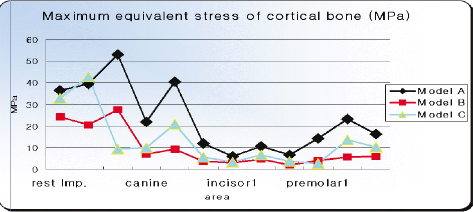
Fig. 6.
Maximum equivalent stress of cortical bone. (Model A: conventional type, Model B: rest implant type, Model C: All-on-Four type)
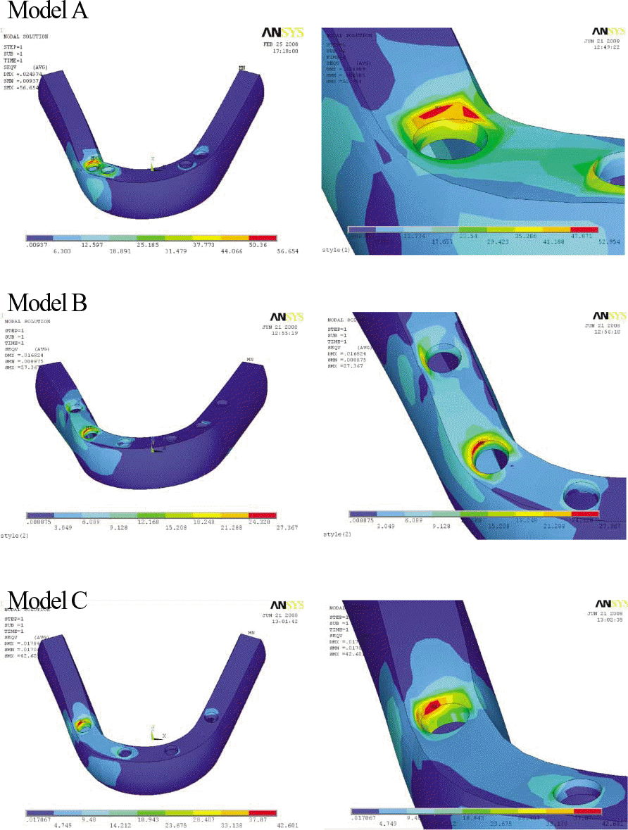
Fig. 7.
Maximum equivalent stress of cancellous bone. (Model A: conventional type, Model B: rest implant type, Model C: Allon-Four type)
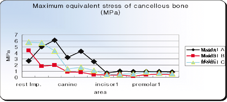
Fig. 8.
Maximum equivalent stress of cancellous bone. (Model A: conventional type, Model B: rest implant type, Model C: All-on-Four type)
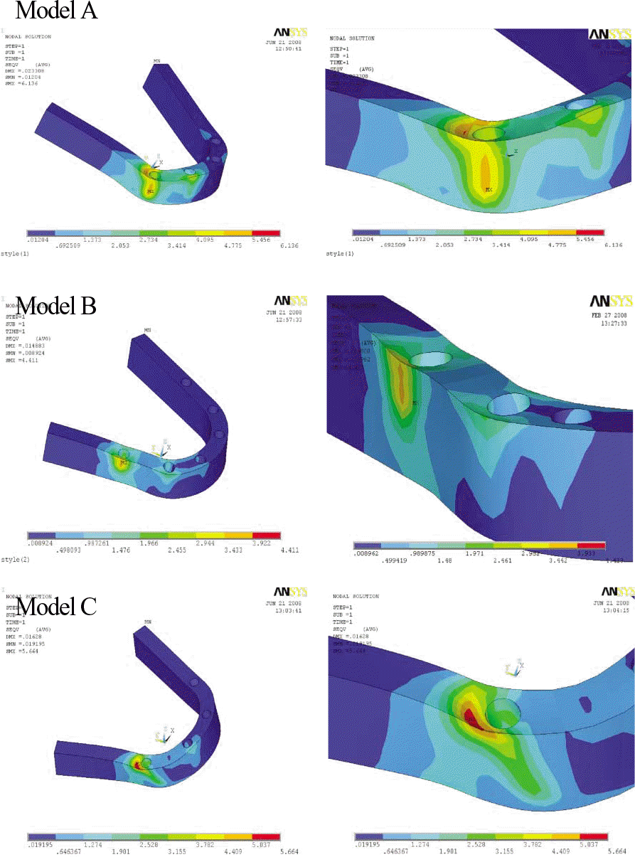
Fig. 9.
Maximum equivalent stress of implants. (Model A: conventional type, Model B: rest implant type, Model C: Allon-Four type)
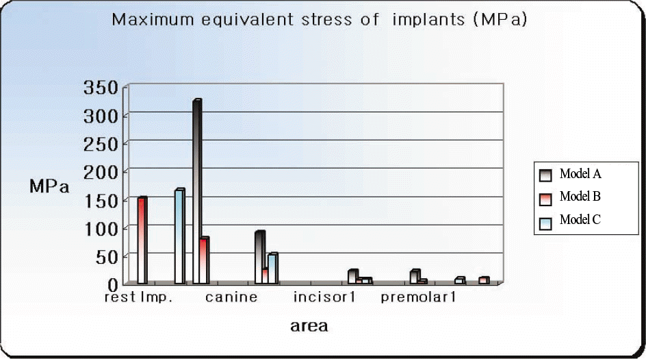
Fig. 10.
Maximum equivalent stress of implants. (Model A: conventional type, Model B: rest implant type, Model C: All-on-Four type)

Fig. 11.
Maximum equivalent stress of prosthesis. (Model A: conventional type, Model B: rest implant type, Model C: Allon-Four type)
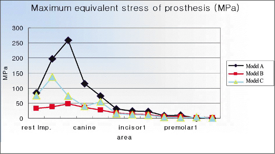
Fig. 12.
Maximum equivalent stress of prosthesis. (Model A: conventional type, Model B: rest implant type, Model C: All-on-Four type)
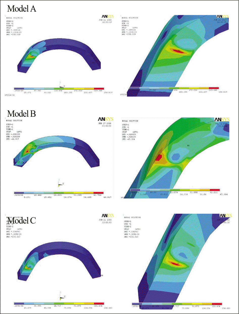
Table I
Number of nodes and elements
| Model | Number of Node | Number of Element |
|---|---|---|
| A | 25675 | 21368 |
| B | 18677 | 15736 |
| C | 22389 | 18933 |
Table II
Mechanical properties of material
| Material | Young's modulus (GPa) | Poisson′ s ratio |
|---|---|---|
| Cortical bone | 20.0 | 0.3 |
| Cancellous bone | 2.0 | 0.2 |
| Titanium | 110.0 | 0.33 |
| Gold alloy | 80.0 | 0.33 |
| Resin composite | 7.0 | 0.2 |
| Resin | 2.7 | 0.35 |
Table III
Maximum equivalent stress of cortical bone (MPa)
Table IV
Maximum equivalent stress of cancellous bone (MPa)
Table V.
Maximum equivalent stress of implants (MPa)
Table VI
Maximum equivalent stress of prosthesis (MPa)


 PDF
PDF ePub
ePub Citation
 Print
Print


 XML Download