

 PDF
PDF ePub
ePub Citation
 Print
Print


Abstract
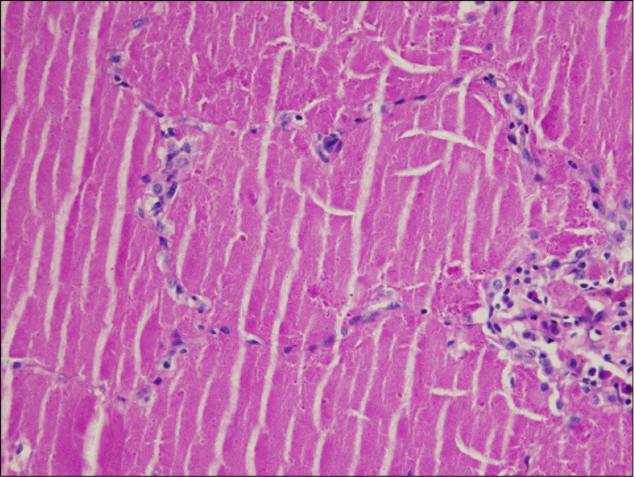
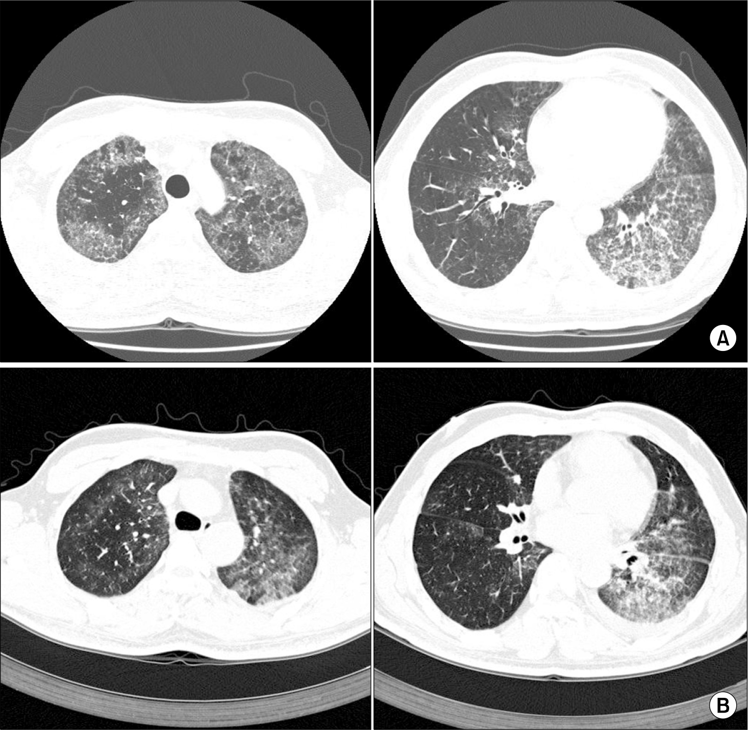
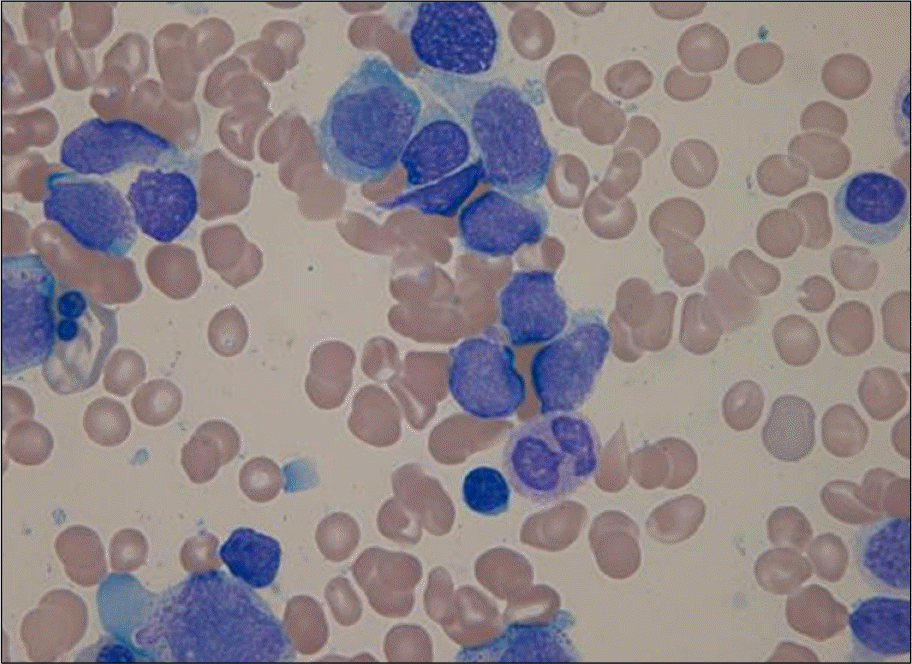
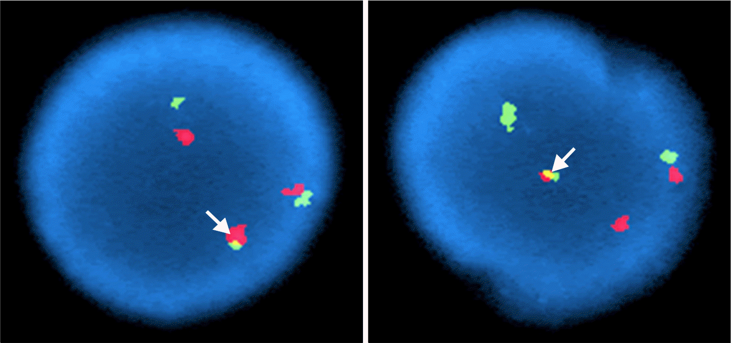
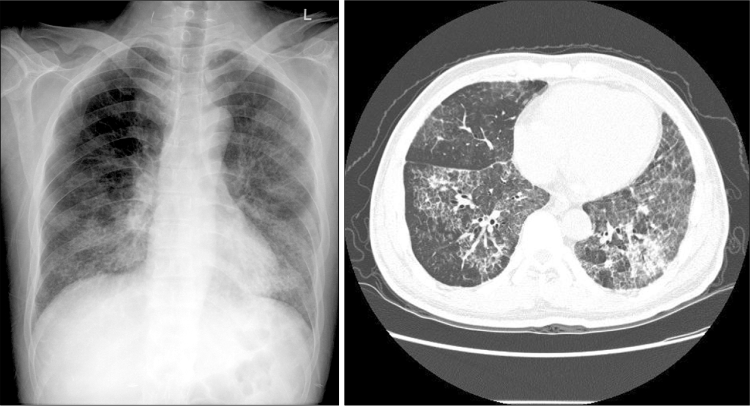
We present the case of a 34-year-old man with acute biphenotype leukemia that co-expressed B-lymphoid and myeloid antigen after the diagnosis of pulmonary alveolar proteinosis (PAP). The diagnosis of PAP was established by Periodic Acid-Schiff reaction staining on the Video Associated Thoracoscope guided lung biopsy and biphenotype leukemia was revealed by immunohistochemical stains of the blasts harvested from the bone marrow biopsy. Supposedly, PAP follows a hematologic malignancy, yet this case shows the reverse sequence.
References
1. Claypool WD, Rogers RM, Matuschak GM. Update on the clinical diagnosis, management, and pathogenesis of pulmonary alveolar proteinosis (phospho-lipidosis). Chest. 1984; 85:550–8.

2. Rosen SH, Castleman B, Liebow AA. Pulmonary alveolar proteinosis. N Engl J Med. 1958; 258:1123–42.
3. Prakash UB, Barham SS, Carpenter HA, Dines DE, Marsh HM. Pulmonary alveolar phospholipiproteinosis, experience with 34 cases and a review. Mayo Clin Proc. 1987; 62:499–518.
4. Trapnell BC, Whitsett JA, Nakata K. Pulmonary alveolar proteinosis. N Engl J Med. 2003; 349:2527–39.
5. Dranoff G, Crawford AD, Sadelain M, et al. Involvement of granulocyte-macrophage colonystimulating factor in pulmonary homeostasis. Science. 1994; 264:713–6.
6. Huffman JA, Hull WM, Dranoff G. Pulmonary epithelial cell expression of GM-CSF corrects the alveolar proteinosis in GM-CSF-deficient mice. J Clin Invest. 1996; 97:649–55.
7. Park MS, Jung SC, Jin MI, et al. A case of pulmonary alveolar proteinosis associated with pulmonary tuberculosis. Tuberc Respir Dis. 2002; 52:411–8.
8. Doki N, Hoshino T, Irisawa H, Sakura T, Miyawaki S. Acute myeloid leukemia complicated with pulmonary alveolar proteinosis at presentation. Rinsho Ketsueki. 2005; 46:522–6.
9. Martin RJ, Rogers RM, Myers NM. Pulmonary alveolar proteinosis: shunt fraction and lactic acid dehydrogenase concentrations as aids to diagnosis. Am Rev Respr Dis. 1978; 117:1059–62.
11. Honda Y, Kuroki Y, Matsuura E, et al. Pulmonary surfactant protein D in sera and bronchoalveolar lavage fluids. Am J Respir Crit care Med. 1995; 152:1860–6.
12. Holbert JM, Costello P, Li W, Hoffman RM, Roqers RM. CT features of pulmonary alveolar proteinosis. AJR AM J Roentgenol. 2001; 176:1287–94.
13. Bonfield TL, Russell D, Burgess S, Malur A, Kavuru MS, Thomassen MJ. Autoantibodies against granulocyte macrophage colonystimulating factor are diagnostic for pulmonary alveolar proteinosis. AM J Respi Cell Mol Biol. 2002; 27:481–6.
14. Cordonnier C, Fleury-Feith J, Escudier E, Atassi K, Bernaudin JF. Secondary proteinosis is reversible cause of respiratory failure in leukemic patients. Am J Respir Crit Care Med. 1994; 149:788–94.
15. Venkateshiah SB, Yan TD, Bonfield TL, et al. An open-label trial of granulocyte macrophage colony stimulating factor therapy for moderate symptomatic pulmonary alveolar proteinosis. Chest. 2006; 130:227–37.
 XML Download