

 PDF
PDF ePub
ePub Citation
 Print
Print


Abstract
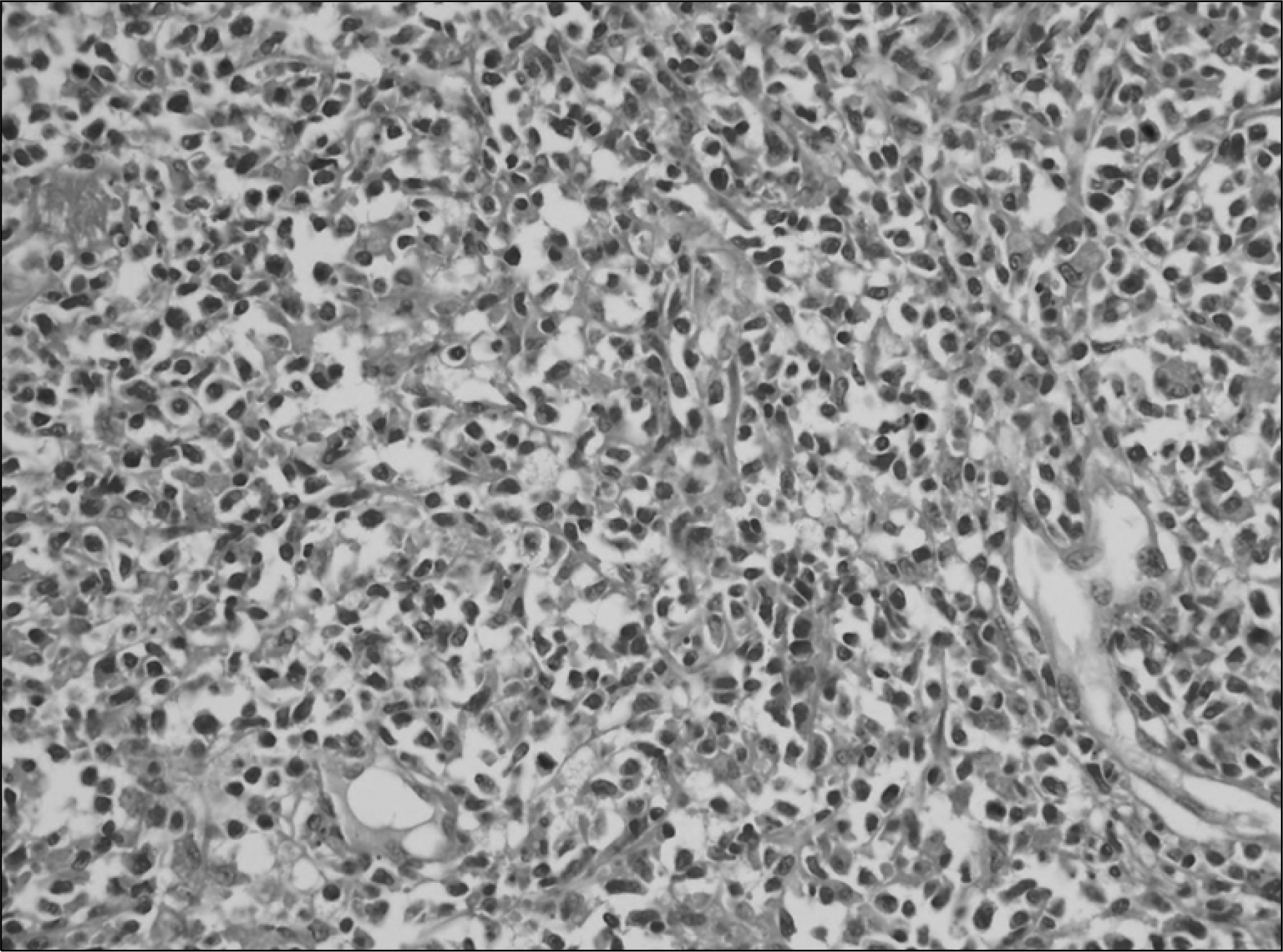
Primary testicular lymphoma account for 1∼5% of all testicular neoplasms. The majority of these tumors are diffuse large B-cell non-Hodgkin lymphoma. Primary NK/T cell lymphoma of the testis is a rare entity. Regardless of treatment, the majority of these patients have exhibited a highly aggressive clinical course and they have died within 1 year because of early dissemination. We report here on a case of the aggressive ‘nasal type’ natural killer (NK)/T cell lymphoma that initially presented as a testicular tumor. A 63-year-old man presented with painless left testicular swelling, and so a left orchiectomy was performed. A biopsy specimen of the testis revealed an extranodal NK/T cell lymphoma of the nasal type. In-situ hybridization for Epstein-Barr virus proved positive. The patient received systemic chemotherapy with 3 courses of a combination regimen.
REFERENCES
1). Ballereau C., Leroy X., Morschhauser F, et al. Testicular natural killer T-cell lymphoma. Int J Urol. 2005. 12:223–4.

2). Ornstein DL., Bifulco CB., Braddock DT., Howe JG. Histopathologic and molecular aspects of CD56+ natural killer/T-cell lymphoma of the testis. Int J Surg Pathol. 2008. 16:291–300.
3). Park BB., Kim JG., Sohn SK, et al. Consideration of aggressive therapeutic strategies for primary testicular lymphoma. Am J Hematol. 2007. 82:840–5.
4). Harris NL., Jaffe ES., Diebold J, et al. The World Health Organization classification of neoplastic diseases of the hematopoietic and lymphoid tissues. Report of the Clinical Advisory Committee meeting, Airlie House, Virginia, November, 1997. Ann Oncol. 1999. 10:1419–32.
5). Siu LL., Chan JK., Kwong YL. Natural killer cell malignancies: clinicopathologic and molecular features. Histol Histopathol. 2002. 17:539–54.
6). Ko YH., Kim CW., Park CS, et al. REAL classification of malignant lymphomas in the Republic of Korea: incidence of recently recognized entities and changes in clinicopathologic features. Hematolymphoreticu-lar study group of the Korean society of pathologists. Revised European-American lymphoma. Cancer. 1998. 83:806–12.
7). Kim CW. Classification of non-hodgkin lymphoma. Korean J Hematol. 1996. 31:3–11.
8). Chiang AK., Chan AC., Srivastava G., Ho FC. Nasal T/natural killer (NK)-cell lymphomas are derived from Epstein-Barr virus-infected cytotoxic lymphocytes of both NK-and T-cell lineage. Int J Cancer. 1997. 73:332–8.
9). Kanavaros P., Lescs MC., Briere J, et al. Nasal T-cell lymphoma: a clinicopathologic entity associated with peculiar phenotype and with Epstein-Barr virus. Blood. 1993. 81:2688–95.
10). Hahn JS., Lee ST., Min YH., Ko YW., Yang WI., Kim GE. Therapeutic outcome of Epstein-Barr virus positive T/NK cell lymphoma in the upper aerodigestive tract. Yonsei Med J. 2002. 43:175–82.
11). Ko YH., Cho EY., Kim JE, et al. NK and NK-like T-cell lymphoma in extranasal sites: a comparative clinicopathological study according to site and EBV status. Histopathology. 2004. 44:480–9.
12). Zeng Q., Cheng F., Gao Q, et al. Primary NK/T cell lymphoma of the testis: A case report and review of the literature. Chinese-German J Clin Oncol. 2007. 6:596–600.
13). Kim YB., Chang SK., Yang WI, et al. Primary NK/T cell lymphoma of the testis. A case report and review of the literature. Acta Haematol. 2003. 109:95–100.
14). Güler G., Altinok G., Uner AH., Sungar A. CD56+ lymphoma presenting as a testicular tumor. Leuk Lymphoma. 1999. 36:207–11.
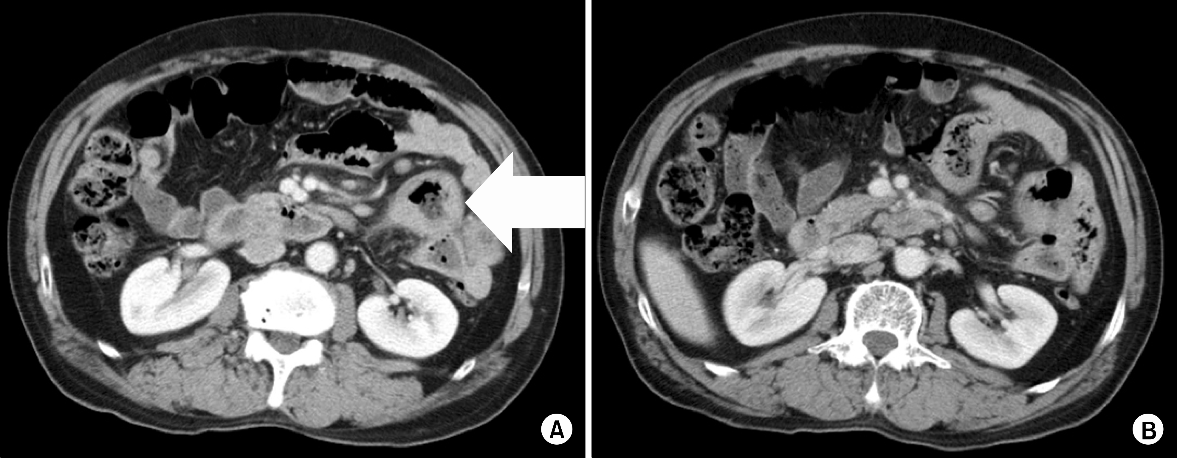
Fig. 1
Postoperative abdominal CT scan finding. (A) The jejunum shows multifocal wall thickening(arrow). (B) Multiple mesenteric lymph nodes are noted.

 XML Download