

 PDF
PDF ePub
ePub Citation
 Print
Print


Abstract
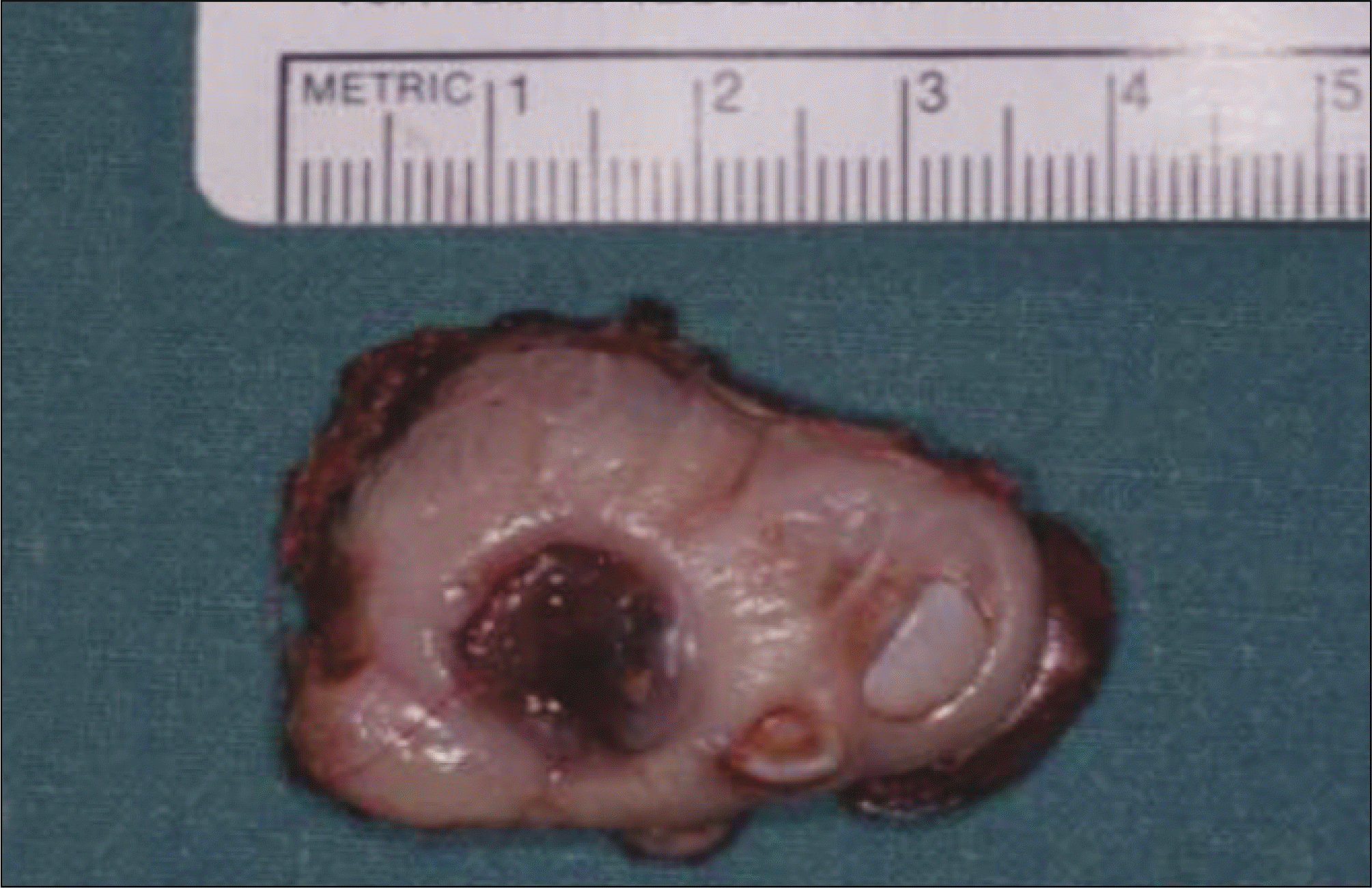
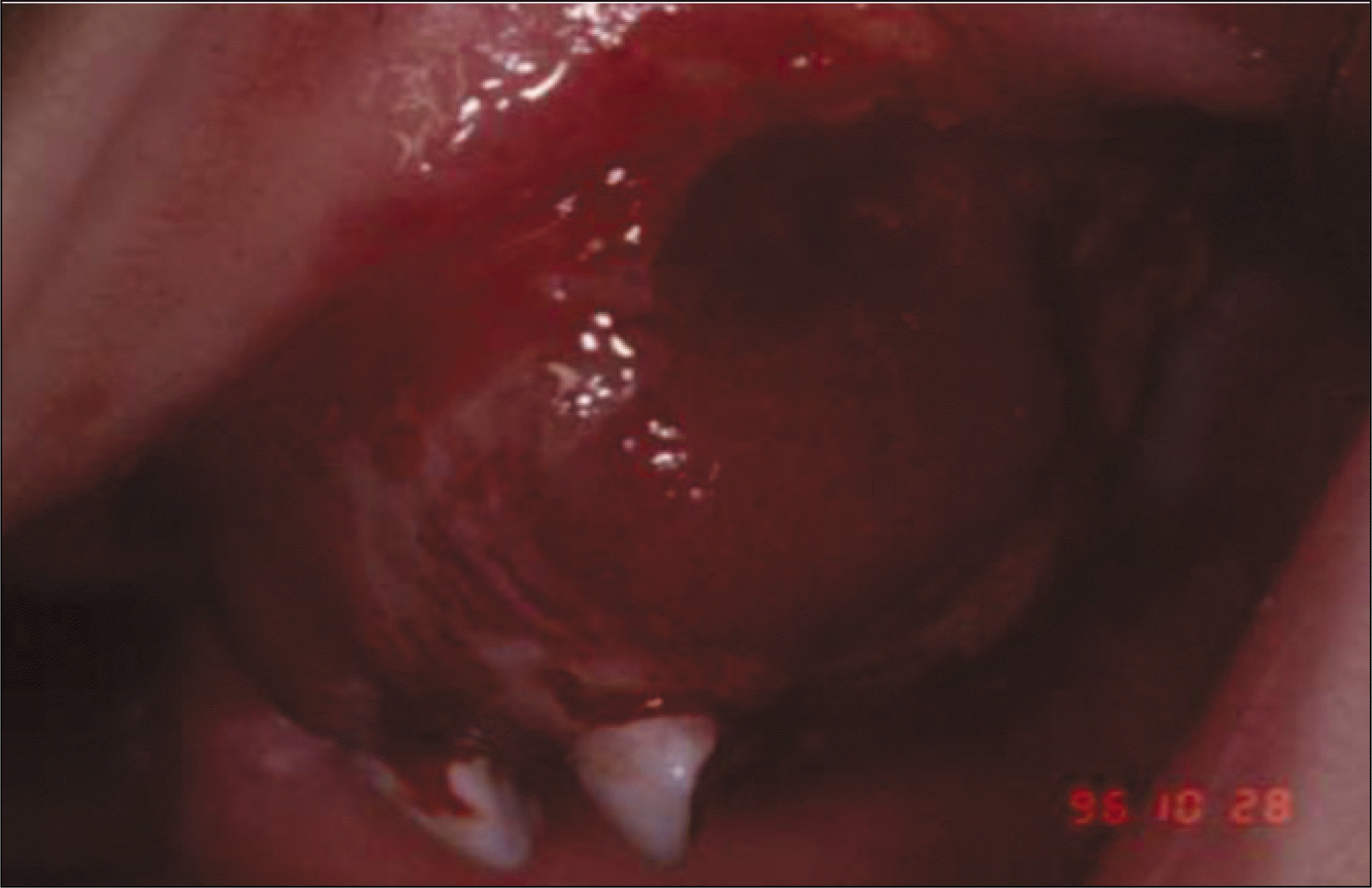
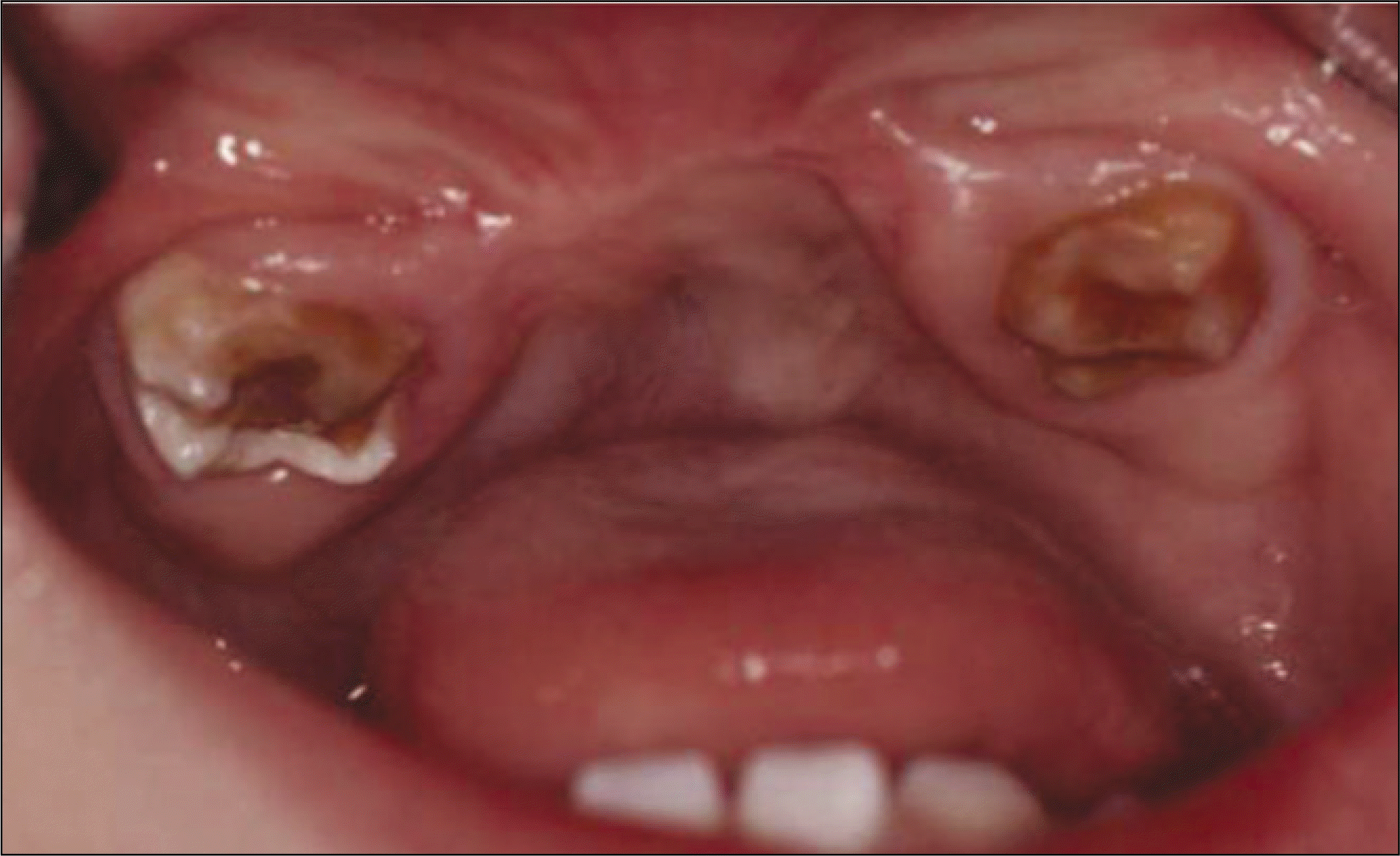
A melanotic neuroectodermal tumor of infancy (MNTI) is a uncommon osteolytic pigmented neoplasm that primarily affects the jaws of newborn infants. Most patients (> 90%) present with the tumor in the first year of life. Approximately 65% form in the maxilla, 11% in the mandible, 5% in the brain and elsewhere. MNTI is normally benign, but up to 15% may recur and a few have metastasized. Approximately 200 cases of MNTI have been reported but only 2 of them presented as multifocal. A case of MNTI in a 7 month old boy was encountered. The chief complaint was maxillary anterior ridge swelling. The incisional biopsy findings were MNTI. Two months after the first operation, mild swelling of another site was observed. The infant was examined periodically since undergoing two procedures with no recurrence. This case demonstrates the possibility of a multicentric MNTI. We report a multicentric MNTI with a review of the relevant literature.
References
1. Johnson RE, Scheithauer BW, Dahlin DC. Melanotic neuroectodermal tumor of infancy. A review of seven cases. Cancer. 1983; 52:661–6.

2. Lopez J Jr. Melanotic neuroectodermal tumor of infancy: review of the literature and report of a case. J Am Dent Assoc. 1976; 93:1159–64.
3. Krompecher E. Zur Histogenese und Morphologie der Adamantinome und sonstiger Kiefergeschwulste. Beitr Pathol Anat Allg Pathol. 1918; 64:165–97.
4. Borello ED, Gorlin RJ. Melanotic neuroectodermal tumor of infancy-a neoplasm of neural crese origin. Report of a case associated with high urinary excretion of vanilmandelic acid. Cancer. 1966; 19:196–206.
5. Koudstaal J, Oldhoff J, Panders AK, Hardonk MJ. Melanotic neuroectodermal tumor of infancy. Cancer. 1968; 22:151–61.
6. Lee CH, Hong SP, Lim CY, Chi JG. Melanotic neuroectodermal tumor of infancy. J Korean Med Sci. 1986; 1:63–7.
7. Navas palacios JJ. Malignant melanotic neuroectodermal tumor: light and electron microscopic study. Cancer. 1980; 46:529–36.
8. Stowens D, Lin TH. Melanotic progonoma of the brain. Hum Pathol. 1974; 5:105–13.
9. Block JC, Waite DE, Dehner LP, Leonard AS, Ogle RG, Gatto DJ. Pigmented neuroectodermal tumor of infancy. An example of rarely expressed malignant behavior. Oral Surg Oral Med Oral Pathol. 1980; 49:279–85.
10. Oruckaptan HH, Soylemezoglu F, Kutluk T, Akalan N. Benign melanotic tumor in infancy: discussion on a rare case and review of the literature. Pediatr Neurosurg. 2000; 32:240–7.
11. Cawson RA, Binnie WH, Barret AW, Wright JM. Oral disease. 3rd ed.London: Mosby;2001.
12. Steinberg B, Shuler C, Wilson S. Melanotic neuroectodermal tumor of infancy: evidency for multicentricity. Oral Surg Oral Med Oral Pathol. 1988; 66:666–9.
13. Jones P, Williams A. A case of multicentric melanotic adamantinomata. Br J Surg. 1960; 48:282–5.
14. Nagase M, Ueda K, Fukushima M, Nakajima T. Recurrent melanotic neuroectodermal tumour of infancy. Case report and survey of 16 cases. J Maxillofac Surg. 1983; 11:131–6.
15. Puchalski R, Shah UK, Carpentieri D, McLaughlin R, Handler SD. Melanotic neuroectodermal tumor of infancy (MNTI) of the hard palate: presentation and management. Int J Pediatr Otorhinolaryngol. 2000; 53:163–8.
16. Hoshina Y, Hamamoto Y, Suzuki I, Nakajima T, Ida-Yonemochi H, Saku T. Melanotic neuroectodermal tumor of infancy in the mandible: report of a case. Oral Surg Oral Med Oral Pathol Oral Radiol Endod. 2000; 89:594–9.
17. Mosby EL, Lowe MW, Cobb CM, Ennis RL. Melanotic neuroectodermal tumor of infancy: review of the literature and report of a case. J Oral Maxillofac Surg. 1992; 50:886–94.
18. Slootweg PJ. Heterologus tissue elements in melanotic neuroectodermal tumor of infancy. J Oral Pathol Med. 1992; 21:90–2.
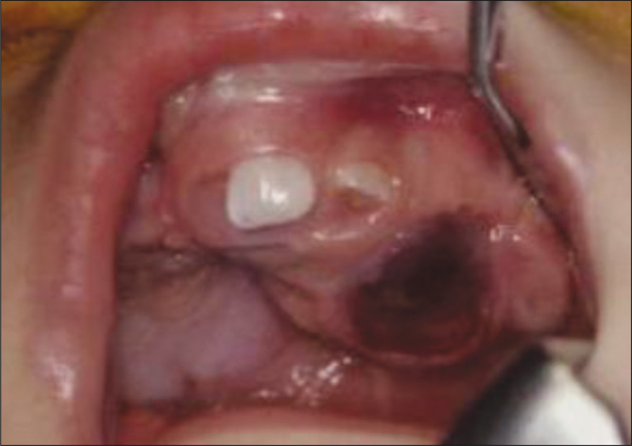
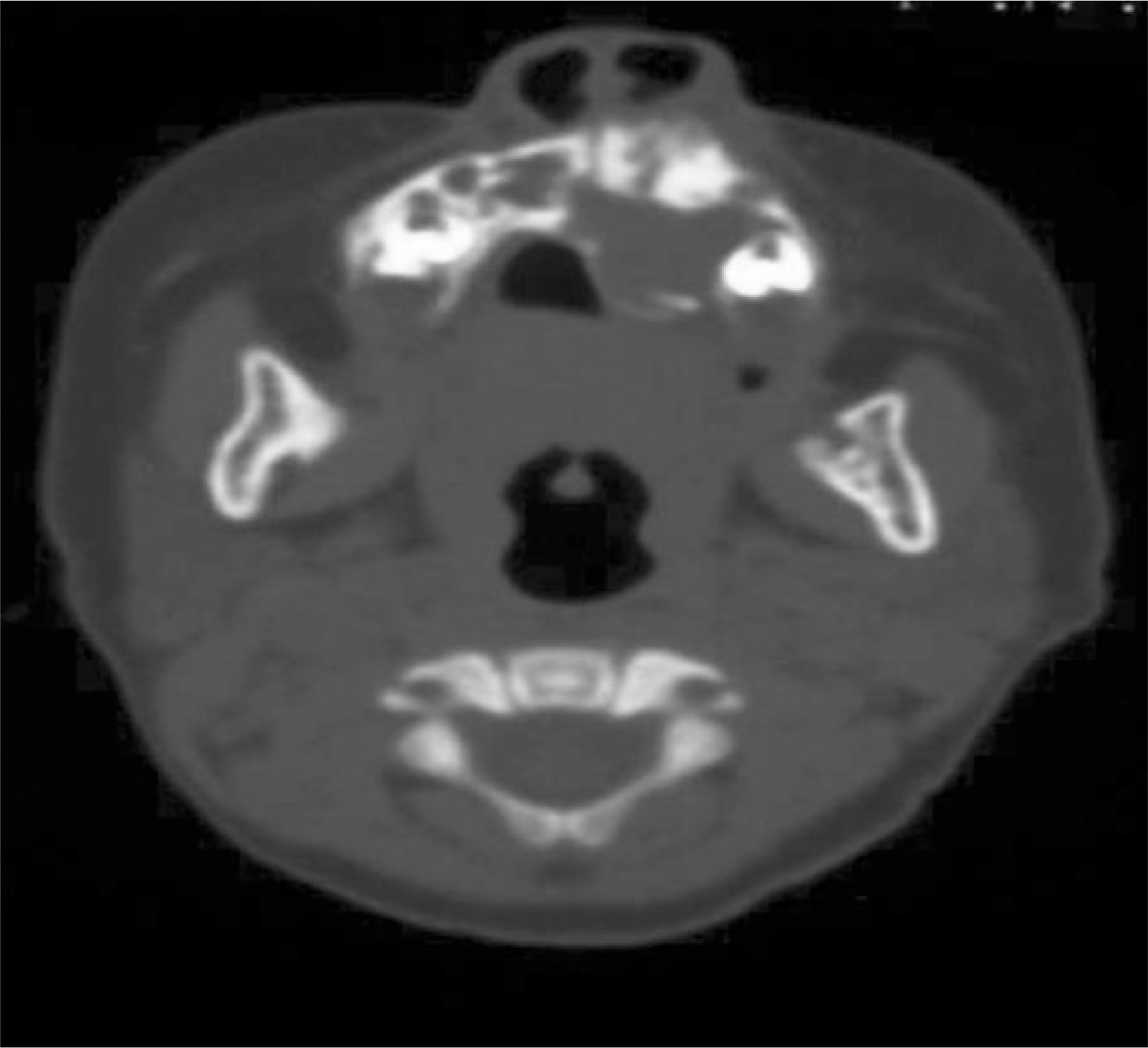
Fig. 2.
Axial computed tomography (CT) of patient showing radiolucent lesion expanding into labial and palatal direction.
 XML Download