

 PDF
PDF ePub
ePub Citation
 Print
Print


INTRODUCTION
Pedicle screw instrumented fusion is used as a safe and effective treatment for adult lumbar spondylolisthesis and other spondylosis.12 However, it is associated with extensive blood loss, a lengthy hospital stay, signifcant cost, and high reoperation rates.34 Standard instrumented fusion requires extensive tissue dissection to expose entry points that provide the lateral-to-medial orientation for optimal screw trajectory. Extensive injury to the back muscles during surgery has been shown to correlate with poor long-term outcomes.56
To overcome these problems, minimally invasive instrumented fusion through small, separate wounds without extensive tissue dissection has been introduced.7 This technique significantly reduces back muscle injury and blood loss, which leads to better trunk muscle performance and faster recovery and rehabilitation.8 However, the potential benefts of minimized tissue disruption, reduced blood loss, and shorter hospital stay must be weighed against the increased rate of neurological complications associated with this technique.9 Moreover, hardware-related complications and pseudarthrosis have been reported in recent studies.1011
Evidence regarding the efficacy of minimally invasive lumbar spinal fusion employing percutaneous pedicle screws exclusively for posterior augmentation has not been synthesized, while plenty of meta-analyses have explored mixed data from studies that utilized conventional pedicle screws for miniopen instrumentation as an alternative to percutaneous pedicle screws. The primary purpose of the current study was to investigate the efficacy of minimally invasive instrumented fusion for adult lumbar spondylolisthesis and other spondylosis. We compared minimally invasive and open pedicle screw instrumented lumbar fusion, especially with respect to 1) pain and functional improvements and fusion rate, 2) complications and subsequent surgeries, and 3) perioperative outcomes (blood loss, hospital stay, operation time, and radiation exposure time).
MATERIALS AND METHODS
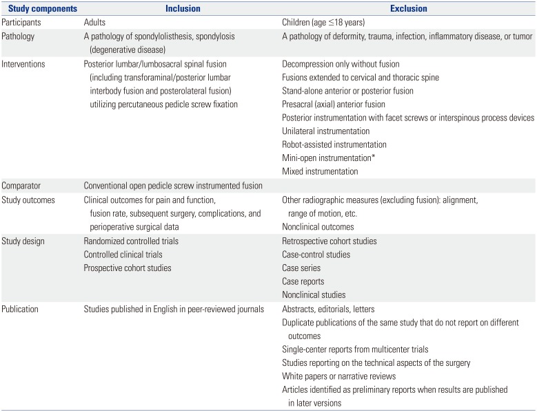
We conducted a thorough and comprehensive review of the literature according to the guidelines for performance and reporting of systematic reviews and meta-analyses outlined in the Meta-analysis of Observational Studies in Epidemiology (MOOSE)12 and Preferred Reporting Items for Systematic Reviews and Meta-analyses (PRISMA).13 This study was exempt from Institutional Review Board review. We searched the literature comparing minimally invasive lumbar spinal fusion with open fusion, including transforaminal lumbar interbody fusion (TLIF), posterior lumbar interbody fusion (PLIF), or posterolateral fusion (PLF), for the treatment of spondylolisthesis and other spondylosis. The literature searches were restricted to randomized controlled trials (RCTs), controlled clinical trials (or quasi-RCTs), and prospective cohort studies published in English. The searches were also limited to studies in which percutaneous pedicle screws were exclusively utilized for posterior spinal fixation in the intervention group. Studies with instrumented conventional pedicle screws instead of percutaneous pedicle screws and a mini-open approach were excluded. We also identified articles with overlapping populations and sought to determine the extent of overlap. In the case of substantial overlap (patients in one article were a subset of those in a larger study), the smaller study was excluded. Our detailed eligibility criteria are listed in Table 1.
For inclusion in the present analysis, studies must have reported the following primary outcomes: postoperative back pain and leg pain improvement measured via a visual analogue scale (VAS); functional improvement measured via Oswestry Disability Index (ODI) score; fusion rate; complications (neurological, hardware-related, and surgical-site complications); subsequent surgeries (revision, removal, reoperation, and supplemental fixation); and perioperative outcomes (blood loss, length of hospital stay, operation time, and radiation exposure time)
Literature search and study selection
Two authors independently performed a comprehensive literature search of PubMed, Embase, and the Cochrane Library database for relevant studies published up to December 2017 using derivatives of the following Medical Subject Headings (MeSH): percutaneous pedicle screw, minimally invasive fusion, minimally invasive arthrodesis, mini-open fusion, miniopen arthrodesis, minimal access fusion, and minimal access arthrodesis. The detailed search strategy is illustrated in Supplementary Table 1 (only online). The reference lists of included articles were also systematically checked to identify additional eligible articles.
One reviewer (SOS) screened titles and abstracts to determine potential inclusion, with a 10% random sample of records independently screened by a second reviewer (SBL). Articles were double blind coded. Inclusion was subsequently confirmed by a team of three reviewers (SOS, SBL, and JWH) who independently checked the full text of all retrieved articles. Uncertainties and disagreements were resolved through team discussion and/or contact with study authors.
Data extraction and analysis
The study reviewers then used a custom data extraction form to extract relevant study data in duplicate. Data elements extracted included methodology data to confirm study eligibility, study design, patient demographics, performed interventions, outcomes of interest, statistical methods, and study results. One reviewer (SOS) then entered extracted data into a spreadsheet (Microsoft Excel 2013, Microsoft Corp., Redmond, WA, USA) with the accuracy of data entry confirmed by the second reviewer (SBL).
We pooled data from each included study and performed meta-analyses (both fixed-effect and random-effects methods) using Comprehensive Meta-Analysis software package Version 2 (Biostat, Englewood, NJ, USA) and STATA Version 14.0 (Stata Corp., College Station, TX, USA). The odds ratio (OR) for the intervention group and the accompanying 95% confidence interval (CI) were calculated for dichotomous outcomes, and the weighted mean difference (WMD) and 95% CI were calculated for continuous outcomes. We reported outcome measures according to the length of follow up: short (<1 year), intermediate (1 to 5 years), and long-term (≥5 years). Pain and functional improvements were analyzed using data from baseline to last follow-up. Fusion rate, complications, and subsequent surgeries were analyzed using data from the last follow-up visit.
The overall quality of evidence for each outcome was categorized as high, moderate, low, or very low according to the Grading of Recommendations Assessment, Development and Evaluation (GRADE) protocol.1213 Five specific domains were used for grading study quality: risk of bias, inconsistency, indirectness, imprecision, and publication bias. We downgraded the evidence by 1 point when fewer than three domains were judged “serious or unclear” or when the study design was not an RCT. We downgraded the evidence by 2 points when four or more domains were judged “serious or unclear.”
Risk of bias
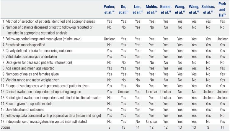
Two independent authors assessed the risk of bias and other major methodological flaws in the included studies using the checklist for RCTs1213 or the checklist for cohort studies by Cowley.14 We defned high-quality studies as those that fulfilled ≥6 of the 12 criteria for RCTs or ≥9 of the 17 criteria of Cowley. We downgraded the quality of evidence by 1 point when risk of bias was serious or when major methodological flaws were noted. Disagreements were resolved by discussion.
Indirectness
We assessed whether the question being addressed in this meta-analysis varied from the available evidence with regard to population, intervention, comparators, or outcomes.
RESULTS
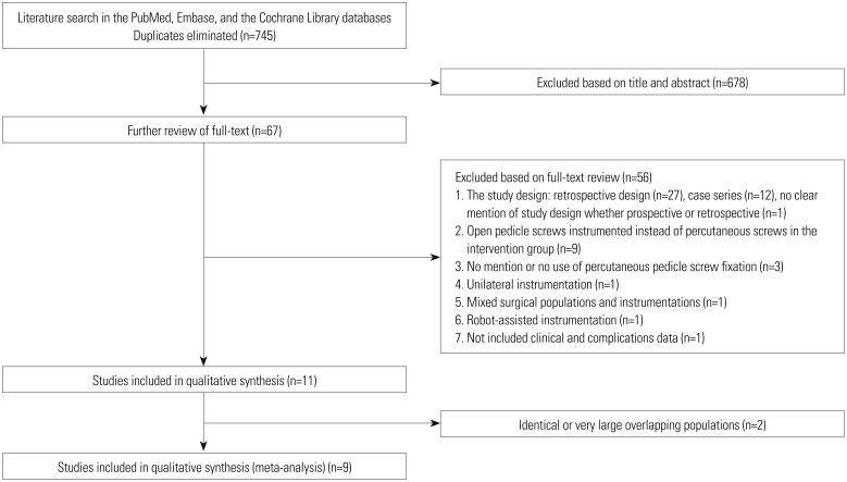
We identifed 745 potentially relevant citations from the electronic database and reference searches after duplicates were eliminated. Sixty-seven studies were selected for full-text assessment after the initial title and abstract screening. Forty articles were excluded because of the study design (27 retrospective, 12 case series, and 1 unclear), and nine (including 1 RCT18) were excluded because open pedicle screws were used in the intervention group instead of percutaneous screws.
A total of 11 studies1920212223242526272829 met the inclusion criteria and two2028 were removed because of overlapping populations: 1) a study population published in 2009 by Peng, et al.28 was determined to be a subset of those in another study published in 2012 by Lee, et al.22 The prior study by Peng, et al.28 overlapped their case-enroll period with the later larger one by Lee, et al.22 Therefore, the study by Peng, et al.28 was excluded. 2) A subset of patients in the study published in 2014 by Wang, et al.20 were judged to be overlapped with those of two other studies published in 201026 and 201125 by the same authors. The other two studies2526 did not have overlapping populations with each other. Consequently, the study published in 2014 by Wang, et al.20 was excluded.
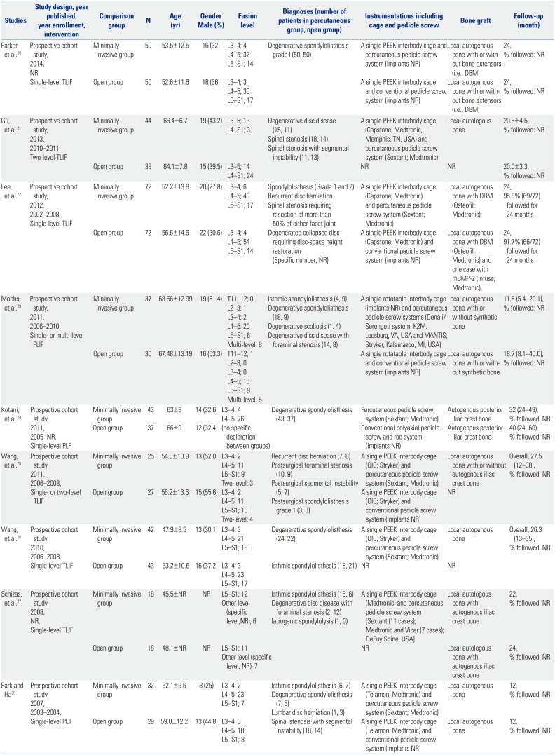
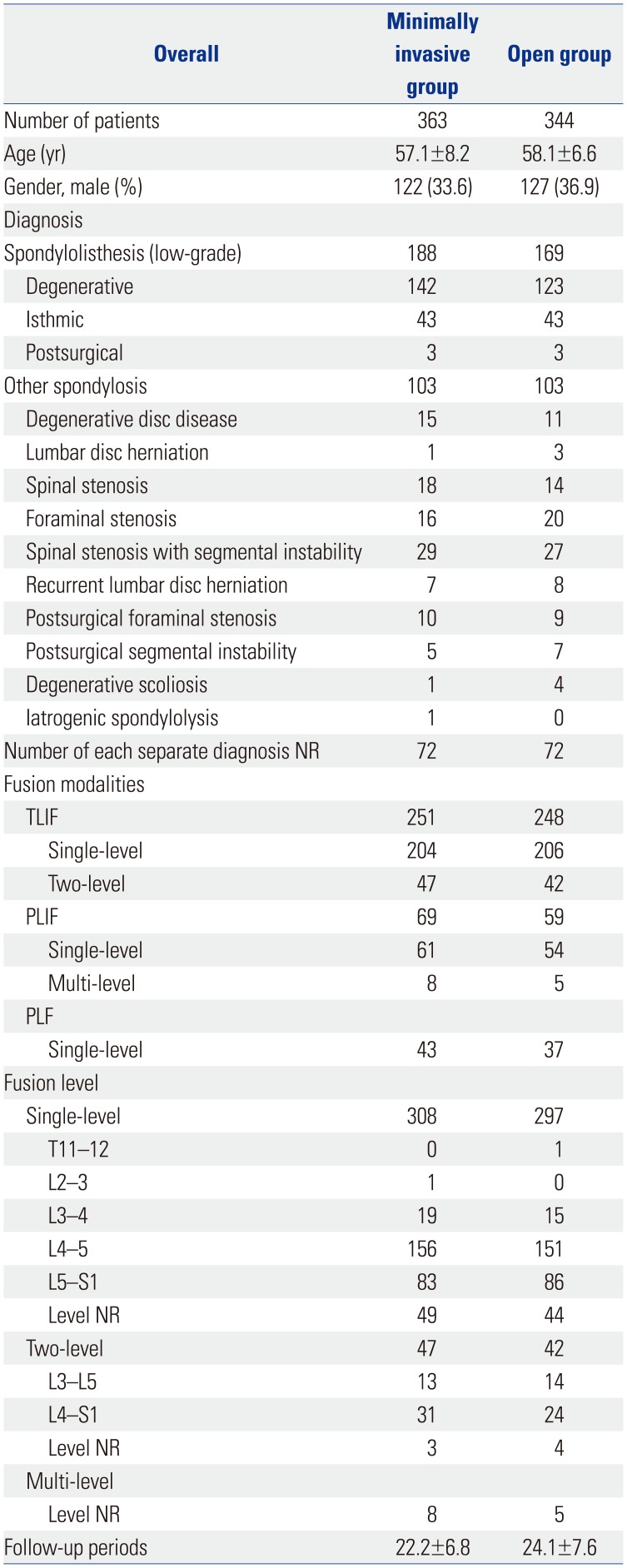
Finally, nine studies192122232425262729 were selected for analysis (Fig. 1). Characteristics of all included studies are summarized in Table 2 and Supplementary Table 2 (only online). A total of 707 participants (363 in the minimally invasive group and 344 in the open group) were included in the nine prospective cohort studies. Mean duration of follow-up was 22.2±6.8 months in the minimally invasive group and 24.1±7.6 months in the open group. Detailed demographic and surgical data at baseline are illustrated in Table 3. The baseline data were similar between the two groups (all p>0.05).
Quality assessment
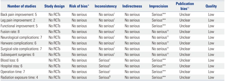
All studies had a Cowley score of 9 or more, and we judged these studies to have a low risk of bias (Table 4). Serious inconsistency was noted in all perioperative outcome measures (blood loss, hospital stay, operation time, and radiation exposure time) with substantial heterogeneity (I2>75%, p<0.1). Serious imprecision was noted in the primary outcome measures for pain and functional improvement, as well as in all perioperative outcome measures (effect size of mean difference crosses 0.5). Publication bias was judged to be unclear because it could not be quantifed due to the small number of studies analyzed.
Based on the GRADE protocol, all studies suffered from methodological flaws (limitations in the design and implementation), leading us to downgrade their quality by 1 point (Table 5). We downgraded the evidence for primary outcomes of back pain, leg pain, and functional improvement by 2 points (low-quality evidence) because of study design and because two domains (imprecision and publication bias) were judged “serious or unclear.” The evidence for primary outcomes of fusion rate, complications, and subsequent surgeries was also downgraded by 2 points (low-quality evidence) because of the design and because one domain (publication bias) was judged “serious or unclear.” Moreover, the evidence for perioperative outcomes (blood loss, hospital stay, operation time, and radiation exposure time) was downgraded by 2 points (low quality evidence) because of the design and because three domains (inconsistency, imprecision, and publication bias) were judged “serious or unclear.”
Pain and functional improvements
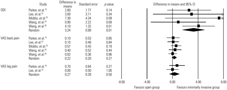
Low-quality evidence from five studies1922232526 revealed that improvement in VAS back pain score was not signifcantly different between the minimally invasive spinal fusion and open fusion groups (WMD, 0.2; 95% CI, −0.2–0.6; p=0.3; mean follow-up, 20.9±7.2 months). Likewise, low-quality evidence from two studies1922 revealed that improvement in VAS leg pain score did not differ significantly between the two groups (WMD, 0.3; 95% CI, −0.5–1.0; p=0.5; mean follow-up, 25.2±2.0 months). In contrast, there was low-quality evidence from five studies1922232526 in which the minimally invasive group had significantly greater improvement in ODI score than the open group (WMD, 3.2; 95% CI, 1.5–5.0; p=0.0003; mean follow-up, 24.2±4.8 months)(Fig. 2). The detailed clinical outcome scores of all included studies are summarized in Supplementary Tables 3 and 4 (only online).
Fusion rate
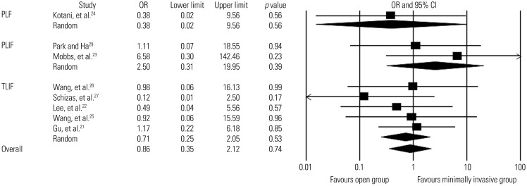
Fusion rates were reported in all studies, with overall rates of 96.7% in the minimally invasive group (351/363 patients; mean follow-up, 22.2±6.8 months) and 97.4% in the open group (335/344 patients; mean follow-up, 24.1±7.6 months) (Supplementary Tables 5 and 6, only online). Eight of nine studies were eligible for the meta-analysis; one study was not eligible because the fusion rate was 100% in both groups.19 We found low-quality evidence from eight studies in which there was no statistically signifcant difference in the overall fusion rate between the two groups (OR, 0.9; 95% CI, 0.3–2.1; p=0.7). Low-quality evidence was obtained from subgroup analyses according to the fusion method (5 studies for TLIF,2122252627 two studies for PLIF,2329 and a single study for PLF24), which revealed no significant difference in the fusion rate between the two groups (Fig. 3).
Complications
The detailed complications of all included studies are summarized in Supplementary Table 7 (only online).
Neurological complications
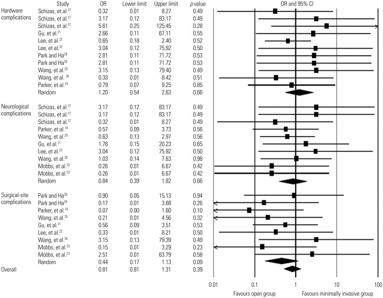
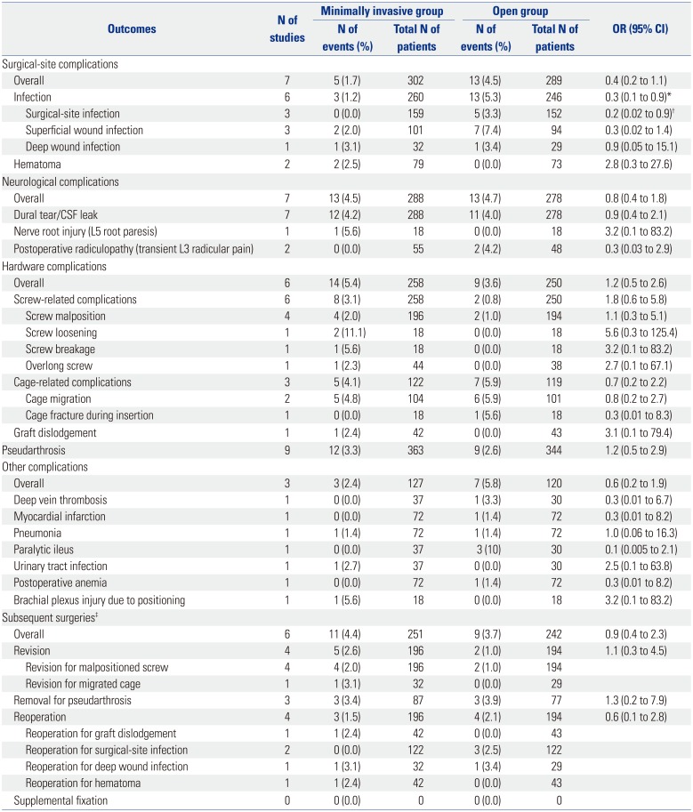
Seven studies19212223252627 addressed neurological complications, including dural tear, CSF leakage, nerve root injury, and postoperative radiculopathy (Table 6). The overall rates of neurological complications were 4.5% in the minimally invasive group (13/288 patients; mean follow-up, 22.3±5.3 months) and 4.7% in the open group (13/278 patients; mean follow-up, 23.5±3.2 months). We found low-quality evidence from these studies that there was no statistically significant difference in the overall rate of neurological complications between the two groups (OR, 0.8; 95% CI, 0.4–1.8; p=0.7) (Figure 4).
Hardware complications
Six studies192122,262729 described hardware-related complications, including screw malposition, screw loosening, screw breakage, overlong screw, cage migration, cage fracture, and graft dislodgement (Table 6). The overall rates of hardware complications were 5.4% in the minimally invasive group (14/258 patients; mean follow-up, 21.8±5.4 months) and 3.6% in the open group (9/250 patients; mean follow-up, 22.3±5.2 months). There was low-quality evidence that the overall rate of hardware complications did not differ significantly between the two groups (OR, 1.2; 95% CI, 0.5–2.6; p=0.7) (Fig. 4).
Surgical-site complications
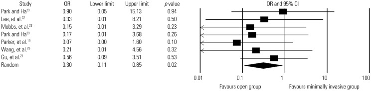
Seven studies19212223252629 reported surgical-site complications, including surgical-site infection, superficial infection, deep infection, and hematoma (Table 6). The overall rates of surgical-site complications were 1.7% in the minimally invasive group (5/302 patients; mean follow-up, 20.9±6.9 months) and 4.5% in the open group (13/289 patients; mean follow-up, 22.6±4.0 months). Low-quality evidence from these seven studies revealed that the overall rate of surgical-site complications did not differ signifcantly between the two groups (OR, 0.4; 95% CI, 0.2–1.1; p=0.1) (Fig. 4). The overall rates of infection (including surgical-site infection, superfcial infection, and deep wound infection) were 1.2% in the minimally invasive group (3/260 patients; mean follow-up, 19.9±6.7 months) and 5.3% in the open group (13/246 patients; mean follow-up, 21.0±5.4 months). There was low-quality evidence from six studies192122232529 in which the minimally invasive group had a significantly lower rate of infection than the open group (OR, 0.3; 95% CI, 0.1–0.9; p=0.02) (Fig. 5).
Subsequent surgeries
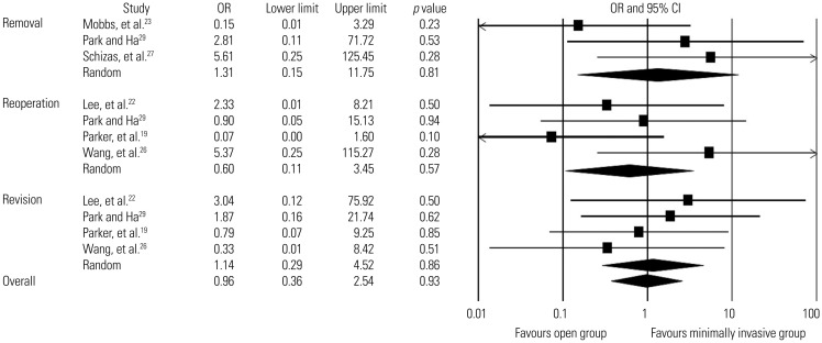
Six studies included information about subsequent surgeries,192223262729 and a total of 493 patients (251 in the minimally invasive group and 242 in the open group) were analyzed (Table 6). We found low-quality evidence suggesting that the overall rate of subsequent surgeries did not differ significantly between the two groups (4.4% in the percutaneous group and 3.7% in the open group; OR, 0.9; 95% CI, 0.4–2.3; p=0.9; mean follow-up, 20.2±6.8 months). Revisions, removals, and reoperations were also analyzed in four studies,19222629 three studies,232729 and four studies,19222629 respectively. The overall rates of revisions were 2.6% in the minimally invasive group (5/196 patients; mean follow-up, 21.9±6.8 months) and 1.0% in the open group (2/194 patients; mean follow-up, 21.9±6.8 months). Overall, 3.4% underwent removals in the minimally invasive group (3/87 patients; mean follow-up, 15.2±5.9 months) and 3.9% in the open group (3/77 patients; mean follow-up, 18.2±6.0 months). The overall rates of reoperations were 1.5% in the minimally invasive group (3/196 patients; mean follow-up, 21.9±6.8 months) and 2.1% in the open group (4/194 patients; mean follow-up, 21.9±6.8 months). Low-quality evidence revealed that the two groups did not differ significantly in rates of revision (OR, 1.1; 95% CI, 0.3–4.5; p=0.9), removal (OR, 1.3; 95% CI, 0.2–11.8; p=0.8), and reoperation (OR, 0.6; 95% CI, 0.1–3.5; p=0.6) (Fig. 6)
Perioperative outcomes
The detailed perioperative outcome data are summarized in Supplementary Tables 8, 9, and 10 (only online).
Blood loss
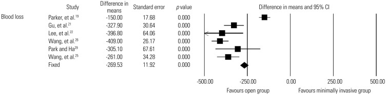
Six studies192122252629 reported estimated blood loss during surgery, with 265 patients in the minimally invasive group and 259 in the open group. Low-quality evidence indicated that the minimally invasive group had significantly less blood loss than the open group (WMD, 269.5 mL; 95% CI, 246.2–292.9 mL; p<0.0001) (Fig. 7).
Hospital stay
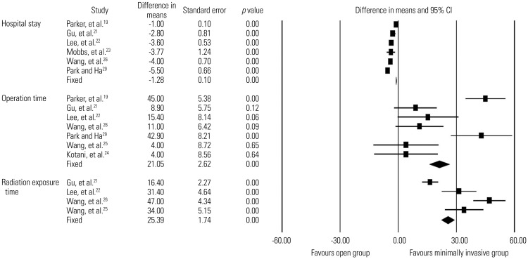
Six studies192122232629 reported length of hospital stay, with 277 patients in the minimally invasive group and 262 in the open group. Low-quality evidence suggested that the minimally invasive group had a significantly shorter hospital stay than the open group (WMD, 1.3 days; 95% CI, 1.1–1.5 days; p<0.0001)(Fig. 8).
Operation time
Seven studies19212224252629 reported operation time, with 302 patients in the minimally invasive group and 289 in the open group. There was low-quality evidence indicating that the minimally invasive group had a significantly longer operation time than the open group (WMD, 21.0 minutes; 95% CI, 15.9–26.2 minutes; p<0.0001) (Fig. 8).
Radiation exposure time
Four studies21222526 reported radiation exposure time, with 183 patients in the minimally invasive group and 180 in the open group. Low-quality evidence suggested that the minimally invasive group had a significantly longer radiation exposure time than the open group (WMD, 25.4 seconds; 95% CI, 22.0–28.8 seconds; p<0.0001) (Fig. 8).
DISCUSSION
This meta-analysis highlights low-quality evidence that indicates minimally invasive lumbar spinal fusion for the treatment of adult spondylolisthesis and spondylosis is more effective than open fusion with regard to functional improvement and reduced infection rate in the intermediate term. The two fusion methods showed similar results in terms of pain relief, fusion rate, complications, and subsequent surgeries, although the evidence was low quality. In addition, we noted low-quality evidence indicating that minimally invasive fusion is associated with decreased blood loss and length of hospital stay, but was less advantageous in terms of operation time and radiation exposure time than open fusion.
Although screw- or cage-related nerve root injuries and secondary radiculopathies were reported, most studies indicated resolution of any neurological deficits and pain with subsequent surgery. These complications also showed an equivalent incidence with open fusion. There were no reports of visceral or vascular injury associated with percutaneous pedicle screw instrumentation. Significant reduction of paraspinal muscle injury via minimally invasive surgery and subsequent preservation of trunk muscle performance are thought to result in greater functional improvement, a lower risk of infection, and better perioperative surgical outcomes (less blood loss, quicker recovery, and shorter hospital stay).
To our knowledge, this study is the first systematic review evaluating the efficacy of minimally invasive lumbar spinal fusion exclusively employing percutaneous pedicle screw instrumentation. The strengths of our study include the exhaustive search strategy, reproducible protocols, and strict adherence to systematic review methodology. The use of standardized and validated data collection and extraction tools limited bias and increased inter-rater reliability.
The major limitations of the present study are the lack of RCTs and the small number of included articles. These weaknesses prevented synthesis of higher-quality evidence. In particular, there were very few studies comparing multi-level instrumented fusion in adult spondylosis patients. Overall, the quality of the evidence was “low” in our comparison of primary outcomes between minimally invasive fusion and open fusion. The magnitude of the effect sizes was small. All studies had attrition bias with no reported number of dropouts, and the mean follow-up duration was less than two years.
Another weakness was variation in the type of arthrodesis (e.g., TLIF, PLIF, or PLF) and bone graft material (e.g., local autogenous bone, autogenous iliac crest bone, or synthetic bone extensor). Variation in preoperative diagnosis (e.g., spondylolisthesis, other spondylosis, and mixed diagnoses) and fusion assessment methods was another drawback of this analysis. Computed tomography (CT) to evaluate screw placement was not routinely performed in all studies. The scarcity of CT imaging data may have led to underestimation of screw malposition, implant loosening, implant breakage, and pseudarthrosis in the reviewed studies.
On intermediate-term follow-up, our results showed low-quality evidence that percutaneous pedicle screw instrumented fusion is more effective at improving ODI score, reducing infection rate, and decreasing blood loss and hospital stay, but less effective at reducing operation and radiation exposure time than open fusion. Furthermore, the two methods were comparable with regard to pain relief, fusion rate, complications, and subsequent surgeries based on low-quality evidence. Several methodological flaws and weaknesses limited the reported results. In particular, there were no well-designed RCTs from which to synthesize high-quality evidence.
The ambiguity in these findings could lead to major alterations of the results derived from our analyses and highlights the need for adequately powered RCTs that will assess the long-term efficacy of minimally invasive lumbar spinal fusion. Future studies should compare subgroups based on fusion modality (e.g., TLIF, PLIF, PLF), spine disorder (e.g., spondylolisthesis, other spondylosis, deformity, trauma), and surgery level (e.g., single or multi-level fusion).
Although the findings are limited by insufficient evidence and lack of adequately powered high-quality RCTs to address this gap in evidence, our results support that minimally invasive lumbar spinal fusion is more effective than open fusion for adult spondylolisthesis and other spondylosis in terms of functional improvement, reducing infection rate, and decreasing blood loss and hospital stay.
 XML Download