

 PDF
PDF ePub
ePub Citation
 Print
Print


INTRODUCTION
Atopic dermatitis (AD) is a chronic and relapsing pruritic inflammatory skin disease, often associated with elevated serum IgE levels and a family history of AD, allergic rhinitis, and asthma.12 The prevalence of AD in industrialized countries has recently increased to 15 to 30% in children and 2 to 10% in adults.3 AD has a complex etiology with genetic, immunological, and environmental aspects. Mutations in the filaggrin gene (FLG) are the most common and significant genetic defects identified to date causing AD, emphasizing the role of skin barrier alterations in AD pathogenesis.1456
FLG was first identified by Dale7 in 1977 as a highly insoluble, histidine-rich protein that was co-purified with keratin intermediate filament proteins in epidermal extracts. FLG monomers have been thought to promote corneocyte compaction by contributing to keratin pattern formation in the lower stratum corneum (SC).4 FLG monomers are proteolyzed into natural moisturizing factors, which are necessary to maintain hydration of the upper SC and acidic pH of the skin surface.1
FLG mutations have been identified as the underlying cause of ichthyosis vulgaris8 (IV; OMIM 146700), which is characterized by dry and scaly skin, palmar and plantar hyperlinearity, and keratosis pilaris.9 Furthermore, FLG mutations have proved to be a major predisposing factor for AD in Europe and Asia.610 Patients with AD who carry FLG mutations have been reported to have more persistent and severe disease, a higher incidence of herpes virus infection, allergic sensitization, and a greater risk of multiple allergies than patients with AD without FLG mutations.15
Some FLG mutations (R501X, R1891X, 3321delA, S1405X, S1515X, W1947X, G2025X, E3070X, Q1701X, Y1767X, S3296X, and K4022X) have been identified in Korea.11121314 This study aimed to examine the spectrum of FLG-null mutations in Koreans with AD to investigate the association between FLG mutations and clinical AD markers and to compare the landscape of Korean FLG mutations with that of other Asian countries.
MATERIALS AND METHODS
Clinical materials
Seventy Korean patients with AD whose parents and all four grandparents were recorded as ethnic Korean were enrolled. Diagnosis of AD was confirmed by experienced dermatologists using AD diagnostic criteria created by Hanifin and Rajka.15 Patients were divided into three groups according to age of onset: early childhood onset (<8 years), late childhood onset (8–18 years), and adult onset (>18 years). AD disease severity was assessed using the SCORing Atopic Dermatitis (SCORAD) index, and patients with AD were grouped into mild (<15 points), moderate (15–40 points), and severe (>40 points) disease gro-ups.16 Peripheral blood samples were analyzed for total serum IgE levels and specific IgE levels for egg, milk, soybean, peanut, fish, wheat, mites (Dermatophagoides pteronyssinus, Dermatophagoides farinae), and cockroach by multiple allergosorbent test chemiluminescent assay (MAST-CLA; Advan-SureTM AlloStation, LG Life Science, Seoul, Korea). Total IgE concentrations ≥200 KIU/L and/or ≥3+ in three categories of the MAST-CLA test were arbitrarily defined as elevated IgE in this study.1718 Associated allergic diseases, including asthma and allergic rhinitis, were determined on the basis of the questionnaire and previous diagnoses by physicians. Patients provided written informed consent, which complied with the principles of the Declaration of Helsinki. This study was approved by the Institutional Review Board (IRB No. 3-2014-0027) of Gangnam Severance Hospital, Seoul.
Mutation analysis
Genomic DNA was extracted from peripheral blood leukocytes with a DNA extraction kit (QIAamp DNA Blood Midi kit, Qiagen, Hilden, Germany). Patients with AD were screened for fourteen FLG mutations that have been identified in Korea, Japan, and China (R501X, 3321delA, S1695X, Q1701X, Q1745X, Y1767X, Q1790X, S2554X, S2889X, S3296X, 3222del4, S1515X, Q2417X, and K4022X) by direct DNA sequencing as described previously.1920
Statistical analysis
Descriptive statistics for quantitative values were expressed as means [±standard deviation (SD)] in accordance with the data distribution. Frequencies and percentages were used to describe categorical variables. We used Fisher's exact test to assess the associations between FLG mutations and AD, as well as AD-associated phenotypes, including age of onset of AD, SCORAD index, allergic AD, family history of AD, and associated allergic diseases like asthma and allergic rhinitis. The strength of associations was estimated by calculating odds ratios and 95% confidence intervals. The level of statistical significance was established at α<0.05. Statistical analyses were performed using SPSS version 19 (SPSS Inc., Chicago, IL, USA).
RESULTS
Clinical features
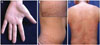
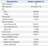
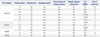
The clinical characteristics of patients with AD are presented in Table 1. A total of 70 patients were enrolled in this study. The mean age was 19.3 years (range 0 to 63, SD=13.14), and 68.6% of patients were male. Thirteen patients had mild AD, 25 had moderate, and 32 had severe AD, as determined by the SCORAD index. Fifty-seven (81.4%) patients had moderate to severe disease. In the AD cohort, 24.3% of patients had hyperlinear palms.
FLG mutations in AD patients
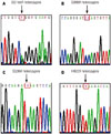
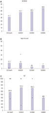
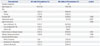
Among the fourteen mutations screened, four, S2889X, S3296X, 3321delA, and K4022X, were identified in our AD patients (Table 2, Fig. 1). Eleven patients had FLG mutations, and all were heterozygous for the mutation. All patients with FLG mutations had moderate to severe AD (Fig. 2). Mutations 3321delA, K4022X, S3296X, and S2889X were carried by six (9.1%), three (4.5%), two (3.0%), and one (1.5%) individuals, respectively. One patient was a heterozygous carrier of two different FLG mutations. This study is the first time S2889X has been identified in Koreans with AD (Fig. 1).
Associations between FLG mutations and AD characteristics
FLG mutations were significantly associated with elevated IgE, palmar hyperlinearity, and a family history of allergic disease (p<0.05) (Table 3). All patients with FLG mutations had high IgE, and were positive for MAST-CLA or moderate to severe AD. Palmar hyperlinearity was present in eight patients (72.7%) with AD and FLG mutations. Eight patients (72.7%) with FLG mutations had a family history of allergic disease. Age of onset was not significantly associated with FLG mutation. AD severity was not statistically significantly associated with FLG mutation (p=0.115).
DISCUSSION
Since two FLG mutations, R501X and 2282del4, were identified in Europeans with AD, many replication studies have reported the prevalence and frequency of mutations in the FLG in individuals with AD.910 New FLG mutations associated with AD have been widely reported in European and non-European cohorts. Previous reports have also shown variations in FLG mutations among individuals with AD in different ethnic groups. The most prevalent FLG mutations are R501X and 2282del4 and have been reported to be present in up to 48% of Europeans with AD.15 The mutation landscape in Asians with AD cohorts has been reported to vary, and the frequency was reported to be much lower than that in Europeans. Although many European countries have a similar FLG mutation landscape, Asian countries have been reported to have distinct FLG mutation landscapes. Among the Asian-specific FLG null mutations identified in Japan, China, Taiwan, and Singapore, only 3321delA was common. In a Japanese AD cohort, the S2554X and S2889X mutations were the most prevalent, followed by 3321delA and S3296X.2122 3321delA and K4671X were the most common FLG mutations in an AD cohort in China.2324 Hsu, et al.25 identified three FLG mutations, 3321delA, Q2417X, and E1795X, in Taiwanese families with ichthyosis vulgaris. The 3321delA mutation was the most prevalent FLG mutation in Singapore.26
In this study, fourteen FLG mutations, R501X, 3321delA, S1695X, Q1701X, Q1745X, Y1767X, Q1790X, S2554X, S2889X, S3296X, 3222del4, S1515X, Q2417X, and K4022X, which previously were reported in Asian AD cohorts, were selected for FLG mutation analysis. We demonstrated that 15.7% of Koreans with AD in our study had four FLG null mutations: S2889X, S3296X, 3321delA, and K4022X. 3321delA (n=6, 9.1%) was the most common FLG mutation in our AD cohort followed by K4022X (n=3, 4.5%). This prevalence is similar to that in China; 3321delA is one of the most prevalent mutations in Chinese patients with AD.24 In a recent study, Park, et al.14 screened 81 Korean AD patients and K4022X was the most common. We also found that K4022X was the second most common mutation in our cohort; therefore, K4022X is thought to be a common mutation in Koreans with AD. There were differences of prevalent FLG mutations and AD-associated phenotype between the previously published two studies. Park, et al.14 reported new FLG mutations in Korean AD patients by analyzing coding exons, whereas we analyzed known mutations. The differences in FLG mutations and AD-associated phenotype may be the result of small sample size in both studies.
S2889X (n=1, 1.5%) and S3296X (n=2, 3.1%), which are common in Japanese patients with AD, were also detected in two Koreans in our study. S2554X, which was the most common FLG mutation in Japan, was not detected in our study. One patient who had two mutations, S2889X and S3296X, had hyperlinear palms, clinical features of IV, and severe AD. Notably, this study was the first to detect the S2889X mutation in Koreans. This study expanded our understanding of the landscape of FLG-null mutations in Koreans with AD.
The frequency of FLG mutation was 31.4% and 26.0% in Chinese patients with AD in previous reports.2324 A Japanese case–control study of eight FLG mutations demonstrated that about 27% of Japanese patients with AD carried at least one FLG mutation.22 The frequency of FLG mutations was 20.2% in Singaporean Chinese patients with AD or IV.26 In our study, the frequency of FLG mutations in Korean patients with AD (15.7%) was similar to that reported by Park, et al.14 The frequency of FLG mutations in Koreans with AD seems to be lower than that in other Asian countries. The low frequency of FLG mutations in our study can be explained by the fact that AD is a multifactorial disease that is affected by genetic and environmental factors. Mutations in other barrier-related genes may contribute to AD in our cohort. In addition, decreased FLG expression in AD as a result of genetic mutations or skin inflammation can induce acquired FLG deficiency. Barrier impairment in AD patients with severe inflammation has been reported to be similar in patients with wild-type and mutant FLG. These FLG alterations due to inflammation can also explain the low FLG mutation frequency in individuals with AD.527 The other important finding of our study is the association between FLG mutations and clinical features of AD in the Korean population. Palmar hyperlinearity and allergic sensitization with increased total IgE levels have been previously reported to be strongly associated with mutant FLG.2628 A significant association between palmar hyperlinearity and FLG mutations was also observed in our study. Previously, associations between FLG mutations and AD severity have been reported.26 In contrast, other groups did not identify an association between FLG mutations and disease severity or skin barrier defects, characterized by high transepidermal water loss.212324 Although AD has been reported to be more severe in patients with FLG mutations, there were no significant associations between FLG mutations and AD severity in our study. The lack of association between FLG mutations and AD severity maybe due to the fact that the SCORAD is a momentary variable that does not reflect overall disease activity.29
Any associated between early-onset AD and FLG mutations is still controversial. FLG mutations are related to early onset and persistent AD.30 However, Meng, et al.31 did not find an association between early-onset AD and the FLG mutation 3321delA. Age of onset was also not significantly associated with FLG mutations in the present study.
In conclusion, our study expands our understanding of the landscape of FLG mutations in Koreans by finding four FLG mutations, of which one has not previously been reported in Koreans with AD. We also found that the frequency of FLG mutations in Korean AD was lower than that of other Asian countries. The FLG mutation spectrum in our cohort was both distinct and partially overlapping with other Asian AD cohorts. We also demonstrated a significant association between FLG mutations and AD phenotype (elevated IgE, palmar hyperlinearity, and a family history of allergic disease).

 XML Download