

 PDF
PDF ePub
ePub Citation
 Print
Print


INTRODUCTION
Thoracic outlet syndrome (TOS) is a condition from compression of the subclavian vessels and/or brachial plexus as the structures travel from the thoracic outlet to the axilla.1 There is some general disagreement among experts on the etiology and pathophysiology of this condition, presumably because of the wide variation in symptoms that manifest in presenting patients, and also because of lack of a definitive gold standard for diagnosis.
Symptoms associated with TOS have traditionally been divided into vascular and neurogenic causes, a distinction based on the underlying structures implicated. Neurogenic TOS (nTOS) typically presents as compression of the brachial plexus that primarily, but not exclusively, involves its lower trunk. Vascular TOS (vTOS) usually involves compression of the vessel, most commonly the subclavian artery or vein, or is secondary to thrombus formation in the vein. Of the two, over 95% of all TOS patients present with nTOS.1
The thoracic outlet has three anatomic compartments: the interscalene triangle, the costoclavicular, and the retropectoralis minor spaces.2 Of these structures, the interscalene triangle between the anterior and middle scalene muscles, which may become spasmodic or swollen, is frequently implicated.3 If nTOS is suspected, electrophysiological nerve studies and anterior scalene muscle blocks provide guidance when screening for patients likely to benefit from surgical decompression of thoracic outlet.4
Patients complaining of vague symptoms of limb fatigue, weakness (not definable to a particular muscle group), subjective sensory complaints, and no accepted electrodiagnostic evidence demonstrative of a neural lesion are not considered to have true nTOS nor any other form of TOS, but should be thoroughly evaluated for some other disorder.5 Especially, possible myofascial pain and other musculoskeletal dysfunctions arising from the scalene muscles can mimic neurogenic compromise of the neural structures potentially involved in TOS.
Since the scalene muscles are not a hard tissue such as a rib, the spasmodic or swollen scalene muscles can irritate brachial plexus and provoke paresthesia in upper extremity, however, it is less likely to damage brachial plexus to the extent that there are abnormal findings in electrodiagnostic test.
We recently observed a significant number of patients not compatible with cervical radiculopathy or true nTOS in electrodiagnostic test, but tenderness of the scalene muscle and upper extremity paresthesia. Nonsurgical techniques to decompress the interscalene space by relaxing the scalene muscles include injections of anesthetic agents, steroids, and botulinum toxin.6 Based on our clinical experience, injection of local steroids with local anesthesia into the anterior and middle scalene muscles reduces upper arm paresthesia in these patients. In these cases, even if the diagnosis of the patients is not true nTOS, and conservative treatment like steroid injection is effective in improving symptoms, unnecessary surgery can be prevented. Moreover, even if the cause of nerve irritation is spasmodic muscle, stretching exercise will be helpful.
To demonstrate that the injection directly affects the spasmodic or swollen muscles, it is necessary to prove that the injection would not directly block brachial plexus. Therefore, we hypothesized that intra-muscular steroid injection or stretching exercise for scalene muscles would decrease pressure on the brachial plexus at the interscalene triangle by relaxing the scalene muscles, which would result in decreased upper extremity paresthesia.
A safe and accurate injection technique into the target muscle has been proven using ultrasound (US) guidance.78 The purpose of this study was to compare the therapeutic effects of intra-muscular steroid injections into the scalene muscle under US guidance on upper extremity paresthesia with those of stretching exercise only.
MATERIALS AND METHODS
Participants
Among patients with suspected nTOS based on clinical examination, without abnormalities in the electrodiagnostic test who visited the outpatient clinic of a university hospital between March 2013 and June 2014, and who met the following criteria were included in this study: 1) a main complaint of paresthesia in the arm, forearm, and/or hand; 2) the visual analog scale (VAS) greater than 4 (on a numeric scale of 0–10) for paresthesia in the arm, forearm, and/or hand; 3) extreme tenderness over the anterior and middle scalene muscles; 4) no weakness in the affected site; and 5) symptoms present for at least 3 months. Exclusion criteria included 1) prior treatment with botulinum toxin, lidocaine, and/or steroid injection of scalene muscles in the affected site; 2) prior treatment with brachial plexus blockade in the affected site; 3) prior scalenectomy or surgery for TOS in the affected site; 4) prior treatment with stretching exercise; 5) cervical radiculopathy or other peripheral nerve compression syndromes in the electrodiagnostic test; 6) medial antebrachial cutaneous nerve conduction abnormality;9 7) previous history of adverse effect of lidocaine or steroid; 8) presence of an unstable medical condition or a known uncontrolled systemic disease; and 9) any conditions or situations that might place the patient at significant risk during the study. Candidates with bilateral symptoms were also excluded to allow symptom improvement to be investigated better.
This study was approved by the Institutional Review Board and human subjects review committee before the study began (4-2014-0717). Written informed consent was obtained from all participants after they were briefed on the purpose and procedures of the study.
Study design
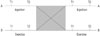
The present study used a single-blind crossover design. Each participant completed one injection and daily exercise program for 2 weeks. For the purpose of the study, we compared the therapeutic effects between 2-weeks after one injection and 2-weeks exercise. One physiatrist (S.C.L.), who was not blinded, examined all patients referred to the clinic for eligibility. The investigator (Y.W.K.) who evaluated the outcome measures was blinded to the group allocation throughout the study, although the physiatrist (S.C.L.) who performed all injections and educated exercise and the participants were not blinded.
The order of exercise and injection was randomly assigned. The randomization sequence for group allocation was prepared by a statistician who was not a co-investigator using a computer random number generator. After randomization, half of all patients received scalene muscle injection before exercise, while the order was reversed for the other patients. To avoid carry-over effects from the previous treatment, each treatment was separated by a one-week rest. The study design is depicted in Fig. 1. Patients were not allowed to take any oral pain-related medicine, engage in physical therapy, or use any intervention except posture correction beginning 2 weeks before and during the study.
US-guided injection
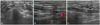
With the patient lying supine, a rolled towel was placed below the neck, and the head was rotated approximately 60° opposite to the injection site, exposing the region of scalene and sternocleidomastoid muscles. Patients with long hair were fitted with disposable head caps. We performed B-mode, real-time ultrasonography with sterile methods using an Accuvix V10 system (Samsung Medison, Seoul, Korea) interfaced with a 5- to 12-MHz linear array transducer around the targeted muscle. A physiatrist with more than 7 years of experience in musculoskeletal ultrasonography carried out US-guided injection procedures. With color Doppler images, a preliminary assessment determined the location of neurovascular bundles. The Doppler setting changed at the vascular level in each subject. The anterior and middle scalene muscles were scanned along the craniocaudal extension and visualized on images through the lower half (Fig. 2A). Then, by rotating the transducer, the long axis of anterior or middle scalene muscle was best visualized on the US image (Fig. 2B and C). Once an adequate approach had been identified, a mark with indelible ink was made on the skin, adjacent to the lateral short axis of the transducer. Approximately 1 mL of lidocaine 1% was injected subcutaneously at the previously marked site. The injections were performed under US guidance with the use of an in-plane method using a 2.6-cm, 23-guage needle. A 0.2 to 0.4 mL volume of saline was injected to confirm the location of the needle tip before injection of triamcinolone. Then, 0.5 mL of 20 mg triamcinolone was injected into each belly of the anterior and middle scalene muscles away from the nerve roots of the brachial plexus.
Stretching exercise
The patients were taught to do self-exercise comprising stretching to alleviate muscle spasm and tightness, and to avoid a posture that might aggravate the symptoms. In a case of a stretch of the right scalene group, with the patient extended the left lateral was flexed and right (ipsilaterally) rotated the neck. An additional stretch could be obtained by using the left hand to passively move the head and neck further in this direction.10 Twenty repetitions were performed, each one held for approximately 15 daily for 2 weeks.
Outcome measures
Gender, age, and symptom duration were documented before the study commenced. Our primary outcome measure was paresthesia in the arm, forearm, and/or hand using a 10-cm horizontal VAS, which varied from no pain (VAS 0) to worst imaginable pain (VAS 10).6 VAS ratings were taken just before and just after each treatment session. Success of treatment was defined as a reduction exceeding 50% in post-treatment VAS compared to pre-treatment, whereas treatment failure was defined as <50% post-treatment VAS reduction compared to pre-treatment. We compared the number of patients with successful treatments between each group.
The sign of brachial plexus block was checked. Whether there were symptoms of unintended brachial plexus block was as follows: 1) partial block, in cases of mild to moderate paresthesia, numbness and/or weakness (focal if involving single trunk; diffuse if multiple trunks), and 2) complete block, in cases of severe diffuse numbness and/or motor deficit of the upper extremity.11 Also, frequencies and severities of adverse events and injection-related complications were monitored throughout the study.
Statistical analyses
SPSS version 21.0 software (SPSS Inc., Chicago, IL, USA) was used for the statistical analyses. Shapiro-Wilk test was used for VAS rating for determining whether or not the distribution was normal, and the results showed no normal distribution (p<0.05 by the Shapiro-Wilk test). Therefore, VASs were analyzed using a repeated measured ANOVA. Within subject factors were treatment (injection versus exercise) and time (T1 and T2). Statistical significance was accepted for p-values <0.05.
RESULTS
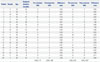
Twenty patients who met the inclusion criteria were recruited to participate in this single-blind, crossover study. Fourteen of the 20 (70%) patients were female. All patients completed the study without follow-up loss. The average age was 45.0±10.5 years and average duration of symptom was 12.2±8.7 months (Table 1).
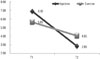
Repeated measures ANOVA showed a significant effect of both treatments [F(1,38)=510.76, p<0.01]. Also, there was a significant interaction between time and treatment [F(1,38)=96.04, p<0.01]. Assessment of treatments is summarized in Table 1. After 2 weeks, there was a significant decrease of VAS after treatment compared with baseline in both groups (6.90 to 2.85 after injection and 5.65 to 4.05 after stretching exercise, p<0.01) (Fig. 3). These findings suggested that pain was diminished in each treatment; however, injection treatment resulted in more improvements than stretching exercise (p<0.01) (Fig. 3). VAS difference of pre- and post-injection was 4.05 and that of pre- and post-exercise was 3.07. The number of patients with successful treatment, whose post-treatment VAS was reduced by more than 50% compared to pre-treatment, was 18 of 20 (90.0%) after injection, whereas the number was 5 of 20 (25.0%) after stretching exercise.
The anterior and middle scalene muscles were reliably identified and the tip of the needle was visualized within the muscle belly on US images in all patients. There were no cases of intravascular needle placement and no instances of infection, hematoma, or allergic reaction. There were no symptoms of unintended brachial plexus block immediately after injection procedure. Nine of the 20 participants experienced focal postinjection pain, but this pain was mild to moderate and self-limited in all and no supplementary treatment was required.
DISCUSSION
This study describes a therapeutic approach for treating scalene muscle-generated upper extremity arm paresthesia. A comparison of treatments (stretching exercise vs. intra-muscular injection) was necessary to exclude an effect of brachial plexus block and to determine the short-term effectiveness of intra-muscular injection on upper extremity paresthesia. We demonstrated that US-guided anterior and middle scalene muscle injection with steroid led to reduced paresthesia and to a high level of successful treatment at short-term follow-up of patients with suspected scalene muscle problems. Stretching exercise of scalene muscle was also effective for upper extremity paresthesia although injection treatment resulted in more improvements.
The definition and acceptance of TOS is one of the greatest challenges in research regarding this clinical entity.12 Some previous studies used clinical criteria of TOS that are predominantly subjective, such as a history of pain and/or paresthesia in the upper extremity, tenderness over the brachial plexus above the clavicle, or a positive elevated arm stress test.16 This diagnosis based on clinical criteria is what some author referred to as "disputed neurogenic" TOS.13 The use of electrodiagnostic studies (e.g., medial antebrachial cutaneous nerve) provides objective evidence of TOS.12 In the present study, we enrolled subjects without abnormalities in the electrodiagnostic studies. The results indicated that patients without abnormal electrodiagnostic findings can be treated with intra-muscular steroid injection and/or stretching exercise without surgical therapy. Although there was no abnormal finding on electrodiagnostic test, it can irritate brachial plexus, if the scalene muscle is spasmodic, tense, or swollen clinically.
Of these interventions to decompress the interscalene space by relaxing the scalene muscle, only botulinum toxin injections have resulted in sustained symptom reduction as demonstrated in case reports.14 However, in previous studies, which suggested steroid injection or stretching exercise is not effective on relieving symptoms, the definition of nTOS was not clear and differed widely between studies. If patients were categorized into with and without abnormal findings on electrodiagnostic studies, different results could be derived.15 Botulinum toxin injections to the scalene muscles did not result in clinically or statistically significant decrease in pain, paresthesia, or dysfunction in a population of subjects with TOS, while other authors have described positive treatment effects of botulinum toxin injections for patients with TOS.6151617 These studies also did not take into account the electrodiagnostic severity; such a consideration may have yielded different results.
In the current study, improvement in the exercise group shows that the effect of injection was not due to blocking of the brachial plexus. A 0.5 mL volume of 20 mg triamcinolone without local anesthesia was injected into each of the belly of the anterior and middle scalene muscles away from the nerve roots of the brachial plexus. Relief from less than 2 mL of local anesthesia for scalene muscle injections in patients with nTOS is not related to blockade of the brachial plexus.11 Considering the muscle relaxation effect of botulinum toxin, the previous studies using botulinum toxin injection also supported that the effect of injection is not by blocking the nerve but by action on scalene muscles.
The exact mechanism provoking scalene muscle upper extremity pain and paresthesia is unclear and may be most likely multifaceted. The actions of the scalene muscles include flexion of the cervical spine when the scalene muscles contract bilaterally and lateral flexion of the cervical spine when the muscles act unilaterally.18 In some cases without obvious structural anomalies at the time of surgery, tonic contraction of the scalene muscles may produce sufficient compression at the interscalene triangle to produce clinical symptoms.3 Spasmodic, swollen, or tense muscle, or myofascial trigger points of the scalenes due to prolonged tonic contraction tend to produce restricted lateral flexion and/or ipsilateral rotation of the neck, entrapment of brachial plexus.
One of scalene muscle dysfunctions is myofascial pain syndrome often resulting from or being perpetuated by acute or chronic overuse of the muscles (e.g., coughing, labored breathing, especially due to chronic obstructive respiratory disease) or motor vehicle accidents.10 However, pathological conditions of the scalene muscles cannot fully be explained by myofascial pain syndrome alone. Injection methods in the current study was not a trigger point injection for myofascial pain syndrome, because we did not use repeated needling technique and observe any local twitch responses. Muscle fibrosis is the most prominent histologic finding upon examination of scalene muscles in nTOS patients, and the proportion of scar tissue present is three times greater than controls.319 However, this was the only surgical finding on true nTOS, and was not compatible with the findings of our patients without abnormal electrodiagnostic study. Chronic muscle strain or injury triggered by repetitive activities and cumulative overwork can lead to scalene muscle dysfunction. Considering the anti-inflammatory action of steroids, steroid injection might be effective in patients with chronic muscle strain.20 In a previous study regarding piriformis syndrome, it was reported that local anesthetic and steroid injections may break the pain and muscle spasm cycle in patients who do not respond to conservative therapy.21 In addition to the effect on inflammation, corticosteroids have also been reported to have a direct stabilizing effect on neural membranes and inhibit C-fiber transmission, thereby reducing ectopic discharges from neural fibers.22 This could help explain how corticosteroids might have antinociceptive effects even in non-inflammatory conditions such as tendinosis. Even though exact mechanism of steroid is unknown our present results, indicate that upper extremity pain or paresthesia, at least without abnormal electrodiagnostic findings, can be treated with intramuscular injection.
Limitations in our study include the small number of participants and the short-term period of follow-up. The long-term effect of intra-muscular steroid injection and stretching exercise remains to be determined. However, the objective of this study was to verify that the effect of steroid injection into a scalene muscle is direct on the muscle itself, but not by brachial plexus block, and not focused to find the long-term efficacy of intramuscular injection or stretching exercise.
One of scalene muscle dysfunctions is myofascial pain syndrome, often resulting from or being perpetuated by acute or chronic overuse of the muscles. Although we did not observe local twitch responses during injection of scalene muscles, it cannot rule out the therapeutic effects of dry needling on upper extremity paresthesia.
US-guided anterior and middle scalene muscle injection with steroid or stretching exercise of these muscles led to reduced upper extremity paresthesia, although injection treatment resulted in more improvement. The results suggest that there might be symptom relief conferred by injection into the muscle alone, not related to blockade of the brachial plexus. Nevertheless of some limitations, intra-muscular injection and stretching exercise of scalene muscles may offer a valuable therapeutic option for a selected group of upper arm paresthesia patients who present with localized tenderness in the scalene muscle without electrodiagnostic abnormalities.
 XML Download