

 PDF
PDF ePub
ePub Citation
 Print
Print


INTRODUCTION
Neurally mediated syncope (NMS) is the most common type of syncope, characterized by abnormal autonomic response with excessive vagal tone and sympathetic withdrawal.12 However, the pathophysiological mechanisms of NMS remain uncertain.23 The head-up tilt test (HUT) is often used to confirm NMS in patients with a suspicious history of NMS. The HUT induces a large gravitational shift of blood away from the chest to the distensible venous capacitance system below the diaphragm. The circulatory adjustments to orthostatic stress lead to an increase in cardiac contractility, heart rate (HR), and vascular tone in order to maintain arterial blood pressure in an upright posture. These adjustments are mediated by the neural pathways of the autonomic nervous system. Arterial and cardiopulmonary reflex changes are also involved in these adjustments.2 However, previous studies did not find any clear evidence of alterations in the arterial baroreflex control of HR in subjects with tilt-induced NMS.4567 Several studies have reported a reduction89 or increase in baroreflex activity,1011 and one study reported that reduced baroreflex sensitivity (BRS) during HUT is an independent value in predicting the recurrence of syncope.12
There are several treatment options of NMS, and tilt training is one of them.1 As results have differed regarding the efficacy of tilt training in preventing recurrence of syncope, tilt training is listed as a class IIb recommendation in current guidelines.1 Moreover, there have been limitations when performing tilt training at hospitals due to hospital admission and medical costs. Therefore, identifying predictors for tilt training response could be useful in terms of clinical management.
This study aimed to assess the role of BRS in predicting tilt training response in patients with NMS.
MATERIALS AND METHODS
Study population
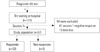
We reviewed our tilt training database, and 111 consecutive patients with recurrent NMS who performed tilt training between March 2006 and March 2014 were identified. The diagnosis of NMS was established based on suggestive clinical history, positive HUT results, and absence of any other cause of syncope. Patients with two consecutive positive responses to HUT (a positive response to the initial diagnostic HUT and a positive response to the first session of tilt training) were enrolled in our study in order to avoid over-diagnosis. We excluded those who had structural heart disease and any other cause of syncope. Among a total of 111 patients, 41 patients with a negative response to the first session of tilt training and 13 patients whose BRS data were lost were excluded. Therefore, 57 patients were ultimately analyzed (Fig. 1). This study received institutional review board approval, and informed consent was waived for this retrospective study.
Diagnostic head-up tilt test
The HUT was performed on patients who had fasted for at least 4 hours. We used the tilt test protocol, which was reported previously.13 After a 10-min resting period in the supine position, the patients were tilted to an angle of 70° for 30 minutes or until symptoms appeared. If a negative response was observed, the intravenous isoproterenol provocation test, which uses incremental doses in order to increase average HR, was performed. Patients were kept in the same 70° upright posture as in the first phase, and isoproterenol was intravenously administered at an initial rate of 1 µg/min. The infusion rate was increased increased by 1 µg/min every 3 minutes to a maximum of 5 µg/ min. A positive HUT response was defined when syncope or presyncope developed in association with hypotension [systolic blood pressure (SBP) <80 mm Hg], bradycardia [sinus arrest (>3 seconds), <45 beats/min in the first phase, or <60 beats/min in the second phase], or both. HR and blood pressure measurements immediately before or during syncope were used to define a positive response to HUT. Faintness without significant hypotension or bradycardia was considered as a negative response to HUT. The tilting table was rapidly lowered to the horizontal position when a positive response appeared or the study endpoint was reached. The syncope was classified on the basis of the previous study:14 type 1 (mixed), type 2 (cardioinhibitory), or type 3 (vasodepressive).
Tilt training
Tilt training was performed during hospital admission. Tilt tests were repeated daily, two sessions per day. Tilt training responders were defined as patients with three consecutive negative responses to HUT during tilt training.
Data acquisition
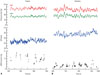
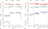
Data recording was initiated at the start of each HUT. During a sequential HUT, beat-to-beat arterial blood pressure was continuously measured non-invasively with a servo-controlled photoplethysmograph (Finometer® PRO; Finapres Medical Systems B.V, Amsterdam, the Netherlands), placed on the midphalanx of the right middle finger (Fig. 2).15 Continuous electrocardiogram data were also obtained during the HUT
Arterial baroreflex sensitivity
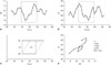
Calculation of cardiac BRS was based on the cross-correlation baroreflex sensitivity (xBRS) method (Fig. 3). The SBP and interbeat interval (IBI) time series were spline interpolated and resampled at 1 Hz. In 10-s windows, correlation and regression slopes between SBP and IBI were computed. Delays of 0- to 5-s increments in IBI were computed, and the delay with the highest positive coefficient of correlation was selected; this optimal delay (tau) was stored. The slope between SBP and IBI was recorded as an estimate of xBRS if the correlation was significant at p<0.01. When these conditions were not met, there was no result for this time segment.1617
Hemodynamic parameters
Mean arterial blood pressure (MBP) was the true integral of the arterial pressure wave over 1 beat divided by the corresponding beat interval. HR was computed as the inverse of the IBI and expressed in beats per minute. Beat-to-beat changes in stroke volume were estimated by modelling flow from finger arterial pressure (Modelflow, Finapres Medical System B.V., Amsterdam, the Netherlands).
Statistical analysis
Hemodynamic data were averaged at fixed 5 min frames within subsequent HUT: 1) 5-min in the supine position before head-up tilt; 2) 5 min after 70° upright tilting of the head-up tilt table; and 3) 5 min before the occurrence of syncope or the end of HUT.
Continuous variables are expressed as mean±standard deviation or median and interquartile range. Categorical variables are expressed as frequency and percentage. To evaluate difference between the study groups, we used Student's unpaired t-test for normally distributed data and the Mann-Whitney U test for skewed data. Categorical variables were analyzed with the chi-square test or Fisher's exact tests using SPSS software (SPSS for Windows, version 20.0, IBM Corp., Armonk, NY, USA). A p value of <0.05 was considered to be significant.
RESULTS
Baseline characteristics of the study population
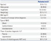
We ultimately analyzed 57 patients (26 males, mean age of 33.9±13.5 years) with NMS and with two consecutive positive responses to HUT (a positive response to the initial diagnostic HUT and a positive response to the first session of tilt training) (Table 1). The vasodepressive type was the most common, and positive responses appeared more frequently in the second phase. Among all patients, 52 obtained three consecutive negative responses to HUT (responders), and five did not reach the target (non-responders). For patients with a response to tilt training, changes in the response to HUT are shown in Fig. 4. Among the non-responder group, three patients obtained two consecutive negative responses to the tilt test, one patient obtained a negative response twice (non-consecutively), and one patient was unable to obtain a negative response during a total of six sessions of tilt training. Baseline clinical characteristics between responders and non-responders are shown in Table 2. The mean age was numerically younger in the responder group (32.9±13.3 years vs. 44.4±13.2 years; p=0.071). The type of syncope, phase of positive HUT response, mean duration of HUT, and total number of tilt training sessions did not differ between the two groups.
Comparison of BRS and hemodynamic parameters
Table 3 shows the BRS and hemodynamic parameters during the first session of tilt training. Univariate analysis showed that BRS in the supine position was significantly higher in the responder group (18.17±10.09 ms/mm Hg vs. 7.99±5.84 ms/mm Hg; p=0.008). The receiver operating characteristic analysis of BRS as a predictor of the non-responder group revealed an area under the curve of 0.846. For non-responders, a BRS cut-off value of 8.945 ms/mm Hg resulted in a sensitivity and specificity of 86.5% and 80.0%, respectively. The proportion of patients with a mean BRS of ≥8.945 ms/mm Hg in the supine position was 86.5% among responders and 20.0% among non-responders (p=0.004). However, BRS values after upright posture and before syncope development did not differ between the two groups. As for hemodynamic parameters, MBP values after upright posture (77.6±10.3 mm Hg vs. 90.0±11.8 mm Hg; p=0.016) and before syncope (70.4±9.3 mm Hg vs. 81.4±12.0 mm Hg; p=0.042) were significantly lower in the responder group. HR and systemic vascular resistance did not differ between the two groups in all phases of tilt training.
Changes of BRS and hemodynamic parameters between first and last session of tilt training
Changes of BRS and hemodynamic parameters were defined as the values of the last session minus the values of the first session during hospital tilt training. Changes in BRS value were not significantly different between the two groups (Table 4). Changes in hemodynamic parameters also did not differ significantly, with the exception of HR after upright posture.
Factors in predicting tilt training non-responders
Univariate analysis showed that the variables associated with tilt training non-responders were a lower BRS value (especially <8.945 ms/mm Hg in the supine position), a higher MBP after upright posture, and a higher MBP before syncope. Table 5 shows the results of the binary logistic regression analysis. MBP after upright posture and before syncope did not remain associated with non-responders in the first and second models that included BRS and clinical factors. In the third model, a BRS value of <8.945 ms/mm Hg in the supine position remained significantly associated with non-responders after correcting for female gender.
DISCUSSION
The main finding of our study was that the tilt training non-responder group had a lower BRS value in the supine position than the responder group. A BRS value of less than 8.945 ms/mm Hg in the supine position was an independent factor in predicting non-responders of hospital tilt training among patients with NMS.
The role of arterial baroreflex function in the pathophysiology of NMS remains unclear.23456789101112 Iacoviello, et al.9 reported that patients with nitrate-induced NMS showed significantly lower BRS values than patients without syncope, and depressed BRS during HUT was reported to be an independent predictor of NMS recurrences.12 However, the study did not evaluate the efficacy of tilt training according the BRS value, and to our knowledge, no studies have performed such an evaluation. The role of tilt training in preventing recurrent syncope is also controversial.1418192021 Several studies performed tilt training at hospitals, while other studies performed tilt training at home or both at home and in hospitals. Although no studies compared the efficacy of tilt training according to the location where tilt training was performed, it is reasonable to suggest that tilt training using a tilt table at a hospital is more effective than self-training at home. Our study estimated the response of tilt training performed at hospitals.
Several studies defined responders to hospital tilt training as patients who obtained two consecutive negative responses to tilt training.2021 However, we defined successful tilt training as three consecutive negative responses to ensure that the tilt training was more effective. In our study, 52 (91.2%) of 57 patients obtained three consecutive negative responses. Among the five patients in the non-responder group, three obtained two consecutive negative responses, and one obtained negative responses twice, though non-consecutively. However, one patient did not obtain a negative response at all during a total of six sessions of tilt training. This indicates that the response to tilt training might differ among patients. The mean BRS value of patients with all positive responses during tilt training was 2.809 ms/mm Hg, and this was the lowest value among our study patients. This implies that a lower BRS value is indicative of a higher possibility of a patient being a non-responder to hospital tilt training. Among the non-responder group, there was only one patient whose BRS value was higher than the cutoff value used when predicting non-responders, and the BRS value of this patient was 17.581 ms/mm Hg. The patient performed five sessions of tilt training, the lowest number of training sessions among the five patients of the non-responder group (two patients performed six sessions, and the other two patients performed eight sessions).
There are several therapeutic options for patients with NMS, including physical counter-pressure maneuvers, tilt training, pharmacological therapy, and cardiac pacing.1 When determining the treatment strategy, we should consider the cost-effectiveness of treatment. Hospital-based tilt training requires a hospital stay and increased medical costs. Therefore, if there are any useful parameters that can be used to predict the response to tilt training before admission, only good candidates could be selected for hospital tilt training. To the best of our knowledge, our study is the first to suggest that BRS may be used in predicting tilt training responses in patients with NMS. Based on our findings, a BRS value of less than 8.945 ms/ mm Hg in the supine position may be useful in predicting nonresponders to tilt training among patients with NMS.
Study limitation
Our study had several limitations. First, this was a retrospective study. Second, a relatively small number of patients were enrolled in our study. Third, the follow-up data were insufficient. We conducted a telephone interview that allowed the study patients to evaluate the recurrence of syncope following hospital tilt training, and 45 (86.5%) of the 52 patients in the responder group and two (40%) of the five patients in the nonresponder group were evaluated. The mean follow-up duration was 46 months. In the responder group, 15 patients (33.3%) experienced a syncopal episode during follow-up, and one of the two evaluated patients in the non-responder group suffered a syncopal episode. Due to the many patients lost to follow-up in the non-responder group and the lack of information regarding home orthostatic self-training after discharge, we were unable to estimate the difference in the recurrence of syncope according to the response to hospital tilt training.


 XML Download