

 PDF
PDF ePub
ePub Citation
 Print
Print


INTRODUCTION
Given concerns regarding late drug eluting stent (DES) thrombosis, imaging modalities have attempted to identify risk factors for the occurrence of stent thrombosis.1234 Of these, late stent malapposition or incomplete coverage of stent struts after DES implantation have been suggested as major potential risks for late adverse clinical outcomes, such as late stent thrombosis.12345 Additionally, several clinical and procedural features after DES implantation have been postulated as potential risk factors for late stent malapposition and incomplete coverage of stent struts. Optical coherence tomography (OCT) has been used to evaluate these risk factors due to its superior resolution over other imaging modalities.467 Nevertheless, little data exists regarding the impact of plaque characteristics or composition of de novo lesions on vascular healing responses, particularly late stent malapposition or incomplete strut coverage, after DES implantation.8910
We hypothesized that quantitative assessment of plaque composition could predict late stent malapposition or incomplete strut coverage after DES implantation. We sought to prove this hypothesis by evaluating the association between pre-procedural coronary plaque composition on virtual histology intravascular ultrasound (VH-IVUS) and strut malapposition or coverage on follow-up OCT after DES implantation.
MATERIALS AND METHODS
Between July 2010 and December 2011, we identified 121 eligible patients (121 lesions) who underwent both pre-procedural VH-IVUS examination on de novo lesions and follow-up OCT after DES implantation from an OCT registry database at our institute. Patients or lesions with the following characteristics were excluded from this study: 1) untreated significant left main coronary artery disease, bifurcation treated with two-stent techniques, in-stent restenosis, graft stenosis, or overlapping DES; 2) ST-elevation myocardial infarction; 3) hemodynamically unstable status or ejection fraction <30%; 4) renal insufficiency with baseline creatinine ≥2.0 mg/dL; and 5) poor intravascular ultrasound (IVUS) or OCT image quality. This study protocol was approved by the Institutional Review Board of Yonsei University College of Medicine. All patients provided written informed consent.
DESs were selected by operators at the time of implantation and included sirolimus-eluting stents (Cypher, Cordis, Miami, FL, USA), zotarolimus-eluting stents (Resolute or Integrity, Medtronic, Santa Rosa, CA, USA), everolimus-eluting stents (Xience, Abbott Vascular, Santa Clara, CA, USA), or biolimus A9-eluting stents (Nobori, Terumo Corporation, Tokyo, Japan or Biomatrix, Biosensors International, Singapore). Each DES was implanted with conventional techniques. Unfractionated heparin was administered as an initial bolus of 100 IU/kg, with additional boluses administered during the procedure to achieve an activated clotting time of 250 to 300 seconds. Dual antiplatelet therapy (aspirin and clopidogrel) was prescribed at least until the follow-up OCT was performed. Quantitative coronary angiography analyses were performed before and after stent implantation and at follow-up using an off-line quantitative coronary angiographic system (CASS system, Pie Medical Instruments, Maastricht, the Netherlands) in an independent core laboratory (Cardiovascular Research Center, Seoul, Korea). Reference vessel diameters and minimal luminal diameters were measured with a guiding catheter for magnification-calibration from diastolic frames in a single, matched view that showed the smallest minimal luminal diameter.
The VH-IVUS examination was performed in the target lesion before pre-dilation using a 20-MHz 2.9 Fr, phased-array IVUS catheter (Eagle Eye, Volcano Therapeutics, Rancho Cordova, CA, USA). After intracoronary administration of nitroglycerin (200 µg), the IVUS catheter was placed distal to the target lesion and then pulled back using a motorized pullback system at 0.5 cm/s. During pullback, gray-scale IVUS was recorded, and raw radiofrequency data were captured at the top of the R wave for reconstruction of a color-coded map by a VH-IVUS data recorder (Volcano Therapeutics). Gray-scale quantitative IVUS analyses of the external elastic membrane, lumen, and plaque and media (external elastic membrane-lumen) were performed according to the Clinical Expert Consensus Document on IVUS.11 VH-IVUS data were analyzed for the entire length of the target lesion covered by DES with 1-mm intervals for volumetric analysis. On the pre-procedural IVUS images, the diseased segment was selected and the references were defined as the most normal-appearing segments within 5 mm proximal and distal to the lesion shoulders. Also, the region of interest, matched frames of the stented segment on follow-up OCT images, was identified based on anatomic landmarks, such as side branches or calcification, aided by angiographic images revealing the IVUS catheter position. VH-IVUS analysis coded tissue as red (necrotic core, NC), white (dense calcium, DC), green (fibrotic, FT), or yellow-green (fibro-fatty, FF).1213 The findings of VH-IVUS analyses were described as absolute volumes and percentages (relative amounts) of plaque volumes.
OCT was performed using two OCT systems (Model M2 Imaging System and C7-XR Imaging System, LightLab Imaging, Inc., St. Jude Medical, St. Paul, MN, USA).69 All of the OCT images were analyzed at a core laboratory (Cardiovascular Research Center, Seoul, Korea) by analysts who were blinded to patient and procedural information. Cross-sectional OCT images were analyzed at 1-mm intervals. Stent and luminal cross-sectional areas (CSA) were measured, and neointimal hyperplasia (NIH) CSA was calculated as the stent CSA minus the luminal CSA.6 Total stent, lumen, and NIH volumes within stented segments were calculated. NIH thickness, which was the distance between the endoluminal surface of the neointima and the strut, was measured inside each strut with a line perpendicular to the neointima and strut.14 An uncovered strut was defined as having a NIH thickness of 0 µm.71415 Stent malapposition was defined as the presence of any malapposed struts.151617 A malapposed strut was defined as a strut that was detached from the vessel wall as follows: Cypher, ≥160 µm; Resolute or Integrity, ≥110 µm; Xience, ≥100 µm; and Nobori or Biomatrix, ≥130 µm.1516171819 The percent of malapposed or uncovered struts in each stented lesion was calculated as: (number of malapposed or uncovered struts/total number of struts in all cross-sections of the lesion)×100.7
Continuous data are presented as mean±standard deviation, and categorical data are presented as number (%). Pearson's two-way test was used to assess the relationships between two quantitative variables. Each pre-procedural plaque component (NC, DC, FT, and FF) was classified into quartiles. Analysis of variance was used to compare the percentage of malapposed or uncovered struts according to each plaque component quartile. Multivariate analysis was performed to determine the independent predictors of late stent malapposition. Variables that reached a p<0.1 in univariate analysis were included in a multivariate forward stepwise regression analysis. Statistical analysis was performed with SPSS software, version 18.0 (SPSS Inc., Chicago, IL, USA), and a p-value <0.05 was considered statistically significant.
RESULTS
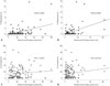
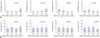
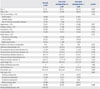
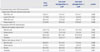
Clinical, procedural, and pre-procedural VH-IVUS characteristics according to the presence of late stent malapposition are summarized in Table 1 and 2. Follow-up OCT characteristics are summarized in Table 3. Pre-procedural absolute total NC and DC plaque volumes were positively correlated with the percentage of malapposed struts (r=0.44, p<0.001, Fig. 1A and r=0.45, p<0.001, Fig. 1B, respectively), and pre-procedural absolute total FT plaque volume was weakly associated with the percentage of malapposed struts (r=0.220, p=0.015). When each relative total plaque volume was classified into quartiles according to the plaque components, the highest NC plaque volume quartile and the highest DC plaque volume quartile had a significantly higher percentage of malapposed struts (p=0.013 for relative NC plaque volume, p=0.020 for relative DC plaque volume) (Fig. 2A). The highest FT plaque volume quartile had a lower percentage of malapposed struts (p=0.040). The lesions with stent malapposition on follow-up OCT had a significantly larger initial absolute total NC plaque volume (30.7±22.3 mm3 vs. 19.5±16.4 mm3, p=0.008) and absolute total DC plaque volume (12.3±14.5 mm3 vs. 5.9±5.4 mm3, p=0.012) (Table 2). Similarly, the lesions with stent malapposition had a significantly larger relative total NC plaque volume (22.1±8.7% vs. 17.4±8.9%, p=0.009) and a larger relative total DC plaque volume (8.6±5.9% vs. 5.9±4.5%,p=0.008) (Table 2). On the pre-procedural IVUS images, among the 37 lesions with late stent malapposition, superficial calcium was observed in 24 lesions (65%) and deep calcium was observed in eight lesions (21%). The remaining five lesions (14%) did not have calcium. Also, the mean calcium length of the 31 calcified lesions was 8.9±8.3 mm, and the proportion of calcium length with regard to total lesion length was 44±30%.
As for the percentage of uncovered struts, there was no significant relationship with absolute total NC plaque volume (r=0.11, p=0.227) (Fig. 1C) or absolute total DC plaque volume (r=0.15, p=0.102) (Fig. 1D). Similarly, there were no significant differences in the percentage of uncovered struts among the relative total volume quartiles of each plaque component (Fig. 2B).
On multivariate analyses, pre-procedural absolute total DC plaque volume was the only independent predictor of late stent malapposition (β=1.12, p=0.002).
DISCUSSION
The main findings of the present study were as follows: 1) preprocedural quantitative assessment of plaque composition by VH-IVUS may predict the degree of late stent malapposition detected by OCT; 2) greater pre-procedural DC plaque burden was associated with greater late stent malapposition and was the only independent predictor for late stent malapposition; and 3) pre-procedural plaque composition was not associated with strut coverage on follow-up OCT after DES implantation.
Until now, there has been little data regarding the association between pre-procedural plaque composition evaluated by VH-IVUS and strut coverage on follow-up OCT after DES implantation. Even though plaque characteristics seem to strongly affect vascular healing responses after DES implantation, no data exists regarding the impact of plaque characteristics on late stent malapposition or coverage of struts, which may be considered potential risk factors for the occurrence of late stent thrombosis.12345 In addition, many previous OCT studies evaluating vascular responses after DES implantation have commonly been limited by a lack of information regarding pre-procedural plaque characteristics of de novo lesions.1617 Given this situation, we sought to investigate the impact of plaque characteristics of de novo lesions on vascular responses following DES implantation using pre-procedural VH-IVUS and follow-up OCT.
Mechanisms of late persistent malapposition have been proposed. Malapposition might be mediated partly by calcified lesions that do not allow for homogeneous stent expansion and could result in the lack of contact of stent struts with the vessel walls.1 Also, as for plaque characteristics affecting the occurrence of stent malapposition, a recent OCT study reported that the presence of calcium was one independent predictor of late persistent stent malapposition after DES implantation.6 Therefore, although we could not classify late stent malapposition into late-acquired and persistent malapposition in the present study, larger DC volumes may be important for late persistent stent malapposition. Other IVUS studies have suggested that late-acquired stent malapposition occurs mainly due to positive remodeling and plaque/thrombus resolution.2021 In the present study, we found that absolute total NC plaque volume is also positively related to the degree of late stent malapposition in univariate analysis. Thus, larger NC volumes may be important for late-acquired stent malapposition. Similar to our study, a previous study reported that a post-stented NC component was associated with the development of late stent malapposition on follow-up IVUS after DES implantation, particularly in patients with acute myocardial infarction and diabetes mellitus.22 However, the present study is different from the previous study in that we assessed the plaque characteristics pre-intervention, not postintervention. Furthermore, we assessed vascular healing responses using OCT, not IVUS, which might weight the importance of this study. Our study showed that pre-procedural plaque composition may play an essential role in formation of late stent malapposition after DES implantation.
Late stent thrombosis after DES implantation is a serious concern because of delayed vascular healing and inflammatory reaction. Uncovered struts on follow-up OCT have been proposed as potential risk factors for late stent thrombosis after DES implantation.45 A previous study showed that coverage of malapposed stent segments is delayed, compared to well-apposed segments, and that the larger the acute malapposition, the greater the likelihood of persistent malapposition and delayed healing at follow-up.4 Thus, we hypothesized that plaque composition, particularly a DC or NC component, may result in delayed strut coverage. However, in the present study, there were no significant associations between the specific plaque components and strut coverage after DES implantation. Considering the results of our previous OCT study, DES type rather than plaque characteristics on VH-IVUS may be the more powerful factor in determining strut coverage.7
This study has several limitations. First, it was not clear whether the malappositions at follow-up were residual or newly acquired because post-procedural intravascular imaging studies were not preformed immediately after DES implantation. Second, this study was based on registry data, and the sample size was quite small. Finally, different types of DES were used.
In conclusion, pre-procedural assessment of plaque composition by VH-IVUS may predict the formation of late stent malapposition detected by OCT at follow-up. Larger pre-procedural DC plaque volumes were associated with late stent malapposition. However, pre-procedural plaque composition was not associated with decreased strut coverage at follow-up after DES implantation.

 XML Download