

 PDF
PDF ePub
ePub Citation
 Print
Print


INTRODUCTION
Idiopathic nephrotic syndrome (NS) is characterized by generalized edema, heavy proteinuria, hypoalbuminemia, and hyperlipidemia, and minimal change disease (MCD) is the most common form of idiopathic NS, accounting for 90% of patients under the age of 10 years and more than 50% of older children.1,2
Although the etiology of idiopathic NS is unknown, several candidate factors have been identified. After the first report of hypersensitivity and NS by Hardwicke3 in 1959, numerous studies have reported an association between NS and allergy.4,5 Several studies have demonstrated elevated serum immunoglobulin E (IgE) levels in NS patients and hypothesized its active role in the pathophysiology, however, a direct relationship between IgE and the pathogenesis of NS is controversial.6,7
Meanwhile, it was proposed that abnormal T cell response and Th2 cytokines are strongly related to the pathogenesis of NS.6,8 Various evidences indicate that lymphocytes derived from an abnormal immune system alter the permeability of the glomerular capillary wall, suggesting that immune dysfunction plays a key role in the pathogenesis of NS.8-11 Activated macrophages and Th2 lymphocytes may be involved in the pathogenesis of NS, and T-cell dysfunction leads to changes in cytokines, causing a loss of negatively charged glycoproteins within the glomerular capillary wall.12,13
Recently, regulatory T cells (Treg) and T-helper17/Treg imbalance has also been suggested as a potential factor in the pathogenesis of MCD and Treg dysfunction and decrease of their related cytokines, transforming growth factor (TGF)-β and interleukin (IL)-10, have been reported.14,15 Especially, TGF-β was suggested as a protective cytokine.14
In this study, we evaluated the clinical characteristics of pediatric steroid-responsive NS patients according to the initial serum IgE levels, and several immune mediators such as Th2 cytokines (IL-4, IL-5) and Treg related cytokines (TGF-β, IL-10) were compared according to their remission status.
MATERIALS AND METHODS
Patients
The study protocol was conducted after approval by the Institutional Review Board of Chungnam National University Hospital, Daejeon, Korea and was carried out after informed consent was obtained from the patients or their legitimate guardians.
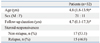
From September, 2004 to July, 2010, the total of 32 patients (21 boys and 11 girls, aged 1.8 to 13.9 years) who underwent prednisolone therapy (17 steroid-sensitive and 15 steroid-dependent) for idiopathic nephrotic syndrome at the Department of Pediatrics, Chungnam National University Hospital were enrolled in this study (Table 1). The median duration of follow-up was 4.7 years. The patients with congenital or secondary renal diseases were excluded. The diagnostic criteria for idiopathic NS were based on the International Study of Kidney Disease in Children.16 Five healthy children (1 boy and 4 girls, aged 6 to 8 years) were included as normal control group.
Study design
The children during their first episode of NS were admitted to the pediatric wards prior to steroid treatment, and clinical data including age, gender, and blood pressure were collected. The blood sample was drawn to evaluate white blood cell count, eosinophil count, platelet, protein, albumin, cholesterol, triglyceride, blood urea nitrogen, creatinine, electrolytes, and various immune mediators, such as complement 3 and 4, antineutrophil antibody, antistreptolysin O and C-reactive protein.
The proteinuria, hematuria and renal functions were screened by spot and 24-hour urine analysis. When MCD was presumed, prednisolone therapy was initiated at a dose of 60 mg/m2/day after confirming a negative purified protein derivative skin test.
All of the steroid responsive NS patients were divided into a normal IgE group and a high IgE group according to the initial serum IgE levels. The patients who showed higher IgE levels, measuring more than upper 2 standard deviation score of the age-adjusted reference range, were defined as high IgE group.17 Remission was characterized by a marked reduction in proteinuria (to <4 mg/m2/hr or urine albumin dipstick of 0 to trace for 3 consecutive days) in association with resolution of edema and normalization of serum albumin to at least 3.5 g/dL. Relapse was defined as recurrence of massive proteinuria (>40 mg/m2/hr, urine protein/creatinine ratio >2.0 mg/mg, or urine albumin dipstick ≥2+ on 3 consecutive days), most often in association with recurrence of edema.2
Measurement of the serum immunoglobulin and cytokine levels
During the nephrotic phase prior to steroid treatment and remission phase, the serum was separated from whole blood and was kept at -70℃ for immune cytokine assay. The serum IgE levels were analyzed by Hitachi optigen allergen specific IgE assay system Korean inhaler panel (Hitachi chemical diagnostic, Inc., Mountain View, CA, USA) based on manufacturer's method. The serum levels of IgG, IgA, and IgM were determined by standard enzyme immunoassay techniques (Seiken, Denka Seika, Nigata-ken, Japan).
The serum IL-4, IL-5 and TGF-β levels were measured by human platinum enzyme-linked immunosorbent assay (ELISA) kit (eBioscience, San Diego, CA, USA). The serum IL-10 levels were detected by human high sensitivity ELISA kit (eBioscience). Briefly, standard dilutions and samples were pipetted into the wells. And the concentrated streptavidin-horseradish peroxidase solution was added to all the wells. After a wash to remove any unbound reagent, a TMB substrate solution was added to the wells and the intensity of the color was measured by a spectrophotometer at 450 µm.
Statistics analysis
The clinical and laboratory information of the NS patients were expressed as a median and range (minimum to maximum) by frequent analysis. The configuration with NS patients between normal IgE and high IgE levels were analyzed by using Mann-Whitney U test and Fisher's exact test. The Kruskall-Wallis test and Scheffe's post hoc comparison were used to compare the cytokines between NS patients with nephrotic and remission phase and healthy controls.
Significant correlation between circulating cytokines and serum albumin levels was assessed by linear regression analysis. The data were analyzed using SPSS version 12.0 for Windows (SPSS Inc., Chicago, IL, USA), and a p value<0.05 was considered significant.
RESULTS
Clinical outcomes according to the initial serum IgE levels
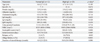
Subjects were divided into two groups; 12 patients (7 boys and 5 girls) who had normal IgE levels and 20 patients (14 boys and 6 girls) who had high IgE levels. The clinical characteristics among the groups were compared by Mann-Whitney U test and Fisher's exact test. At the time of initial diagnosis, the data of their age, gender, and the laboratory findings of blood chemistry such as white blood cell count, eosinophil count, serum protein, albumin, cholesterol, IgG, IgA, IgM, immune mediators, and 24-hour urine protein/creatinine did not show any significant differences between the two groups (Table 2). However, the duration of clinical remission was significantly shorter in the normal IgE group than in the high IgE group (p=0.005). The relapse rate (p=0.003) and the concomitant allergic diseases (p<0.001) were more frequent in the high IgE group. The median (range) time to the initial remission was 8.5 (7.0-12.0) days in the normal IgE group and 12.5 (8.0-32.0) days in the high IgE group (p=0.005) and the median (range) duration of total steroid therapy was 4.0 (2.5-22.0) months in the normal IgE group and 54.0 (3.0-108.0) months in the high IgE group (p=0.006) (Table 2). Total follow-up durations of two groups were same.
Comparison of serum cytokines according to the serum IgE levels
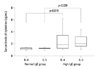
In the normal IgE group, the median (range) initial serum levels of each IL-4 and IL-5 were 1.3 (1.0-1.4) and 1.3 (0.9-1.4) pg/mL, respectively. In the high IgE group, the serum levels of IL-4 and IL-5 was 1.9 (1.0-6.2) and 2.0 (1.1-4.4) pg/mL, respectively. Initial serum levels of IL-4 (p=0.018) and IL-5 (p=0.039) were significantly higher in the high IgE group than in the normal IgE group (Fig. 1). However, the serum levels of IL-10 and TGF-β showed no difference between them (data are not shown).
Comparison of serum IgE levels according to the remission state
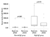
There were no changes in the median (range) levels of serum IgE according to the treatment state (nephrotic or remission phase) between the normal and high IgE groups (Fig. 2).
Changes of serum cytokines according to the disease activity
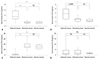
To explore the disease activities and the roles of cytokines, the serum levels of IL-4, IL-5, IL-10, and TGF-β were examined in both NS patients and healthy controls. The serum levels of IL-4 were significantly higher in the nephrotic phase of NS than in the remission phase (p<0.001) and control group (p<0.001). However no difference was found between remission phase of NS and control group (p=0.991) (Fig. 3A).
The serum levels of IL-5 were higher also in the nephrotic phase patient than those of remission phase of NS (p=0.051) and controls (p=0.028), but there were no significant differences between the patients with remission phase and control group (p=0.858) (Fig. 3B). The serum levels of IL-10 did not show any differences between both groups (Fig. 3C).
The serum levels of TGF-β in the nephrotic phase of NS patients were significantly lower than those of remission phase (p<0.001) and control group (p<0.001). However, there was no statistically significant difference between patients with remission phase and control group (p=0.576) (Fig. 3D).
Correlation between serum albumin and cytokines
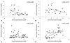
Linear regression analysis showed that the serum levels of IL-4 had a negative correlation with that of albumin (R2=0.259, p=0.004) (Fig. 4A), while the serum levels of IL-5 and IL-10 did not show any correlations with that of albumin (R2=0.170, p=0.113 and R2=0.001, p=0.571, respectively) (Fig. 4B and C). On the other hand, the serum level of TGF-β had a positive correlation with that of albumin (R2=0.501, p<0.001) (Fig. 4D).
DISCUSSION
The pathogenesis of idiopathic NS is not thoroughly understood. However, the hypothesis was put forward in 1974 that idiopathic NS is an immune disorder with increased levels of lymphocyte-derived permeability factors. Since then, numerous lines of evidence have been presented, suggesting that immune mediators play a pivotal role in its pathogenesis.6-8,10,18
Although a pathogenic relationship remains to be confirmed, the serum levels of IgE show a close relationship to NS, and many studies have shown a strong association between idiopathic NS and atopic disorders.4,5 IgE levels were significantly higher in NS patients with atopy than in nonatopic patients, especially during the relapse phase of NS compared with the remission phase, implicating IgE as a barometer of disease severity and giving it prognostic value.6,19 In our present study, in a comparison of the clinical characteristics of NS patients with normal IgE and those with high IgE, the high-IgE group required a significantly longer time to reach their initial remission, was more susceptible to frequent relapse and comorbid allergic diseases, and required longer steroid therapy to recover from the nephrotic phase than the normal-IgE group. Serum IgG levels were lower in NS patients, whereas the levels were not different between the normal- and high-IgE groups. It is not certain whether the increased levels of IgE in idiopathic NS are pathogenic or co-incident, nevertheless, our results suggest that the regulation of total IgE production correlates with the disease activity and outcome of NS.20 However, it should be noted that part of the nephrotic children had persistently normal serum IgE levels, indicating different etiologies in the pathogenesis of NS.
The signals for IgE synthesis are delivered by IL-4 or IL-13. In relapsed NS, activated T cells increase the secretion of IL-4 and IL-13, which could induce isotype switching in IgE. IL-5 also promotes the production of IgE, whereas IFN-γ inhibits the secretion of IgE.13,21,22 In our study, high-IgE group displayed significantly elevated serum IL-4 and IL-5 levels. Thus, we suggest that the increased production of IL-4 and IL-5 in idiopathic NS may indicate that these cytokines are involved in the enhanced production of serum IgE. Furthermore, allergic treatment alone could not induce the remission of NS, suggesting that the elevated serum IgE in some patients can not cause their NS, but rather reflects allergic responses to the perturbation of their humoral immunity.4,23
In order to evaluate immunologic pattern and their modulating serum cytokines and to analyze the correlation with disease activity in children with idiopathic NS, we evaluated initial serum levels of IL-4, IL-5, IL-10 and TGF-β and their kinetics during treatment with or without relapse. A number of reports have shown that activated T cells of idiopathic NS are driven toward the Th2 phenotype.13
IL-4 is the most important Th2 cytokine which mediates IgE synthesis.24 In NS, IL-4 can act on glomerular visceral epithelial cells by binding IL-4 receptor, and plays a role in glomerular permeability.25 This suggests that, regardless of atopy, IL-4 may be one of the cytokines associated with the pathogenesis of proteinuria and also be a predictive factor for disease severity in NS. In our present study, the serum levels of IL-4 were higher in the nephritic phase of NS than the remission phase and control group and were correlated with serum albumin levels. However, this seems to be variance with the results in many previous studies that serum IL-4 levels decreased during relapse but increased in nephritic patients with long-term remission.26
Several authors have reported that serum IL-5 levels in the nephrotic phase were higher than in the remission phase and Ohtomo, et al.27 reported that suplatast tosilate, which suppresses IL-4 and IL-5 production, was effective for steroid reduction in idiopathic steroid sensitive NS.13,23 We also found that serum IL-5 levels were higher in the nephrotic phase than in the remission phase with steroid treatment. However, because other hither-to-uncharacterized factors might have been needed to induce idiopathic NS, further studies should be conducted to clarify the effects of IL-4 and IL-5 on idiopathic NS.
Recently, regulatory T cells (Treg cells) have been described as subsets distinct from Th1 and Th2 cells. Treg cells expressing the forkhead/winged helix transcription factor (Foxp3) have an anti-inflammatory role and maintain tolerance to self-components through direct contact with cells or by releasing anti-inflammatory cytokines such as IL-10 and TGF-β.28,29 Treg cell dysfunction has been found in several kidney disease animal models.30-32 A reduction in Treg cells induces local tissue inflammation, which may be related to the pathogenesis of proteinuria and the propagation of idiopathic NS.33 In relapsed idiopathic minimal NS, Treg cells have an impaired capacity to suppress T-effector-cell proliferation.14 Liu, et al.15 reported that Th17/Treg ratios were increased along with increased proteinuria and decreased albumin levels in adult patients with minimal change NS.
TGF-β has been shown to regulate both immunogenic and immunosuppressive response, depending on the cellular environment.34 Therefore, overall systemic effects of TGF-β must be understood within the context of other collaborative networks involving other immunoregulatory signals.
TGF-β is often associated with the progression of kidney disease, and exhibits fibrogenic and proinflammatory properties in the kidney.35-37 Proteinuria predominantly involves albumin, which triggers the positive feedback loop for TGF-β expression, subsequently inhibiting albumin endocytosis.38 Prolonged proteinuria gives rise to increased TGF-β, which modulates the expression of the extracellular matrix, leading to glomerulosclerosis and interstitial fibrosis, and retarding the progression to renal failure.39
On the other hand TGF-β may promote the expression of Foxp3 and induce the differentiation of Treg cells that which originate from CD4+CD25-T cells.40,41 TGF-β is the inhibitor of Th17 differentiation in humans,42 and it also inhibits the vascular permeability factor released by T cells in normal subjects and patients with minimal change disease.9 In the present study, we found that TGF-β and IL-10 concentrations decreased in minimal change NS patients and positively correlated with circulating Treg frequencies, which suggested that TGF-β and IL-10 may play a protective role, partly through inducing the production of Treg cells, and act as one of the effective factors of Treg cells.15
IL-10 is also pleiotropic cytokine that can exert either immunosuppressive or immunostimulatory effects on a variety of cell types and plays a central role in controlling inflammatory processes.43 Previous studies have shown that IL-10 is involved in the positive feedback loops with TGF-β and acts at local sites of inflammation, whereas TGF-β is involved in the systemic immune response related to humoral lymphoproliferation.44,45
In this study, the initial serum levels of lL-10 and TGF-β did not differ between the normal- and high-IgE groups, whereas the initial serum TGF-β levels in the nephrotic phase of NS were lower than in the remission phase or in the healthy controls, and differed significantly according to the degree of proteinuria and serum albumin level. However, serum lL-10 levels were not significantly different between the groups. The results of this study suggest that insufficient levels of TGF-β in the nephrotic phase of NS may aggravate the inflammatory responses and contribute to the induction of proteinuria. To better understand the nature, regulation, and function of Treg cells and their cytokines in idiopathic NS, further studies should be carried out.
 XML Download