

 PDF
PDF ePub
ePub Citation
 Print
Print


INTRODUCTION
The causative agent of scrub typhus, Orientia tsutsugamushi, is obligate intracellular bacteria proliferating within eukaryotic cells.1 Several studies on the effect of interferon on the growth of rickettsia have been conducted, and interferon-γ has been reported to inhibit the growth of rickettsia in vitro; however, its direct effect on recovery from acute infection and prevention of recurrence has not yet been clearly clarified.2,3
Supplemental administration of corticosteroids for rickettsial diseases is still controversial, and definitive data to substantiate its beneficial or detrimental effects are not sufficient. Physicians can be confronted with the dilemma of initiating immunosuppressive corticosteroid treatment in a critically ill patient, for which the tentative diagnoses include immunologically mediated anemia and/or immunity-mediated thrombocytopenia or severe rickettsial disease.4
Indeed, in some studies, the simultaneous administration of corticosteroid to severe rickettsia-infected patients with concomitant multiorgan impairment has been reported to be of help to the improvement of the clinical course or reduction of mortality.4-6 But in a clinical course, the direct effects of drugs and many diverse factors may be involved and it is difficult to evaluate the pure effect of drugs in clinical studies. The possibility that corticosteroids could stimulate the proliferation of pathogens in some infectious diseases could be a limiting factor to the additional administration of corticosteroids.4
Therefore, we objectively evaluated the effects of the administration of corticosteroids on the growth rate of Orientia tsutsugamushi in an L929 cell line infected in vitro.
MATERIALS AND METHODS
Cell culture
Murine L929 cells provided by the American Type Culture Collection (CCL1, Rockville, MD, USA) and kept at the Department of Microbiology and Immunology at Seoul National University were obtained and used. L929 cells were cultured in Dulbecco's modified Eagle's medium (DMEM, Gibco BRL, Grand Island, NY, USA) containing 10% (vol/vol) fetal bovine serum (FBS), penicillin (100 unit/mL), streptomycin (100 µg/mL), and 2 mM L-Glutamine in a 37℃ incubator maintained 5% CO2. When the cells grew and covered 80-90% of the bottom of culture flasks, they were either infected with rickettsia or a subculture was performed.
Rickettsia
In 6-well plates (Corning Glass Works, Corning, NY, USA), 106 cells per well were added, cultured for 24-36 hr, and the L929 cells formed a monolayer that was infected with Orientia tsutsugamushi Gilliam at a density of 0.5 particle per cell. Subsequently, DMEM medium was added, containing 5% FBS, the cells were incubated in a 35℃ incubator maintained at 5% CO2, and the cell proliferation level was examined.
After more than 90% of cells were infected, the cultured L929 cells were collected using a scraper and the collected cell suspension was disrupted by glass dounce (tight piston, Wheaton Inc., Millville, NJ, USA). The disrupted cell solution was centrifuged at 500×g for 5 min, cell debris was removed, and the supernatant was collected. The supernatant was centrifuged at 1,000×g for 10 min. Precipitates were collected and resuspended in DMEM medium, aliquoted as 1 mL, stored at -80℃ in liquid nitrogen, and used in the experiments.
Antibody and indirect immunofluorescence staining
Anti-Orientia tsutsugamushi Gilliam mouse serum was obtained by injecting C3H mice 2-3 times with rickettsia at 2-week intervals and sampling after 4 weeks. Using an anti-Orientia tsutsugamushi Gilliam mouse serum with the titer higher than 1 : 640 and FITC-conjuated goat anti-mouse IgG (Cappel Laboratories, Cochranville, PA, USA) diluted 1 : 100 in PBS, rickettsia-infected cells were stained by indirect immunofluorescent antibody method and examined under a fluorescent microscope (microscope trinocular, Olympus, Tokyo, Japan). Infection levels of the L929 cells were assessed and flow cytometry (FACSCalibur, Becton Dickinson, San Jose, CA, USA) was used to evaluate infected cell proliferation.
Addition of corticosteroids
To evaluate the effect of corticosteroids on the proliferation of cells themselves, 1 mL DMEM medium containing dexamethasone (Sigma Chemical Co., St. Louis, MO, USA) at final concentrations of 101, 103, 105, and 107 pg/mL was added to the L929 cells cultured as a monolayer in six wells and by varying the culture period, cell viability level was measured by MTT assay and hemocytometer.7
To examine the effects of rickettsia infection, rickettsia suspension was added to L929 cells, cultured at 35℃ for 1 hr, and under the infected conditions, 1 mL DMEM medium with dexamethasone (101 to 107 pg/mL) was added and cultured for six days.
Interferon-γ administration
Monolayers of L929 cells in 6-well plates (Corning Glass Works, Corning, NY, USA) received maintenance DMEM medium containing an appropriate amount of interferon-γ (Serotec, Oxford, UK) and were incubated for 24 hrs. After aspiration of the medium, the cells were inoculated with the rickettsia at multiplicity of infection and absorbed for 1 hr at 35℃. Unabsorbed rickettsiae were aspirated off, and each culture was fed with 1 mL of maintenance medium containing interferon-γ. Untreated control cultures were inoculated with rickettsia and treated with maintenance medium alone. Cultures were incubated at 35℃ and processed for flow cytometry at various intervals.8
Preparation of samples used for flow cytometry
The fixation and staining of cells infected with rickettsia was performed as suggested in previous studies.8,9 Summarizing briefly, after rickettsial infection and addition of corticosteroid, cells cultured for a certain time were washed with PBS, PBS was added to cell precipitates obtained by the dissociation of cells using 0.25% trypsin-0.1% etylene-diamine tetraacetate-PBS, centrifuged, and resuspended to 106 cells/mL. Cell precipitates obtained by centrifugation at 5,000 rpm for 15 sec were resuspended with 4℃ PBS 100 µL and -30℃ methanol 900 µL was added and kept at -30℃ for more than 15 min. These cells were stored at -30℃ until staining. As described above, the fixed cells were stained by the indirect immunofluorescent antibody method, washed with PBS, and used for flow cytometry analysis.
Flow cytometry measurement
Rickettsia particles within L929 cells appearing as green fluorescence were measured by EPICS Elite flow cytometer (Coulter Corp., Hialeah, FL, USA) and 20,000 cells were analyzed to find the fluorescence level of each sample. The excitation wave length was 488 nm, and data were presented as fluorescence intensity, the cellular frequency of its log scale.
RESULTS
Cellular morphology of L929 cells after Orientia tsutsugamushi infection and the addition of corticosteroids
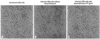
Considering the results of previous studies regarding morphological changes of mouse fibroblasts prior to and after Orientia tsutsugamushi infection, uninfected L929 cells, L929 cells infected with Orientia tsutsugamushi, and L929 cells treated with corticosteroids after infection were cultured.10 At 24-hr intervals between 24 and 96 hrs, the gross morphology of the L929 cells was examined by phase contrast microscopy. No differences were detected in the number of cells or morphology according to the status of Orientia tsutsugamushi infection or corticosteroid doses (Fig. 1).
Alteration of growth of L929 cells according to corticosteroid concentration
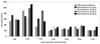
To examine the effect of corticosteroids on the proliferation of L929 cells to be infected Orientia tsutsugamushi, cells were cultured for 10 days with various concentrations of corticosteroids and the number of cells was checked. The results showed that during 1-7 days of culture, cell proliferation was not affected by the presence or absence of corticosteroid, nor was it influenced by the dosage of corticosteroid (Fig. 2). After day 3, it was observed that cell proliferation decreased markedly because cells were in a fully saturated state.
Effect of corticosteroid on Orientia tsutsugamushi growth?
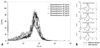
Corticosteroid concentration was raised from 101 pg/mL to 107 pg/mL by 10-fold. The growth level of Orientia tsutsugaobmushi was evaluated by flow cytometry and the results showed that differences in the growth of Orientia tsutsugamushi with or without the addition of corticosteroids as well as added dosages were not detected (Fig. 3). To evaluate the reliability of our results, experiments that assessed the effects of interferon on the growth of Orientia tsutsugamushi by flow cytometry reported in other studies were used as the control group in our study and, similar to the previously reported results, reduction of the growth of Orientia tsutsugamushi in response to interferon-γ administration was confirmed (Fig. 4).8
DISCUSSION
Orientia tsutsugamushi is classified as one species; however, diverse serotypes are present. Among these, the standard serotyes are Gilliam, Karp, and Kato, with various serotypes having been reported in different countries. In Korea, Gilliam, Karp, and Boryung are the major serotypes, and the Boryung serotype has been isolated most abundantly.11 As shown by recent clinical studies, the clinical manifestation of patients and laboratory results are differ greatly and opinions suggesting that this is due to serological differences have been reported. In the literature, all previous reported deaths were caused by the Gilliam serotype. This suggests the possibility that the severity of clinical manifestation is influenced by serotype.12,13 The Gilliam serotype associated with severe rickettsial disease was used in our experiments because the use of corticosteroids was considered in cases suspected to be severe rickettsial disease. Although it was not shown in our results, similar results have been obtained in experiments using the Karp serotype.
For the treatment of scrub typhus, administration of 100 mg of doxycycline twice a day for 3-7 days is recommended.14 This treatment has been reported to have excellent results, and the mortality rate has decreased noticeably from 50% before use of antibiotics was introduced. Recently, in the northern area of Thailand, Orientia tsutsugamushi pathogen showing resistance to doxycyclin was isolated and showed a tendency of delayed response to antibiotics, but this did not raise mortality.15 Although not frequently encountered, other substitutions could not be suggested for severe rickettsial diseases besides antibiotic and conservative treatments. In some patients, progression to severe infection with involvement of major organs (such as resulting in encephalitis, myocarditis, kidney injury, and interstitial pneumonitis) occurred before the antibiotics took effect. Administration of corticosteroid to such patients showed inconsistent results.16 In severe rickettsial disease with immune mediated thrombocytopenia and hemolytic anemia, it is difficult for physicians to decide to use corticosteroids at the immunosuppressive prednisolone dose (2.0 mg/kg).4
In terms of the use of corticosteroids for bacterial diseases, the most investigated area is septic shock. Prior to the 1940s, corticosteroid treatment had been administered empirically to severe septic shock patients. Upon summarizing the results of clinical studies conducted for the objective evaluation of effects, its effects remian unclear.17,18 Despite these results, in catecholamine-dependent septic shock, low-dose corticosteroid treatments have been continuously investigated because of relative adrenal function deterioration, peripheral steroid resistance, etc. Recently, effects on the improvement of symptoms, disease duration, and mortality have been reported.19 However, in severe sepsis, short-term administration of high-dose corticosteroids is not recommended except in cases such as typhoid fever, pneumocystitis carinii pneumonia in acquired immune deficiency syndrome, or bacterial encephalomeningitis in children.20 The outcome of treatment with corticosteroids in bacterial infections is difficult to discuss completely. Time of administration, dosage, maintenance time of corticosteroids as well as antibiotics, causative pathogens, and characteristics of the infectious disease should be considered.
In rickettsial diseases, administration of corticosteroids is frequently applied to Rocky Mountain Spotted Fever (RMSF) that shows ocular symptoms including uveitis, ocular hemorrhage, exudate retinosis, etc.21 In addition, it has been reported that in severe RMSF infection accompanying initial endotoxemia, hyperazotemia, or hypoproteinemia, cases in which cortisone and antibiotics were administered together had better treatment outcomes than those cases treated with antibiotics alone.5 There have been animal-modeled experiments to show the theoretical basis for this. Because of the similarity of diseases between humans and dogs, Rickettsia rickettsii infected animal models with the using dogs is a proper methodology for human infection research.22 Based on recent animal-modeled research on corticosteroid adjuvant therapy, it has been reported that concurrent use of corticosteroids and doxycycline confers no clinically relevant detrimental effects.4
However, when we tried to apply positive treatment results from RMSF to scrub typhus, there was one serious limitation. We were not able to control the direct risk of corticosteroid for the growth of Orientia tsutsugamush, because it could cause adverse effects. Although one important limitation of our study is that in vitro experiments on the effects of dexamethasone on cells in culture cannot be extrapolated to the complex in vivo situation, our results do have meaning in showing that corticosteroid itself does not increase the growth of Orientia tsutsugamush, and also in reporting that the concurrent use of anti-inflammatory or immunosuppressive doses of corticosteroid in conjunction with antimicrobials did not lead to adverse influences on the course of scrub typhus, because it could not induce bacterial growth. Recently, there have been some research into the positive effects of corticosteroid in rickettsial disease.6,23 After this study, more clinical research is needed, such as animal model experiments or various experiments to find the proper dosages, times and medication duration for corticosteroids as treatment for serious rickettsial disease.
 XML Download