

 PDF
PDF ePub
ePub Citation
 Print
Print


INTRODUCTION
Cardiac myxoma is a rare disease with an overall incidence of about 0.5/million/year1 and accounts for approximately 70 percent of all primary cardiac tumors.5 Several studies of cardiac myxoma involving large sample sizes have been reported in the past decade.2-4 Recently, well developed preoperative diagnostic techniques, especially echocardiography, have enabled earlier detection than was previously possible. Therefore, the number of patients who undergo surgery has increased, and long-term results have improved. This report reviews patients who underwent surgery for cardiac myxoma in our center and the long-term results of treatment.
MATERIALS AND METHODS
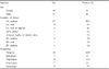
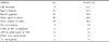
Seventy-four patients underwent surgery for cardiac myxoma in the Yonsei Cardiovascular Center between August 1980 and February 2005. The medical records were retrospectively reviewed and some patients were contacted by telephone for follow-up. During this period, cardiac myxoma was the most common primary cardiac tumor in our center, comprising 81.4% of all primary cardiac tumors. Twenty-one patients (28.4%) were male and 53 (71.6%) were female (Table 1), and the mean age was 50.4 ± 15.0 years (range, 7 to 80 years). The locations of tumors were: left atrium in 70 patients, right atrium in three patients, and right ventricular septum in one patient. Preoperative symptoms were dyspnea, which occurred in 44 patients (59.5%), an embolic event in 8 (10.8%), palpitations in 9 (12.2%), chest pain in 8 (10.8%), and neurologic symptoms such as dizziness or syncope in 5 (7.1%). One patient, who underwent an emergency Fogarty embolectomy of both iliac arteries due to acute embolism, was found to have myxoma of the heart and underwent surgery for cardiac myxoma. In another patient, cardiac myxoma was found incidentally during a procedure for a ventricular septal defect and aortic regurgitation. Twenty patients (27.0%) were greater than functional class III of the New York Heart Association. Forty-two patients were diagnosed by transthoracic echocardiography only, while thirty-two patients required both transthoracic and transesophageal echocardiograms for more accurate anatomic descriptions. All patients over 40 years of age underwent a coronary angiogram to rule out coronary disease. Only one patient who showed shortness of breath was diagnosed as having coronary artery obstructive disease. The mean duration from onset of symptoms to diagnosis was about nine months (range, 1 week to 180 months). Chest X-rays showed abnormalities in 21 patients (28.4%), including cardiomegaly, pulmonary edema, and pleural effusion. Electrocardiography showed abnormalities in 36 patients (48.6%), including atrial fibrillation, left atrial enlargement, and right axis deviation. These abnormalities are frequently seen in mitral valve stenosis. Median sternotomy was performed in all patients, and the operative procedure was performed under moderate hypothermia and an arrested heart with cardiopulmoary bypass (Table 2). The mean cardiopulmonary bypass time and aortic cross clamp time were 100.4 ± 37.1 and 64.8 ± 29.8 minutes, respectively. The size variation of tumors was from 1 × 1 × 1 cm to 12 × 5 × 3 cm. In 70 cases, the tumor was located in the left atrium, of which 60 cases (81.1%) were derived from the septum, five cases (6.8%) from the free wall, three cases (4.1%) from both the left atrial septum and the free wall, and one each from the orifice of the left pulmonary vein and the anterior leaflet of the mitral valve. If the tumor was located in the left atrial side, the vent sucker was inserted after opening of the left atrium and aortic cross clamping. In three cases, the tumor was in the right atrium, with two of those involving the right atrial septum and one involving the free wall. The remaining case involved a tumor on the septal wall of the right ventricle. One patient had two separate tumors in the left atrium, with sizes of 8 × 4 × 3 cm and 6 × 4 × 3 cm. Another patient, whose sister had also undergone surgery for cardiac myxoma at another hospital, had three separate tumors in the left atrium, with sizes of approximately 2 × 1.5 × 1 cm. For resection of the tumors, 38 patients (54.3%) required a biatrial approach, 26 patients (37.1%) required a right atrial approach, and 10 patients (14.3%) required a left atrial approach. Where possible, tumors were resected with the en bloc method for complete resection and visual examination was then performed to prevent postoperative embolic events. For repair of a defective area, autologous pericardium or an artificial patch was used in 52 patients (70.3%), and direct closures were possible in 20 patients (27.0%). The myxoma located in the septum of the right ventricle was resected incompletely due to severe attachment with the septal wall. The following associated procedures were performed: mitral annuloplasty in three cases, tricuspid annuloplasty in three cases, coronary artery bypass graft with mitral annuloplasty in one, aortic valve replacement with patch repair of a ventricular septal defect in one, and mitral valve replacement in one.
RESULTS
The mean durations of stay in the intensive care unit and hospital were 2.3 ± 1.3 days and 11.9 ± 7.6 days, respectively. There were no operative mortalities but seven cases (9.5%) of postoperative complications: two cases of atrial fibrillation and one case each of mitral regurgitation, perioperative myocardial infarction, pneumonia, sepsis, and wound complication (Table 3). All patients recovered from their postoperative conditions except for the 59 year-old patient with mitral regurgitation. The cause of this regurgitation was thought to be incidental injury to the mitral valve leaflet; this patient underwent mitral valve replacement with a tissue valve on the first postoperative day. All patients underwent an echocardiogram before discharge to evaluate the possibility of residual tumor. During follow up echocardiography was performed as required by the physician according to patients' symptoms. All patients received follow-up for a mean duration of 105.7 ± 73.6 months. There were six (8.6%) late deaths; however, there was no evidence that these deaths were directly related to cardiac myxoma. In 13 patients electrocardiographic abnormalities that were present before surgery had disappeared. The patient who underwent mitral valve replacement on the first postoperative day needed a second mitral valve replacement 12 years later due to severe mitral regurgitation of the tissue valve. There was no increase in tumor size during the 14 month follow-up period in the patient whose tumor was incompletely resected due to severe attachment with the right ventricular septum. The remaining 73 patients, including the late deaths, showed no evidence of tumor recurrence clinically or on echocardiograms (between 7 days to 282 months postoperatively) during the follow-up period.
DISCUSSION
About 90 percent of primary cardiac tumors are known to be benign tumors.6 Cardiac myxoma comprises about 70 to 80 percent of primary cardiac tumors and are the most commonly operated on. Since cardiac myxoma exhibits rapid growth most patients show advanced symptoms at the time of surgery.2 However, if the tumor is resected completely, the recurrence rate is very low.2,4 Possible risk factors for recurrence of cardiac myxoma include incomplete resection, intracardiac implantation, embolization, and a reserve of tumor precursor cells in the subendocardium.3 It has been reported that preoperative symptoms are most commonly embolism or heart failure due to obstructive lesions, and emergency surgery is required in cases showing embolic symptoms.3 The risk of embolism is higher for polypoid or multilobular shaped tumors than round ones.7,8 Therefore, if a polypoid or multilobular shaped myxoma is suspected on preoperative echocardiography, urgent surgical intervention is necessary to prevent an embolism. Recent studies on the resection of cardiac myxoma with normal cardiac tissues indicated that there was no difference in recurrence rates between patients who underwent an en bloc resection and resection with only endocardial tissue attached with the tumor.9 However, the type of resection depends largely on factors such as the location of the tumor and the shape of tumor stalks. In the present study, repair of a large defective area with autologous or artificial patches after tumor resection was necessary in 48 cases, and one case required re-operation due to excessive resection of the mitral valve tissues. Incomplete resection may result in cases where the tumor is attached to the mitral valve or conduction tissue, or occurs in the ventricular septum. Endocardial resection of the tumor is considered acceptable if postoperative complications are expected due to excessive resection of tissue, or if the tumor has a narrow based stalk attached to the endocardium.
Centofanti and associates reported that four-chamber examination after tumor resection for the removal of remaining tumor tissues or debris is not warranted due to the recent development of perioperative transesophageal echocardiography.9 However we feel that, whenever possible, a thorough visual examination is beneficial for the prevention of postoperative embolism. In the majority of our cases the biatrial approach was used, allowing more thorough examinations. As reported by Jones and associates,10 the advantages of the biatrial approach are: direct visualization allowing a more clearly defined tumor pedicle, minimal manipulation of the tumor, adequate margins of excision, inspection of all heart chambers, and secure closure of the atrial septal defect. Familial myxoma accounts for 5% of cases, and it is known to recur even after several decades.2,11 Although one of our cases was suspected to be familial myxoma, there was no evidence of recurrence during the follow-up period. Thorough examination of the patient's past history and family history was needed in this case, and regular follow-up was essential. In our study there was one case of incomplete resection due to severe infiltration to the right ventricular septum. Gawdzinski and associates reported a similar case of right ventricular myxoma, and performed endocardial decortication of the right ventriclular septum and a tricuspid valve replacement due to tricuspid valve involvement.12 However, the patient underwent re-operation after three months due to severe thrombotic tricuspid valve obstruction and recurrence of the tumor. In the present case, there was no increase in tumor size and no thrombotic evidence in the final follow up echocardiogram performed 10 months after the operation. However, thorough follow-up is important for this young patient.

 XML Download