

 PDF
PDF ePub
ePub Citation
 Print
Print


INTRODUCTION
Aneurysms of the deep femoral artery are very rare because of their anatomical features and the characteristics of the arterial wall.1-4 Most patients with a deep femoral artery aneurysm have few symptoms until it expands, and once this happens, rapid enlargement is noted. However, they have a high rate of rupture in comparison with other peripheral arterial aneurysms, and local pressure symptoms caused by the aneurysm could precipitate nerve and vein compression and thrombosis in some cases. To establish the diagnosis, color Doppler ultrasound, multi-detector computed tomographic (CT) scanning, and angiography are all extremely useful. Multi-detector CT scanning or angiography is especially recommended to avoid missing other aneurysms and occlusive arterial lesions. In our case, a multi-detector CT scan confirmed the diagnosis of a deep femoral artery aneurysm and revealed no evidence of aneurysms or occlusive lesions in the other arteries.
CASE REPORT
A 58-year-old man was admitted to the hospital, with a 3-day history of acute swelling of the right lower limb. He had undergone aortic valve replacement surgery due to severe aortic regurgitation at an outside facility 7 years before and was being treated with warfarin (5 mg daily, adjusted to maintain a prothrombin time of 2.0 INR). He denied any history of previous trauma, fractures, intravenous drug abuse, or recent infections. Physical examination revealed an 8 by 10 cm, tender, pulsatile mass in the right femoral area and swelling in the right lower limb. Both popliteal and pedal pulses were palpable. Apart from a prolonged prothrombin time (2.11 INR), blood chemistry parameters were unremarkable.
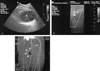
A color Doppler ultrasound showed a huge deep femoral artery aneurysm posterior to the superficial femoral vein, measuring approximately 8.5 × 9.0 × 7.5 cm (Fig. 1a). The deep femoral artery aneurysm compressed the superficial femoral vein resulting in venous stasis with suspicious soft thrombus formation in the right lower extremity veins (Fig. 1b). A multi-detector CT scan (GE CT LightSpeed Ultra 16, GE, Milwaukee, Wisconsin, USA) revealed a large pseudoaneurysm arising from the deep femoral artery without evidence of aneurysms or occlusive lesions in the other arteries (Fig. 1c).
An emergent operation was performed, during which an 8-cm diameter aneurysm, originating from the deep femoral artery, was identified posterior to the superficial femoral artery and vein. The superficial femoral vein was compressed by the aneurysm but there was no thrombus inside the vein. After clamping of the deep femoral artery proximal and distal to the aneurysm, the aneurysm was resected with ligation of the deep femoral artery. After resection of the aneurysm, venous flow of the superficial femoral vein was restored.
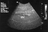
After surgery, the patient experienced rapid resolution of symptoms. A follow-up color Doppler ultrasound and multi-detector CT scan showed normal flow in the superficial femoral and popliteal arteries and veins without evidence of an aneurysm (Fig. 2). The postoperative course was uneventful and the patient was discharged without complication.
DISCUSSION
Isolated aneurysms of the deep femoral artery are rare and account for only 0.5% of peripheral aneurysms and 1-2.6% of femoral aneurysms.1-4 The reason for its rarity is primarily due to its anatomical characteristics. Several muscles cover the deep femoral artery and it has a rich muscle layer with few elastic fibers.4-6 Most patients with a deep femoral artery aneurysm have few symptoms until it expands, and once this happens rapid enlargement is noted. However, they have a high rate of rupture in comparison with other peripheral arterial aneurysms, resulting in emergent surgical procedures with a high amputation rate.7,8 In one series, one in three ruptured deep femoral artery aneurysms resulted in amputation.1 Additional complications include acute expansion with local pressure symptoms such as femoral nerve neuropathy or phlegmasia from venous occlusion and acute lower limb ischemia secondary to thrombosis or embolization.1-8
Diagnosis of deep femoral artery aneurysms is almost impossible on a clinical basis, owing to the rarity of the disease and difficulty of determining if a pulsating mass in the groin or thigh pertains to one or to another of the femoral arteries.1-9 To establish the diagnosis, color Doppler ultrasound, multi-detector CT scanning, and angiography are all extremely useful. Multi-detector CT scanning or angiography is especially recommended to avoid missing other aneurysms and occlusive arterial lesions. Angiography is the best tool for defining the site and length of arterial involvement. In the case of a thrombosis, however, angiography can miss even a large aneurysm. Furthermore, it is an invasive procedure. The availability of highly sensitive and non-invasive tools for diagnosis of peripheral arterial aneurysms and occlusive diseases would be of great clinical benefit in order to reduce the number of patients undergoing angiography, even in the absence of significant lesions. Catalano et al. compared multi-detector CT scanning with digital subtraction angiography in evaluation of the infrarenal aorta and lower-extremity arterial system.10 They concluded that multi-detector CT scanning appears consistent and accurate in the assessment of patients with peripheral arterial disease. The multi-detector CT scan in our case confirmed the diagnosis of a deep femoral artery aneurysm and demonstrated that there were no aneurysms or occlusive arterial lesions in any other arteries.
The significant morbidity associated with an emergent operation and the uncertainty of the natural history of deep femoral artery aneurysms have led to recommendations that deep femoral artery aneurysms should be treated operatively once diagnosed. The type of operation largely depends on the need or advisability of restoring flow continuity along the course of the deep femoral artery.9 In our case, because of the difficulty in handling the distal deep femoral artery and the lack of evidence of ischemic change, we performed aneurysmectomy without revascularization. However, we believe that bypass grafting of the deep femoral aneurysms should be performed because this disease usually occurs in patients of advanced age with atherosclerotic lesions.
In conclusion, an aneurysm of the deep femoral artery can compress the superficial femoral vein resulting in venous stasis and it should be taken into consideration as a differential diagnosis of deep venous thrombosis. Multi-detector CT scanning is a useful, non-invasive technique to establish the diagnosis of a deep femoral artery aneurysm and reduces the likelihood that other aneurysms and occlusive arterial lesions will be missed.
 XML Download