

 PDF
PDF ePub
ePub Citation
 Print
Print


A giant cell tumor of soft tissue is rare but grossly and histologically similar to its bony counterpart, a giant cell tumor of bone, and presents as a brown fleshy tumor. This type of tumor is most frequently found in the thigh, trunk, and upper extremities (1). In addition, degenerative changes, including stromal hemorrhaging, foci of the hemosiderin deposition, necrosis, metaplastic bone formation, and aneurysmal bone cyst-like changes, may be seen (2).
We report a case of a giant cell tumor in the soft tissue of a thigh with atypical sonographic and magnetic resonance image (MRI) findings, which suggest hemorrhaging and cystic degeneration.
CASE REPORT
A 23-year-old woman presented with a palpable mass in her right lateral thigh, which was first diagnosed a year before, became noticeably larger over the last 2 to 3 months. The mass was non-tender and felt a little warm to the touch. The mass lesion was not soft at first; however, it became softer as it grew in size. The woman had no prior history of injury and a clinical examination revealed that the mass lesion was not tender and did not display any discharge or drainage sinus.
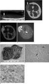
The initial sonography revealed a well-circumscribed large cystic mass located in the subcutaneous tissue. It consisted of two cysts; one large main cyst and another daughter cyst. The main cyst was predominantly composed of hypoechoic fluid and debris in the dependent portion within it. Inside the cyst, a hyperechoic nodular lesion was observed. The daughter cyst adjacent to the main cyst also contained debris-like materials (Fig. 1A). A Doppler sonographic examination showed no evidence of a vascular signal, and MRI obtained two months later showed a high signal intensity cystic lesion with a wall of low signal intensity on both the T1- and T2-weighted images. The size of the mass was measured to be about 5.5×4.8×2.4 cm. The daughter cyst had a fluid content at slightly lower signal intensity than the main cyst on a T2-weighted image. A nodular lesion within the main cyst was also observed as low signal intensity on a T2-weighted image, which was similar to the sonographic finding. The solid portion of the mass was directly adjacent to the cysts, and was contiguous with the peripheral wall of the smaller cysts (Figs. 1B, C). With gadolinium enhancement, the solid portion and cystic wall diffusely enhanced in a similar fashion (Fig. 1D). The preoperative differential diagnoses, based on the imaging findings (US and MRI), were a complicated epidermoid cyst, cystic change of a neurogenic tumor, and a parasitic cyst such as a hydatid cyst.
A surgical excision of the mass was performed, and the mass was revealed as a brownish, smooth and myxoid nature. Furthermore, the mass showed extensive cystic change and a focal solid nodule in the cystic wall. A microscopic examination revealed that the mass was composed of a mixture of abundant giant cells and mononuclear cells with diffuse interstitial hemorrhaging and cystic degeneration (Figs. 1E, F). The immunohistochemical results revealed that the mass had a diffuse positive stain for CD68 (Fig. 1G) and a focal positive stain for S-100; however, it was negative for desmin, which is consistent with a giant cell tumor.
DISCUSSION
A giant cell tumor of soft tissue has been described to be a well-circumscribed mass with a dark brown or red color. The previously reported mean size of the benign tumors is on average 3.1 cm (3). In this case, the whole size was much larger than this because of the predominantly large cystic component. Unlike the tumors cited in the literature, which were predominantly solid with often noted smaller cystic regions, the cystic component of the tumor was predominant in our patient. Histologically, a giant cell tumor of soft tissue is similar to a giant cell tumor of bone, demonstrating the multinodular aggregates of round to spindle-shaped neoplastic cells admixed with numerous uniformly scattered osteoclast-like multinucleated giant cells, microscopically (1). The diffuse interstitial hemorrhaging within the cellular regions of the tumor may be present as non-endothelial lined blood-filled cystic spaces, which are somewhat similar to the type present within the aneurysmal bone cyst (3). Immunohistochemically, CD 68 immunoreactivity is frequently strong and diffuse in multinucleated giant cells, whereas they are focal in mononuclear cells. On rare occasions, the S-100 immunoreactivity of stromal cells has been reported, but these cells appear to lack desmin (2). Similarly, our patient also showed typical histological and immunohistochemical features of giant cell tumor, aside from the prominent cystic feature; however, the scattered dilated, blood-filled spaces and distinctive large cysts without any lining epithelium or endothelium were the most characteristic features of this case. Our patient also showed typical histological and immunohistochemical features of a giant cell tumor except for the prominent cystic feature; however, the mixtures of the mononuclear spindle cells and multinucleated osteoclast-like giant cells with diffuse interstitial hemorrhaging and the diffuse positive staining for CD68 were the most characteristic features of this case (1-3).
MR image of a giant cell tumor of bone generally indicates solid components with low to intermediate signal intensity for both the T1- and T2-weighted MR images (4). In addition, previous reports have indicated that a low to intermediate signal intensity appearance may be caused by hemosiderin deposition or high collagen content (5). The aneurysmal bone cyst components in the giant cell tumor of bone are found in 19-39% of lesions (6). In some cases, bone cyst elements may have more aggressive expansile cystic components and fluid levels (4). As a result, these areas show variable signal intensity on the T1-weighted MR images and increased signal intensity on the T2-weighted MR images as a function of the hemorrhage stage (7). Contrast-enhanced MR imaging revealed that the cystic areas enhance with a thin, delicate peripheral and septal pattern. In contrast, the solid regions enhance diffusely, reflecting the hypervascular tissue (4).
A giant cell tumor of soft tissue is characterized as the soft-tissue equivalent to a giant cell tumor of bone and is an extremely rare tumor. Only a few case reports including imaging findings have been published anecdotally. Simple radiographs reveal that these giant cell tumors do not show any mass-like opacity or bony erosion (8). The MRI indicated that the giant cell tumor appears to be a mass without a clear tissue plane and is as low in signal as muscle on T1-weighted images. However, the signal intensity did increase on the inversion recovery images, although not intensely. In addition, the results indicate a diffuse enhancement with gadolinium. Similar to the simple radiographs, the higher signal intensity is confined to soft tissue with no noted bone abnormalities (9). Moreover, no evidence of prominent cystic components was noted in a literature review.
A physical examination indicated that the lesion was suspected to be cystic in nature; therefore, an US was performed initially. The lesion's main component was cystic, with only a small portion as a solid mass. MRI showed signal intensity typical of a giant cell tumor on a contrast enhanced image as well as on T1- and T2-weighted images. However, due to the prominence of the cystic component, we initially considered a complicated epidermoid cyst or cystic change of a neurogenic tumor. The nodule and debris-like materials leading to a fluid-fluid level within the main cyst on US was probably a hematoma which was eventually dissolved. A giant cell tumor of soft tissue with hemorrhaging and, large cystic degeneration is unusual and to the best of our knowledge, has not been reported previously.
In conclusion, we have described a histologically proven giant cell tumor of soft tissue with hemorrhaging and cystic degeneration in the soft tissue of the thigh. The diagnosis of the preoperative US and MRI was difficult to ascertain due to a predominant cystic component; however, the recognition of this unusual finding of giant cell tumor of soft tissue may help radiologists in considering giant cell tumors of soft tissue with cystic degeneration among the differential diagnoses of a soft tissue mass with a cystic component.
 XML Download