

 PDF
PDF ePub
ePub Citation
 Print
Print


Hemodialysis patients require a functioning vascular access to achieve long-term survival and an optimal quality of life. Depending on age when kidney failure occurs, between 23% and 51% of patients will live at least an additional 10 years after starting dialysis (1). Because the number of vascular access sites is limited, the preservation of each site for as long as possible is important for the long-term management of these patients.
Arteriovenous fistulas (AVFs) have several advantages over synthetic grafts including: better patency, fewer infections, lower incidence of vascular steal syndrome, and reduced morbidity related to the surgical procedure during shunt creation (2). The clinical practice guidelines of the National Kidney Foundation Dialysis Outcomes Quality Initiative (NKF-DOQI) has recently recommended that, ultimately, 40% of patients undergoing hemodialysis should have an arteriovenous fistula (3). However, it is also known that although the thrombosis rate for AVFs has been reported to be approximately one sixth that for synthetic grafts (4), a majority of autogenous fistulas eventually fail; this contributes significantly to morbidity and hospitalization for these patients.
Traditionally, the standard therapy for a thrombosed AVF has been surgical repair. Surgical thrombectomy with repair of the underlying stenosis or creation of a new more proximal arteriovenous anastomosis are procedures commonly performed (5). However, the results of surgical thrombectomy and revision are not satisfactory, with reported success rates varying from 28% to 73% (6-8). Because only a minority of thrombosed AVFs can be salvaged surgically following a thrombosis (9), many surgeons have recommended that salvage procedures should not be attempted on thrombosed AVFs. Therefore, additional management options are needed.
Recent studies suggest that percutaneous treatment is effective in for salvaging thrombosed native AVFs (10-19). There are a variety of percutaneous methods available to remove the clot formation from the fistulas. Current approaches include: mechanical thrombectomy with various devices, manual thromboaspiration, balloon maceration of the clot, bolus or pulse-spray infusion of thrombolytic agents, or a combination of any of these methods. Reported technical success rates range from 73% to 100% and primary and secondary patency rates at 6 months were 18%-81% and 44%-84%, respectively (10-19).
We present here our experience with percutaneous treatment in restoring the function of failed AVFs, in which pulse-spray pharmacomechanical thrombolysis was used as the primary mode of therapy, and report on the efficacy, safety and durability.
MATERIALS AND METHODS
From June 2001 to July 2005, fourteen patients with thrombosed native AVFs who underwent percutaneous restoration for 20 episodes of thrombosis were included. One patient experienced six episodes of thrombosis in a forearm AVF and another patient experienced two episodes of thrombosis in a forearm AVF. The included patients were 10 men and four women who ranged in age from 27 to 73 years with a mean age of 53.8 years. The etiologies of end-stage renal disease requiring hemodialysis were diabetes mellitus (n = 7), chronic glomerulonephritis (n = 2), Immunoglobulin A nephropathy (n = 1), Henoch-Schonlein purpura (n = 1), Alport syndrome (n = 1), polycystic kidney disease (n = 1), and hypertension (n = 1).
All fistulas were side-to-end AVFs with a distal forearm radio-cephalic Brescia-Cimino fistula in 12 patients, a mid arm radio-cephalic AVF in one patient and an upper arm brachio-cephalic AVF in one patient. Fistula location was on the left side in 10 patients and on the right side in four patients. Fistula duration ranged from two to 57 months with a mean of 25.9 months from the time of creation (median, 27.5 months). All patients underwent elective treatment within 72 hours of clotting. Before intervention, written informed consent detailing the planned procedure was obtained.
Salvage Procedures
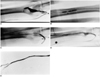
Puncture sites were anesthetized with 2% lidocaine hydrochloride. All punctures were made using a 22-guage single-wall vascular access needle or a micropuncture set (Cook, Bloomington, IN). The direction and site of the initial puncture was determined by clinical examination and/or ultrasound of the fistula. A tourniquet was applied for passive vein congestion to facilitate venous puncture by palpation. In 17 of 18 thrombosed forearm AVFs, initial puncture was made in the outflow cephalic vein of the fistula: seven retrograde venous punctures (Fig. 1), three antegrade venous punctures, and seven both retro- and ante-grade venous punctures. The seven initial retro- and ante-grade venous punctures were made directly into the occluded segment of the thrombosed AVF where a long segmental occlusion from the anastomosis to the outflow vein near the elbow was suspected. One thrombosed forearm AVF was initially accessed by a brachial artery puncture because the vein of the fistula was immature and not punctured. One thrombosed upper arm AVF, where a thrill was palpated on the venous limb close to the anastomosis, was accessed by an antegrade venous puncture. One thrombosed mid arm AVF, where a long segmental occlusion from the anastomosis was suspected, was initially accessed by a brachial artery puncture. After a 6 or 7 Fr vascular sheath(s) was/were introduced in a retrograde, an antegrade, or a criss-cross fashion, a 0.035-inch hydrophilic wire (Terumo, Tokyo, Japan) was manipulated across the occluded segment with passage of the wire into a radial artery or downstream outflow vein. Three cases of thrombosed forearm AVFs needed an additional puncture of the brachial artery to cannulate the AV anastomosis because the wire did not thread the occluded segment near or at the anastomosis in a retrograde fashion. Two upper or mid arm fistulas needed an additional retrograde venous puncture; to treat a separate stenosis at the anastomosis in one (Fig. 2) and to perform aspiration thrombectomy with a 7Fr curved catheter for residual thrombus within an aneurysmal venous segment in the other. After evaluating the arterial inflow and venous outflow of the AVFs with careful injection of contrast medium through the catheter or sheath, we treated all occluded fistulas, except one, by pharmacomechanical thrombolysis. One fistula presenting with less than 1 cm of occlusion in the cephalic vein close to the anastomosis was treated by clot maceration by percutaneous balloon angioplasty alone.
Pharmacomechanical thrombolysis was performed with initial urokinase administration and subsequent clot maceration using a balloon catheter (Bluemax, Boston Scientific, Watertown, MA; Powerplex extreme, Cordis, Miami, FL). After placement of a multi-side port infusion catheter (Cook, Bloomington, IN) in the thrombosed segment, 100,000 IU to 750,000 IU of urokinase (25,000 IU/mL in a normal saline solution) was administered using the pulse-spray technique (forceful injections of 12,500 IU of urokinase every 20 seconds with a 1-mL tuberculin syringe). The dosage of urokinase was determined at the discretion of the treating radiologist and was based primarily on the length of the occlusion. Namely, 3,000 IU to 5,000 IU of heparin (70 IU/kg) was injected either intravenously before thrombolysis or as a mixture with urokinase. In the long segmental occlusions, an infusion catheter was initially placed at the venous end of the occluded segment and repositioned to the arterial end of the occluded segment after thrombolysis of the venous segment had been accomplished. In the five thrombosed AVFs where initial or additional puncture of the brachial artery was needed, pharmacomechanical thrombolysis and subsequent balloon dilation were performed through the brachial artery access.
Balloon angioplasty was performed to macerate residual thrombi and treat all underlying stenoses. The size of the balloon used was determined by the size of the adjacent vein distal to the area to be dilated. Typically, lesions near the AV anastomosis were dilated with 4-6 mm balloon catheters. Lesions of the venous outflow of the fistula were dilated with 6-10 mm balloon catheters. Under fluoroscopic control, the angioplasty balloon was routinely inflated up to 20 atmospheres for about 2 minutes until the balloon waist disappeared. Resistant stenoses were treated by prolonged balloon inflation for 5-10 minutes, up to 25 atmospheres or by using a balloon with a 1-mm larger diameter. If necessary, the procedure was repeated until the control angiogram revealed a satisfactory reduction in residual stenosis. The acceptable degree of stenosis was less than 30% of the adjacent vessel lumen. Two cases of elastic recoils unresponsive to balloon inflation were treated with a 5-mm cutting balloon (Boston Scientific, Donegal, Ireland). No stents were placed.
After angioplasty, complete angiography was performed from the arterial anastomosis to the right atrium. For adherent or residual thrombi within the aneurysmal venous segment persisting after initial urokinase injection and subsequent balloon angioplasty, additional aspiration thrombectomy with a 7 Fr curved guiding catheter (Vistabrite tip guiding catheter, Cordis, Miami, FL) was performed. After the procedure, the introducer sheaths were kept for immediate hemodialysis.
Definition and Data Analysis
Technical success was defined as the restoration of flow in the fistula with a residual stenosis of less than 30% diameter. Clinical success was defined as the resumption of normal dialysis for at least one session after percutaneous treatment. Complications were classified as major or minor according to the criteria of the Society of Cardiovascular and Interventional Radiology published guidelines (20). Minor complications were defined as problems requiring no or nominal therapy and having no clinical consequences. Major complications were defined as problems that required additional therapy with clinical consequences. Primary patency was defined as the interval following intervention until the next access thrombosis or repeated intervention. Secondary patency was defined as the interval after intervention until the access was surgically declotted, revised, or abandoned.
Medical records and hemodialysis charts were reviewed to obtain information for follow-up and access site complications. Follow-up information for four patients who had dialysis performed in other hospitals, at the time of this investigation, was obtained by telephone. The feasibility of our technique was assessed by a retrospective analysis. Primary and secondary patencies were calculated by using a Kaplan-Meier analysis.
RESULTS
Salvage of the thrombosed AVFs with flow restoration was achieved in 15 (75%) of the 20 episodes in 10 (71%) of 14 patients. Immediate hemodialysis after the procedures resumed in all 15 cases that achieved technical success. The function of one fistula was restored by balloon angioplasty alone, that of 11 fistulas was restored by initial urokinase injection and subsequent balloon angioplasty, and that of three fistulas by initial urokinase injection and subsequent balloon angioplasty and aspiration thrombectomy. Technical failures occurred in five forearm fistulas. Four failures were attributed to the inability of to crossing the occluded segment even with the use of ante- and retrograde approaches. Two of these cases occurred in immature AVFs never used for hemodialysis having fistula ages of two and three months, respectively. One patient, who had a short segmental occlusion at the anastomosis, refused further participation in the trial through a brachial artery access following failure to cross the occluded segment through an initial retrograde venous puncture.
After balloon dilation, there was one episode of minor venous rupture which immediately healed without any treatment. No patient complained of clinical respiratory distress during or immediately after the salvage procedure. There were no major complications related to the procedure.
Including the initial technical failures, primary patency rates at six and 12 months were 64% and 55%, respectively and secondary patency rates at six and 12 months were 71% and 63%, respectively (Fig. 3). The length of follow-up ranged from two to 45 months (mean, 19.3 months; median, 11 months). Early re-thrombosis was seen the next day after pharmacomechanical thrombolysis in one forearm AVF which re-occluded four more times during 11 months of follow-up. At all episodes, the function of the AVF was successfully restored by percutaneous pharmacomechanical thrombolysis. Another forearm AVF was re-thrombosed at a 10 month follow-up. However, it could not be salvaged by percutaneous treatment because a tight occlusion prohibited the guide wire from passing. One patient died of pneumonia at 45 months after the procedure and another patient underwent kidney transplantation at 39 months after the procedure.
DISCUSSION
Currently, purely mechanical and pharmacomechanical thrombolysis are the two mainstays of percutaneous treatment for thrombosed dialysis fistulas and grafts. Based on the results for treating the thrombosed dialysis grafts, the procedure times of mechanical thrombectomy were shorter than those of pharmacomechanical thrombectomy, but there were no significant differences in the technical success, complication, and primary patency rates between two groups (21-23). On the other hand, devices for mechanical thrombectomy, except for balloon angioplasty and manual thromboaspiration, are more expensive and have the potential to damage the vein wall (23,24). Although it is still unclear which method is better, the administration of a lytic agent, such as urokinase, has been shown to reduce the clot burden in the fistula. In addition, lytic agents address both small pulmonary emboli which may be dislodged by mechanical maceration and preexisting pulmonary emboli (25).
The present study used pulse-spray pharmacomechanical thrombolysis as the primary mode of therapy for the percutaneous treatment of thrombosed native fistulas. A technical success rate of 75% (15 of 20) was achieved with primary and secondary patency rates of 64% and 71% at six months and 55% and 63% at 12 months, respectively. Published studies using mechanical thrombectomy as the primary therapeutic method for removing clots in thrombosed AVFs utilize various mechanical thrombectomy devices, manual thromboaspiration, and balloon maceration of clot (12-16, 18). These studies have reported technical success rates of 76%-100% with primary patency rates of 18%-81% at six months and assisted primary or secondary patency rates of 60%-80% at six months. Pharmacomechanical thrombolysis as a primary therapeutic method for removing clots in thrombosed AVFs has not been thoroughly investigated. Schon and Mishler (17), in a study of 16 thrombosed AVFs, used pharmacomechanical thrombolysis with a bolus injection of low dose urokinase or tissue plasminogen activator (tPA) in conjunction with external manual maceration of clots; the reported technical success rate was 94%. Patency rates were not reported. In another study by Zaleski et al. (10), initial anastomotic angioplasty, bolus urokinase infusion for 1 minute, and balloon maceration of the clot were used for pharmacomechanical thrombolysis in 17 thrombosed fistulas. They reported a technical success rate of 82% with primary patency rates of 71% at six months and 64% at 12 months and secondary rates of 100% at six and 12 months (excluding initial failures). Rajan et al. (19) used a method of pharmacomechanical thrombolysis similar to ours; they treated 25 thrombosed AVFs by pulse-spray injection of urokinase or recombinant tPA in conjunction with balloon maceration or a thrombectomy device to treat the clot, and reported a technical success rate of 77% with primary and secondary patency rates of 28% and 44% at six months and 24% and 44% at 12 months, respectively.
Our study has a slightly lower technical success rate compared to that of previous studies (10-19). Of our five technical failures, one resulted from the patient's refusal and two failures developed in immature fistulas which were not included in the previous studies. Thus, if these three technical failures are excluded, our technical success rate becomes 88% and is comparable to prior reports. The primary and secondary patency rates of this study are better when compared to those of Rajan et al. (19) who used a technique similar to ours.
As in previous studies for thrombosed AVFs, the technical failures in this study are attributed primarily to the inability to cross the occluded segment. Once this problem could be overcome, percutaneous restoration was successful in all 15 cases. In addition, if we exclude the five technical failures, primary and secondary patency rates are excellent; 90% and 100% at six months and 77% and 88% at 12 months, respectively.
Thrombosed native AVFs have several features that make them difficult to work with these include: a thin venous wall that is difficult to palpate and is usually transfixed without resistance, an irregular anatomy that makes it frequently impossible to localize the anastomosis clinically, an underlying stenosis that occurs anywhere from the feeding artery to the central veins and is frequently very tight and difficult to traverse, deceptive collaterals, a great variety in clot volume, and more frequent aneurysms than in synthetic grafts that usually contain thick layers of old wall-adherent thrombi (12). Therefore, percutaneous removal of clots is more difficult to perform in standard native fistulas than synthetic grafts. Despite this difficulty, the present study, as well as others, show comparable technical success rates and much higher primary patency rates than those (technical success rate, 85%; primary patency rates, 40% at 3 months) recommended in the NKF-DOQI guidelines for dialysis graft thrombolysis (3). Because there are neither uniform methods nor reporting standards for treating thrombosed fistulas, a variety of approaches have been used in this and previous studies. However, we believe that a variety of methods are acceptable as long as they achieve effective, safe and durable results in treating underlying stenosis and removing all thrombus formation.
The more aneurysmal portions of the fistula are somewhat resistant to thrombolysis simply because not all of the clots in these segments are immediately exposed to the lytic agents. For our three patients with residual thrombus formation within the aneurysmal venous segment, which did not respond to urokinase infusion and conventional balloon angioplasty, we used a catheter-directed thromboaspiration. Zaleski et al. (10) treated the residual thrombus within the aneurysmal venous segment in five fistulas by using a compliant occlusion balloon catheter.
During an 11 month follow-up, one patient who had a right forearm AVF has experienced long segmental occlusion, on six occasions, due to severe stenosis of the outflow vein near the elbow region. Successful salvage was achieved each time. It was deemed important to maintain the fistula as long as possible because he had a history of left arm AVF not salvaged.
Reported procedure-related complications include puncture site problems such as local hematoma and subcutaneous pseudoaneurysm, venous rupture or dissection, arterial embolism, bleeding or hematoma at remote site, and symptomatic pulmonary embolism (20, 26). Some of these problems were followed with observation and others required surgery. One minor venous rupture after balloon dilation which immediately healed without any treatment occurred in our study. There were no major procedure related complications. There were no cases of pulmonary embolism.
Percutaneous treatment has several advantages over surgical treatment: dialysis can be resumed immediately without the need for placement of a temporary access catheter, thrombolysis/thrombectomy can be performed on an outpatient basis, and future access sites are not compromised. In the present study, percutaneous restoration was performed on an outpatient basis in 18 of 20 cases and during hospitalization in two cases, due to pneumonia in one and scheduled bilateral nephrectomy for polycystic kidney disease in the other.
The limitations of this study are the relatively small sample size and an inherent selection bias due to retrospective data collection. A larger prospective randomized study is needed to assess primary patency more accurately and to establish definitely the most technically successful method.
In conclusion, percutaneous treatment that uses pulsespray pharmacomechanical thrombolysis as the primary mode of therapy provides an acceptable technical success rate and excellent access patency in the salvage of failed AVFs, and exceeds the rates recommended by NKF-DOQI guidelines for thrombosed grafts. Thus, pulse-spray pharmacomechanical thrombolysis should be considered as an option to restore the function of failed AVF before a surgical procedure.


 XML Download