

 PDF
PDF ePub
ePub Citation
 Print
Print


The coherent steady-state free precession (SSFP) pulse sequence is increasingly being used in cine imaging for evaluating the cardiac function. Compared to the spoiled gradient echo MRI, which has usually been performed for cardiac cine imaging, the SSFP imaging sequence provides a higher signal-to-noise ratio (SNR) by recycling the residual transverse magnetization, and there is less of an inflow effect by using a very short repetition time. A few studies have recently reported that contrast-enhanced (CE) cine-SSFP imaging could be used for estimating the viability of infarcted myocardium with or without the simultaneous evaluation of the ventricular function (1). Since the contrast of the SSFP sequence imaging is based on the ratio of T2 to T1, T1-shortening contrast-enhancement is shown as hyperenhancement, which is similar to that in the delayed-enhanced T1-imaging. In this issue of the Korean Journal of Radiology, Kim et al. (2) have reported that the CE-cine-SSFP sequence could be used for differentiating the acutely infarcted myocardium from chronic myocardial scar with a sensitivity of 95.8%. This was a new approach for evaluating the age of myocardial infarction (MI) by using MRI.
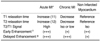
The most important pathologic changes of infarcted myocardium that influence the signal intensities (SI) on the T1- and T2-weighted imaging (WI) are the edema in acute MI and the fibrosis in chronic MI. The T1 and T2 relaxation times are directly correlated with the water content and they are inversely correlated with the collagen content (3) (Table 1). The abnormal signal intensity of infarcted myocardium on the T2-WI is usually conspicuous compared to that on the T1-WI. Therefore, to evaluate the age of an infarct, the T2-WI could be added to the infarct MRI (4). T2-WI reveals a high signal intensity of edema, which inevitably accompanies acutely infarcted myocardium and it is intensified by reperfusion therapy. The abnormal signal intensity of infarcted myocardium on the cine-SSFP images should reflect the T2 SI more than the T1 SI. However, the low SI of the infarcted myocardium that was seen in the chronic MI of this study is in disagreement with those findings of Abdel-Aty et al., in which the T2 SI of the infarcted myocardium in chronic MI showed as isosignal intensity compared to the non-infarcted myocardium (4). This discrepancy may be due to the usage of contrast-enhancement in Kim et al's study.
Hyperenhancement of infarcted myocardium on the delayed contrast-enhanced (DCE) MRI has been well established for evaluating an infarct's location and size. However, there is the same degree of hyperenhancement of the infarcted myocardium on the DCE MRI regardless of the infarct's age. The low SI of infarcted myocardium in chronic MI could be accounted for by the slow enhancement of fibrosis in the relatively early phase compared to the DCE, which occurred two minutes after contrast administration in Kim's study, as contrasted with the enhancement of the surrounding normal myocardium. Although the concept of early enhancement, which is different from the first-pass perfusion, has not been well established in cardiac imaging, dynamic enhancement has already been used as a diagnostic imaging technique for the radiological examination for various pathologies; this was recently recognized to be different from the delayed enhancement in cardiac imaging and this has been applied to amyloid cardiomyopathy (5).
We can suggest another possible explanation for the low SI of infarcted myocardium in chronic MI on the CE-cine-SSFP imaging: it is a fat tissue related artifact from the lipomatous metaplasia of chronic scar (6). In one study, large amounts of adipose tissue were demonstrated on the histology of 4- to 18-month old transmural infarcts that were experimentally produced in dog hearts (7). In addition, considering the frequent findings of profound hypo-SI of thin infarcted scars on the cine-SSFP imaging without contrast-enhancement, a fat tissue related artifact is more likely to induce the hypo-SI rather than the low-degree enhancement during the early phase of the CE-cine-SSFP imaging. The shape and degree of hypo-SI of the infarcted scar in this study strongly suggested a phase cancellation artifact in the cine imaging with a low spatial resolution, which can be seen at any interface containing both fat and water regardless of the encoding direction. To exclude artifacts due to fat tissue, fat suppression techniques, swapping encoding directions or increasing the receiver bandwidth can be tried. The pre- and early post-contrast enhanced imagings should be compared to elucidate what the SI change in the CE-cine-SSFP imaging is, in addition to performing T1-and T2-WI for further explanation.
In this study, the high sensitivity of the CE-cine-SSFP imaging for differentiating acute MI from chronic MI was in contrast with the low sensitivity of measuring the end-diastolic wall thickness (EDWT), which was done for the same purpose. Wall thinning is a result of remodeling of the infarcted myocardium, and it is a manifestation of chronic MI. An EDWT < 6 mm, which was used as a cut-off value for the diagnosis of chronic MI in this study, was originally obtained from the measurement of chronic transmural scar done at autopsy. A study showed that a EDWT < 5.5 mm on the MRI obtained with the gradient echo sequence, which was calculated as the mean minus 2.5 standard deviations (SDs) of a healthy control group, was a useful cut-off value for the diagnosis of non-viable myocardium (8). When comparing the cine-SSFP sequence to the cine turbo gradient echo sequence, the 'mean-2.5 SDs' of the EDWT was 3.8 and 5.3 mm, respectively (9). Theoretically, 3.8 mm of EDWT with the cine-SSFP imaging can be tried as a cut-off value for the diagnosis of viability. This accounts for why the sensitivity of the 6 mm cut-off value was that low. In addition, wall thinning had been used as an index for the determination of viability, and not for the differentiation of acute MI from chronic MI, because chronic MI confined to the subendocardial layer does not necessarily demonstrate wall thinning.
From a practical point of view, is differentiating acute MI from chronic MI clinically important? The authors suggest that localizing the acute infarcted myocardium would be difficult when there is a preexisting chronic myocardial infarction, and ECG and coronary angiography are not able to localize the acute lesion. How many patients with MI have difficulty with the treatment planning and follow-up after treatment due to the coexistence of acute MI and chronic MI at the first diagnosis? In a discussion on this matter, Sechtem et al. (10) commented that differentiation of acute and chronic MI had a limited application for those patients with multiple previous ischemic events in rare instances when catheter identification of the culprit lesion was not possible. Even though the differentiation is not so clinically significant, it is true that the CE-cine-SSFP pulse sequence provides information about T2/T1 SI, the contrast-enhancement, the wall motion and the wall thickness all at once, and it may be possible to substitute this for the T2-WI for differentiating acute MI from chronic MI as a time-effective method.
 XML Download