

 PDF
PDF ePub
ePub Citation
 Print
Print


INTRODUCTION
Charcoal was originally used for localizing a non-palpable tumor mass (1), however, it is being increasingly used for localizing suspicious metastatic cervical lymph nodes or recurrent tumors in the thyroid bed after surgery (2).Although, charcoal is known to be safe and stable when injected subcutaneously for preoperative tumor localization, it may cause foreign body reactions and granuloma, if it remains in situ for more than 6 months (1). Until recently, charcoal granuloma in the neck was not reported in the literature. Under the approval of the Institutional Review Board of our hospital, we reported a case of a charcoal granuloma that was suspected to be a recurrent tumor on ultrasonography (US) and 18F-fluorodeoxyglucose (FDG)-positron emission tomography/computed tomography (PET/CT), but was correctly identified after US-guided core biopsy.
CASE REPORT
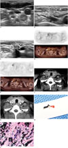
A 47-year-old woman was referred to our department for US-guided biopsy due to a persistent hypermetabolic nodule in the right level IV nodal station. She was diagnosed with invasive ductal carcinoma of the right breast and cervical lymph node metastasis at the right level IV (TNM stage; T2N3M0). She had undergone breast-conserving surgery and excision of a metastatic lymph node (Fig. 1A) 3 years prior. Before excision of cervical lymph node metastasis, US-guided localization had been performed using charcoal. The surgical specimen of the cervical lymph node revealed metastasis with perinodal infiltration. After surgery, she received chemotherapy and radiotherapy (up to 50.4 Gy) for the right breast and the right lower cervical area.
On her first follow-up FDG-PET/CT (Fig. 1D), which was performed 6 months after completion of treatment, a focal hypermetabolic lesion (maximum standardized uptake value [SUVmax] = 4.0) was noted at the lateral aspect of the previous operative bed in the right level IV. US-guided biopsy was ordered with suspicion of recurrent tumor. On US, the lesion was irregular and hyperechoic with an indistinct margin (Fig. 1B). US-guided fine needle aspiration was performed using a 25-G needle. Cytological examination revealed some atypical cells favoring the diagnosis of a benign lesion.
The second follow-up PET/CT examination, performed 1 year after the first follow-up PET/CT scan, showed a persistent hypermetabolic lesion (SUVmax = 4.3) at the same site (Fig. 1E). On chest CT, the lesion was ovoid, measured 9.8 mm in its maximal longitudinal diameter, and had an indistinct margin from the adjacent sternocleidomastoid muscle (Fig. 1F). On pre-contrast CT, the image noise and suspicious beam hardening artifact related to overlapped clavicles and shoulder girdles, rendered identification of the lesion difficult. However, the lesion seemed to be slightly hyperdense relative to the adjacent sternocleidomastoid muscle (mean attenuation: 89-100 Hounsfield unit) and did not significantly enhance after contrast administration (Fig. 1G). Since there was no evidence of recurrence at any other site of the body except the right side of the neck, and given the conflicting results of the previous cytological and PET/CT or chest CT examinations were, we decided to closely monitor and follow-up again 6 months later.
Six months later, US showed a change in the echogenicity of the nodule with no change in size, but a slight change in shape (Fig. 1C). The echogenicity of the nodule decreased, and posterior acoustic shadowing appeared newly. As the morphology of the nodule changed, we could not rule out the possibility of a recurrent tumor. Thus, we decided to perform a core-needle biopsy, as the previous US-guided fine-needle aspiration did not yield sufficient tissue for an accurate diagnosis. US-guided core-needle biopsy was performed thrice using an 18-G semi-automatic biopsy needle (TSK Stericut; TSK Laboratory, Soja, Japan). The gross specimens were dark-pigmented soft tissue fragments (Fig. 1H). Microscopically, these dark-pigmented specimens were foreign body granulomas that comprised extensive deposits of charcoal and multinucleated giant cells, as well as fibrosis; no tumor cells were detected (Fig. 1I).
DISCUSSION
Patients who undergo surgery for malignant lesions in the neck may subsequently show benign conditions mimicking recurrent tumors. These include reactive lymph nodes, traumatic neuroma, postoperative fibrosis, and suture granuloma (3, 4, 5). Charcoal granuloma should also be considered, in the differential diagnosis of such cases.
Charcoal is originally used for localization of the non-palpable breast mass (1). Charcoal is known to be a biologically inert material when injected subcutaneously and may remain in situ for up to 60 days without triggering a foreign body reaction (1). However, in an experimental study, charcoal particles were ingested by macrophages and very slowly carried away from the injection site, causing inflammatory processes (6). Charcoal particles that remained in the surgical bed might have facilitated an inflammatory reaction during the healing process after surgery in our patient. Our patient also received radiotherapy after excision of the metastatic lymph node, possibly resulting in synergistic effects leading to fibrotic charcoal granuloma.
The US features of charcoal granuloma in our report were characterized by ill-defined irregular shaped lesion with initial hyperechogenicty that changed over time, and posterior shadowing. Indistinct margin and posterior shadowing of the charcoal granuloma in our patient was in accordance with the finding of charcoal granuloma in the breast (7). Charcoal granulomas were hypoechoic according to a previous report (7). Conversely, our lesion was hyperechoic at first, and decreased in echogenicity over time. Initial hyperechogenicity of our lesion might be a helpful finding for differentiating a tumor recurrence from a non-tumorous lesion, such as a charcoal granuloma. Tumor recurrence was reported to be hypoechoic to markedly hypoechoic in the surgical bed in the post-thyroidectomy neck (8). Although the size of the nodule was not helpful to differentiate recurrence from benign postoperative change at one time point (8), the interval change of the nodule size could become one of the important imaging findings to differentiate recurrence from benign lesion. In our case, the shape and echogenicity of the lesion was changed over time, however, the lesion did not significantly increase in size during a follow-up, which suggested a benign status. CT and FDG-PET imaging findings in our patient were consistent with those of charcoal granulomas in the peritoneal cavity related to intraoperative, intraperitoneal chemotherapy (9). The lesions showed high attenuation on precontrast CT with no remarkable enhancement on post-contrast scans. Higher attenuation of the lesion compared to the adjacent muscles might be attributed to the high concentration of carbon particles (9). FDG-PET/CT showed hypermetabolism of the charcoal granulomas. Both suture granulomas and charcoal granulomas can present hypermetabolic lesions due to increased utilization of glucose by activated multinucleated giant cells (9, 10). Therefore, such foreign body granulomas can be a common cause of false positive findings in the evaluation of tumor recurrence in the postoperative neck.
Ultrasonography is the first line of imaging follow-up in patients with neck dissection or thyroid surgery. Possible differential diagnoses of charcoal granuloma include recurrent cancer and suture granuloma. Suture granulomas as a result of the knotted suture materials after thyroid surgery, usually had characteristic irregularly shaped, multiple or paired central or paracentral echogenic foci > 1 mm, and heterogeneous hypo- to isoechoic lesions on US (4). Tumor recurrence showed hypoechoic nodular lesion. Although US alone is not easy to distinguish tumor recurrence from other benign lesions, initial hyperechogenicity might be a feature of a non-tumorous condition. CT may be helpful to differentiate the 2 entities because of the hyperattenuation of the charcoal granuloma. Following imaging workup, US-guided fine-needle aspiration biopsy is generally indicated for the evaluation of recurrent nodal metastases. However, as in our patient, charcoal granuloma with charcoal materials may not be diagnosed properly with fine needle aspiration cytology alone. Cases suspected of charcoal granuloma are better diagnosed by US-guided core-needle biopsy.
In summary, we reported US, CT, and FDG-PET/CT findings of a charcoal granuloma in the neck after excision of a metastatic lymph node resulting from breast cancer. These findings included an irregular, heterogeneous hyperechoic nodule with subsequent decreased echogenicity and development of posterior acoustic shadowing on US, a high attenuating nodule on precontrast CT and a hypermetabolic lesion on PET/CT. Charcoal granulomas should be included in the differential diagnosis of incidentally detected mass-like lesions at the site of presurgical localization using charcoal.
 XML Download