

 PDF
PDF ePub
ePub Citation
 Print
Print


INTRODUCTION
F-18-fluorodeoxyglucose (FDG) positron emission tomography/computed tomography (PET/CT) is used to stage and restage various malignancies like head-neck tumors, lymphoma, lung, breast, colorectal and gynecological carcinomas (1). Widely utilized in oncology, PET/CT is based on the principle of combining simultaneously acquired anatomic and metabolic images. FDG, a radioactive glucose analogue, is the most frequently used tracer in oncological studies. Although malignant cells, owing to their high metabolic activity, consume glucose at a higher level as compared to normal there is still the possibility that benign pathologies and even normal cells may display FDG uptake, setting the stage for false-positive PET/CT scans (2, 3). An other condition that may lead to a false positivity is the FDG hot-clot artifact that appears due to injection technique. Microemboli, developed secondary to the agglutination of FDG by erythrocytes may lead to a transient obstruction of small pulmonary arterioles. In such case, PET images disclose areas of focal FDG uptake with high intensity in the lung parenchyma, whereas the CT images display no lesions at the same spots (4). Since a misdiagnosis of lung metastasis may lead to an inappropriate staging, it is important to distinguish hot-clot artifacts; rescanning is recommended in cases of doubt. In this report, we present two cases with incidental focal FDG uptake in the lungs in association with normal CT scans, both shown to be hot-clot artifacts by subsequent rescanning.
CASE REPORTS
Case 1
A 53-year-old female with squamous cell esophageal carcinoma underwent a PET/CT with the presumptive diagnosis of metastasis in the right lung. Whole body PET/CT images of both cases were acquired 60 minutes after FDG injection using a dedicated full-ring PET/CT scanner (Biograph 6; Siemens Medical Systems, Erlangen, Germany). Non-enhanced CT images with a section width of 3 mm were acquired at 130 kV and 90 mA (mean). The PET scan was obtained immediately after the CT scan and 5-7 bed positions with an acquisition time of 4 minutes for each were used. CT-based attenuation corrections were performed for the PET images and reconstruction was carried out using an iterative reconstruction algorithm.
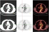
The patient was found to have pathologically increased metabolic activity in the pre- and subcarinal lymph nodes and an area of irregular wall thickening at the mid-distal esophagus. On the other hand, the 12 × 16 mm diameter parenchymal lesion evident at the posterior aspect of the right upper lobe did not display pathologically elevated metabolic activity. Still, there was focal and intensely elevated FDG uptake with maximum standardized uptake value (SUV-max) of 28.8 at the medial aspect of the anterior right upper lobe. CT images disclosed no lesion at this spot (Fig. 1). The scan was repeated with a lower dose CT protocol (110 kV and 50 mA) confined to the thoracic region to reduce the whole body radiation dose. This time, the above defined FDG uptake was no longer visible (Fig. 1) proving that FDG hot-clot and lung metastasis was ruled out.
Case 2
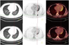
A 23-year-old male with stage-II Non-Hodkin's lymphoma underwent PET/CT scanning to evaluate the response to therapy and was found to have profound regression on whole body imaging. But there was a focal and dense FDG uptake (SUV-max of 17.7) at the superior segment of the lower lobe of the left lung, which was not evident on the previous PET/CT scan. The CT images displayed no lesion at the same spot (Fig. 2). A repeated scan using a lower dose CT protocol (110 kV and 50 mA) confined to the thoracic region verified the presumptive diagnosis of FDG hot-clot artifact (Fig. 2).
DISCUSSION
In addition to the attenuation correction by CT imaging, PET/CT scanning enables an anatomical correlation of sites with metabolic activity evident on PET images. The absence of metabolic activity at lesions evident on CT images is a frequent occurence and may be due to various reasons like low FDG affinity, response to therapy, small nodular size that falls below the PET resolution threshold or a partial-volume effect. On the other hand, focal metabolic activity on PET images without a CT counterpart is a confusing situation. Hot-clot artifact is one of various reasons for such a finding. The underlying mechanism of this artifact is the agglutination of FDG by erythrocytes during FDG injection. Microemboli secondary developed to the adhesion of FDG to concentrated erytrocytes lead to occlusive plugs in the pulmonary arterial system and create the focal FDG uptake with high metabolic activity on PET images (5), but the CT images display no parencymal nodules. Microemboli develop most frequently due to blood aspiration into the injector, but paravenous injection and high speed injection may also be possible causes. The tiny blood clots lodge in the distal capillary lung bed very similar to macroaggregates of albumin lodging in the lung parenchyma (6). A misalignment between PET and CT image planes is another reason for mismatches between findings on these two components of PET/CT imaging. The lung is the organ that frequently displays the misalignment of PET and CT images. A misalignment may cause attenuation correction artifacts. PET images without attenuation correction and fusion images may be used to identify those artifacts. A misalignment due to breathing may occur in the lower lung lobes, diaphragm and upper abdomen. Shallow breathing is recommended to achieve an optimal image fusion during the PET/CT acquisition (7).
Both cases presented in this report had a focal incidental FDG uptake at the lung parenchyma without accompanying lesions on the CT images. Moreover, non-attenuation corrected PET images disclosed a focal FDG uptake in both patients. There was no shift compatible with a misalignment of anatomic landmarks on the fusion images. The subsequent CT images disclosed no parenchymal lesion. The FDG uptake disappeared in both patients on a repeated FDG PET/CT scan the following day.
This finding was regarded as hot-clot artifact in both cases. The level of FDG uptake was considerably high in our cases; the SUV-max was 28.8 in the first and 17.7 in the second one.
There are three important points in the diagnosis of a FDG hot-clot artifact:
1) A single spot or multiple spots of focal pulmonary FDG uptake without accompanying CT findings.
2) A high level of visually and quantitatively metabolic activity at the involved foci.
3) Disappearence of the defined uptake at rescanning (8).
There is scanty of knowledge regarding FDG hot-clot in the literature (4, 5, 9). Ha et al. (5) reported on three cases in whom one patient had five spots of uptake. Karantanis et al. (4) noted a FDG uptake progression to more peripheral lung sites on repeated scanning 30 minutes after the first scan, thus excluding a real parenchymal lung lesion.
The authors believe that there are more data needed to evaluate the contribution of late scanning to the differentiation of real lesions from artifacts in PET/CT imaging.
It is important to recognize FDG hot-clot artifacts in FDG PET/CT imaging because an erroneous staging may lead to mistreatment in oncological patients. One should pay attention to the injection technique and avoid extravasation or blood aspiration into the injector to avoid such artifacts.
 XML Download