

 PDF
PDF ePub
ePub Citation
 Print
Print


INTRODUCTION
Gross hematuria is not an uncommon presentation to the emergency department (ED). Nevertheless, hematuria secondary to vesical varices is an unusual presentation. No definitive treatment has been established for bleeding vesical varices. In symptomatic vesical varices, surgical devascularization, laser sclerosis, and coagulation are often only temporarily effective. As another treatment option, percutaneous transheapatic obliteration (1) and endoscopic injection of Histoacryl could be performed.
We report a male patient who had a history of bladder substitution with ileal segments and had been treated by balloon-occluded percutaneous transhepatic obliteration of vesical varices that presented with recurrent gross hematuria.
CASE REPORT
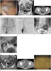
A 50-year-old male presented to the ED with recurrent episodes of gross hematuria. 30 years ago, he was admitted to our hospital and underwent left nephrectomy and orthotopic bladder substitution with ileal segments after radical cystectomy due to renal tuberculosis in the left kidney. This patient was readmitted in October 2009 with gross hematuria and underwent transurethral cystoscopic coagulation of varices in the urinary bladder (Fig. 1A). On arrival to our ED, he was initially treated with saline bladder irrigation and blood transfusion. In view of his history of recurrent gross hematuria, a contrast enhanced computed tomography (CECT, Siemens Medical Systems, Erlangen, Germany) scan of the abdomen and pelvis was performed and showed dilated blood vessels along the right side of the bladder wall with drainage into the superior mesenteric vein (SMV). Additional varix was not presented in the CT images (Fig. 1B, C).
Arterial portographies via the superior mesenteric artery showed dilated veins draining into the SMV in the right pelvic area (Fig. 1D). Percutaneous transhepatic portography (PTP) was performed by catheterization of the right anterior branch of the intrahepatic portal vein. PTP showed vesical varices with drainage into the SMV. The 10 mm diameter balloon catheter with a length of 40 mm (COOK Inc., Bloomington, IN, USA) was then advanced over the wire into the SMV. A retrograde venogram was obtained while the outflow tract was occluded with the balloon (Fig. 1E, F). A Renegade microcatheter (Boston Scientific Corporation, Natick, MA, USA) was advanced through the balloon catheter into varices using 0.14 Transcend microwire (Boston Scientific Corporation, Natick, MA, USA). The balloon catheter was inflated in the SMV to prevent the migration of 5% ethanolmaine oleate (Grelan Pharmaceutical, Tokyo, Japan)-lipiodol mixture (1 : 1). About 10 mL of the mixture was injected via the microcatheter until the entire variceal complex was opacified. After approximately 15 minutes, complete occlusion of the varices was observed during deflation of the balloon catheter (Fig. 1G). The access tract was embolized with one detachable microcoil (Interolocking detachable coil, Boston Scientfic Corporation, Natick, MA, USA) on completion of the procedure. There were no immediate complications and the patient's symptoms improved. After 6 weeks, CECT showed well obliterated vesical varices and the patient did not complain of gross hematuria (Fig. 1H, J). Follow-up cystoscopy showed the disappearance of vesical varices (Fig. 1J).
DISCUSSION
Gross hematuria secondary to vesical varices is an unusual presentation. Vesical varices secondary to portal hypertension are rare since the bladder wall is not the usual collateral route for venous splanchnic blood flow (2). Vesical varices may occur in patients with portal hypertension in circumstances where the normal splanchnic collaterals fail to develop due to prior obliteration from treatments such as surgery, sclerotherapy or ligation (3). The second probability is when the anatomy of the venous drainage of the bladder is altered from surgery such as bladder augmentation with a bowel segment. In our case, he underwent left nephrectomy and orthotopic bladder substitution with ileal segments after radical cystectomy due to renal tuberculosis in the left kidney, 30 years ago.
Varices secondary to portal hypertension have been known to occur in ectopic areas like duodenum, jejunum, ileum and anorectal regions. These varices in the gastrointestinal tract have been treated with endoscopic injection sclerotherapy, endosopic band ligation, balloon-occluded retrograde transvernous obliteration, transjugular intrahepatic portosystemic shunts, percutaneous transhepatic obliteration, and surgical procedures such as segmental resection and ligation (4).
Percutaneous transhepatic obliteration of sclerosants is a less invasive procedure when compared to surgical procedure and hence may be the only means of actively arresting bleeding vesical varices in a patient who is unfit or unsuitable for surgery. The potential disadvantage of sclerosants injection is its embolization which could lead to infarcts at distant sites or even fatality. Embolization could be prevented by balloon occlusion of drained vein.
In conclusion, the possibility of vesical varices needs to be considered in the care of patients with recurrent hematuria, especially, those associated with portal hypertension. Balloon-occluded percutaneous transhepatic obliteration could be a treatment option for vesical varices, especially, in a patients who is unfit or unsuitable for surgery.
 XML Download