

 PDF
PDF ePub
ePub Citation
 Print
Print


INTRODUCTION
Biliary magnetic resonance (MR) imaging using a hepatocyte-specific MR imaging contrast agent has been reported to be useful for detecting bronchobiliary fistula and active bile leakage after laparoscopic cholecystectomy (1-3). Nevertheless, to the best of our knowledge, there have been no reports describing biliary MR imaging using contrast agent in patients with biliary injury after radiofrequency ablation (RFA). This paper describes the first case of biliary peritonitis after RFA for hepatocellular carcinoma diagnosed by MR imaging with gadolinium ethoxybenzyl diethylenetriaminepentaacetic acid (Gd-EOB-DTPA, a hepatocyte-specific MR imaging contrast agent).
CASE REPORT
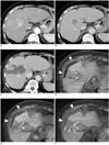
An 80-year-old female with liver cirrhosis from hepatitis C was presented to our hospital with elevated serum alpha-fetoprotein. Liver dynamic CT consisting of two phases was performed with a four-channel multi-detector row CT (LightSpeed QX/i; General Electric Medical Systems, Milwaukee, WI, USA). Two-phasic scanning was started with a 35-delay for the arterial phase, and a 90-second delay for the portal venous phase, after the beginning of contrast medium injection. The patient had a hepatic mass in segment VIII, which showed marked enhancement in the arterial phase and low attenuation compared to liver parenchyma in the portal venous phase, which was diagnosed as typical hepatocellular carcinoma (Fig. 1A, B). RFA was the most appropriate treatment as opposed to surgery, and was performed using three bipolar RF electrodes (CelonPOWER system; OLYMPUS Winter & Ibe GmbH, OLYMPUS, Japan) with a 30-mm exposed tip. Contrast-enhanced CT in the portal venous phase obtained seven days after RFA revealed an ablated tumor with sufficient margin (Fig. 1C). The patient had a good course and was discharged two weeks after RFA therapy.
Two months later, the patient had sudden upper abdominal pain, and underwent a dynamic contrast-enhanced CT scan. The scan demonstrated marked enhancement on the surface of the liver, fluid collection around the liver, and dirty omentum. Biliary peritonitis was suspected. The next day, MR imaging with Gd-EOB-DTPA (Primovist, Bayer-Schering, Berlin, Germany) was performed to evaluate biliary leakage from the liver.
MRI examination was performed with a 1.5-T clinical unit (Signa EXCITE EchoSpeed Plus; General Electric Medical Systems, Milwaukee, WI, USA) using a TORSO coil. Liver Acquisition with Volume Acceleration sequence was used with the following parameters: TR/TE/FA: 5.3 ms/2.3 ms/12°, 4.6-mm thickness without gap, and 320 × 192 matrix. A bolus of 0.025 mmol/kg body weight Gd-EOB-DTPA was injected intravenously by a power injector at a rate of 1 mL/s. Enhanced T1-weighted images were acquired 20, 60, and 300 minutes after the administration of Gd-EOB-DTPA (Fig. 1D-F). Enhanced T1-weighted MR imaging acquired at 300 minutes showed increased signal intensity in the fluid collection around the liver than in the image acquired at 20 minutes. Linear high signal areas to match the RFA puncture route were also demonstrated, suggesting a biliary leakage route from the bile duct to the fluid collection around the liver. Biliary peritonitis was diagnosed. Percutaneous drainage for fluid collection around the liver was performed, and the collected fluid was confirmed as bile.
DISCUSSION
Gadolinium ethoxybenzyl diethylenetriaminepentaacetic acid is a bolus-injectable MRI contrast agent that allows for combined dynamic and hepatocyte-specific imaging in one examination. Approximately 50% of the Gd-EOB-DTPA is excreted into the biliary system (1, 2). Excluding patients with severe liver dysfunction, it has been reported that T1-weighted images obtained 20 minutes after Gd-EOB-DTPA injection show intense enhancement of the bile duct (1, 2). The extravasation of the contrast material is visualized as a hyperintense area in the T1-weighted image, and it has been reported in some cases that Gd-EOB-DTPA-enhanced T1-weighted MR imaging can be used to evaluate the presence and exact location of bile leakage caused by cholecystectomy (3).
To our knowledge, there has been no report showing the usefulness of imaging for detecting bile leakage after RFA on MR imaging, CT, or hepatobililary scintigraphy. The incidence rate of biliary system complications of RFA, such as biloma, is a few percent, including mild cases, and many cases require no treatment (4-6). Biliary peritonitis is a rare complication caused by RFA, and it develops within one day after RFA (4). In the patient examined, the clinical course was atypical, and peritonitis could be diagnosed by CT, but it was difficult to definitively diagnose RFA-induced biliary peritonitis with CT. This case showed a characteristic finding in the T1-weighted image obtained 300 minutes after Gd-EOB-DTPA injection, in that extravasation of the contrast material was visualized as a marked hyperintense area. In addition, an extra-biliary enhanced area was observed along the route of RFA puncture, suggesting the pathway of bile leakage.
To our knowledge, there has been no report showing the usefulness of imaging for detecting bile leakage after RFA on MR imaging, CT, or hepatobililary scintigraphy. The incidence rate of biliary system complications of RFA, such as biloma, is a few percent, including mild cases, and many cases require no treatment (4-6). Biliary peritonitis is a rare complication caused by RFA, and it develops within one day after RFA (4). In the patient examined, the clinical course was atypical, and peritonitis could be diagnosed by CT, but it was difficult to definitively diagnose RFA-induced biliary peritonitis with CT. This case showed a characteristic finding in the T1-weighted image obtained 300 minutes after Gd-EOB-DTPA injection, in that extravasation of the contrast material was visualized as a marked hyperintense area. In addition, an extra-biliary enhanced area was observed along the route of RFA puncture, suggesting the pathway of bile leakage.
To clearly detect the extravasation of contrast material, the optimal acquisition timing after Gd-EOB-DTPA administration should be investigated with consideration of the liver function and location of bile leakage. When the liver function is normal, the bile duct is favorably visualized within 30 minutes in Gd-EOB-DTPA-enhanced T1-weighted MR imaging (2). Reportedly, favorable images of the biliary tract can be acquired at about 2-3 hours after injection (7). However, the extravasation of contrast material may be poorly visualized even at 2 hours after injection in patients with severe hepatic cirrhosis (8). Moreover, the visualization level may vary depending on the location and severity of the bile duct injury. The leakage of bile after cholecystectomy was evaluated by enhanced T1-weighted MR imaging performed about 30 minutes after MnDPDP administration in one report (3), and cholecystectomy-induced bile leakage was observed at 30 minutes after the administration of a contrast agent with hepatobililary scintigraphy or CT cholangiography after the administration of biliary intravenous contrast medium in other reports (9, 10). These studies suggest that the presence and exact location of bile leakage can be evaluated in images obtained within a relatively short time after the administration of a contrast agent, because a large volume of bile is leaked through the large bile duct. Also the extrahepatic bile duct lacks a protective system in extrahepatic bile duct injury against the leakage of bile like the liver parenchyma, so bile leakage is readily developed, and the extravasation of contrast material can be detected in an image acquired in only a relatively early phase after contrast agent injection. In contrast, when the intrahepatic duct is injured, the volume of bile leakage is usually small, and the liver parenchyma plays a role in protecting against the leakage of bile, so the extravasation of contrast material may be overlooked in an image acquired in a relatively early phase after contrast medium injection. Although no report has discussed these points, images at several points in time after the administration of a contrast agent may be required to detect the slight extravasation of contrast material in patients with intrahepatic duct injury.
In the patient examined, since intrahepatic bile duct injury was assumed, images were acquired at 3 points in time (20, 60, and 300 minutes after Gd-EOB-DTPA administration) to detect bile leakage. The image acquired at 300 minutes was the most useful for detecting bile leakage. In enhanced T1-weighted MR imaging performed about 300 minutes after the administration of Gd-EOB-DTPA, visualization of the common bile duct was poor, while an increase in the signal intensity of fluid collected with leaked bile was observed, and the pathway of bile leakage was clearly demonstrated as a hyperintense area. In images acquired at 20 and 60 minutes, the common bile duct was well visualized, but the extravasation of contrast material was poorly observed because of the small volume of leaked bile, which made it difficult to definitively diagnose bile leakage. When intrahepatic bile duct injury is predicted, it may be necessary to evaluate images acquired at several hours after the administration of Gd-EOB-DTPA.
In conclusion, the usefulness of T1-weighted MR image obtained 300 minutes after the intravenous injection of Gd-EOB-DTPA for biliary peritonitis after RFA has been reported. It is important to decide on the acquisition timing with consideration of the predicted location of bile duct injury.
 XML Download