

 PDF
PDF Citation
 Print
Print


INTRODUCTION
Gastroesophageal varices are present in approximately 50% of patients with cirrhosis. Their presence correlates with the severity of liver disease; while only 40% of Child A patients have varices, they are present in 85% of Child C patients (1, 2). Variceal hemorrhage occurs at a yearly rate of 5-15%, and about 20% of cirrhotic patients with acute variceal bleeding die within 6 weeks (3-5). Although variceal bleeding ceases spontaneously in 40-50% of patients, the incidence of early rebleeding ranges between 30% and 40% within the first 6 weeks, and about 40% of all rebleeding episodes occur in within the first 5 days (4, 6).
Gastric varices (GV) bleed less frequently than esophageal varices and are the bleeding source in approximately 10-30% of patients suffering from variceal hemorrhage (7). However, gastric variceal bleeding tends to be more severe with higher mortality. Additionally, a high proportion of patients, from 35% to 90%, rebleed after spontaneous hemostasis.
Although GV have long been recognized, our understanding of their natural history has lagged far behind that of esophageal varices. Recently, new endoscopic treatment options and interventional radiological procedures have broadened the therapeutic armamentarium for GV. This review provides an overview of the classification and pathophysiology of GV, which have direct consequences for management; an introduction to current endoscopic and interventional radiological management options for GV; and details of a practical approach to endoscopic variceal obturation (EVO) using N-butyl-2-cyanoacrylate.
Classification and Pathophysiology of GV
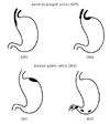
GV are heterogeneous entities in terms of the direction of blood flow, as well as their location and shape. GV are categorized into 4 types based on the relationship with esophageal varices, as well as by their location in the stomach: gastroesophageal varix (GOV) type 1, GOV type 2, isolated gastric varix (IGV) type 1, and IGV type 2 (Fig. 1) (8). GOV type 1 is the most common type, accounting for 74% of all GV. However, the incidence of bleeding is highest with IGV type 1 followed by GOV type 2. These two types of varices are classified as fundal varices and are the most challenging varices to treat.
The wall stress of the varix is ultimately what determines whether it ruptures or not. Wall stress is defined by the equation σ = p (r/w), in which σ is the wall stress, p is the transmural pressure (the portal pressure), r is the radius, and w is the wall width. Clearly, if an increase in the transmural pressure occurs, this can lead to both an enlargement of the varix (r) and a decrease in varix wall thickness (w) (9, 10). Thus, small increments in portal pressure can lead to an exponential increase in the wall stress (σ), precipitating rupture. This explains in part why GV, which are usually larger than esophageal varices, can rupture, despite possessing thicker walls and lower portal pressures than esophageal varices (11).
Overall, the most important predictor of hemorrhage is the size of varices, with the highest risk of first hemorrhage (15% per year) occurring in patients with large varices (12). Other predictors of hemorrhage are decompensated cirrhosis (Child B or C) and the endoscopic presence of red wale marks (12).
Management of Bleeding GV
Diagnostic endoscopy should be performed for acutely bleeding patients as soon as possible to determine the site of bleeding. Even patients with portal hypertension and documented varices can bleed from other sources than varices (13, 14). When a varix is identified and no other source of bleeding is evident, the varix should be regarded as the source of bleeding and should be treated to prevent rebleeding.
The optimal management of bleeding GV remains controversial due to a lack of large, randomized, controlled trials. Although endoscopic variceal band ligation is the undisputed gold standard therapy for bleeding esophageal varices, this approach has been less successful for the treatment of bleeding GV (15). Treatment options for GV include radiological intervention; transjugular intrahepatic portosystemic shunt (TIPS) or balloon-occluded retrograde transvenous obliteration (BRTO) (16) and endoscopic treatment; tissue adhesive injection; thrombin injection; sclerotherapy; or band ligation.
TIPS placement has been demonstrated to be useful in reducing rebleeding rates from GV despite that GV tend to bleed at lower portal pressures (11). However, TIPS seemed to be less effective for GV treatment than for esophageal varices (17). In another study, although TIPS insertion led to a successful reduction in the mean hepatoportal gradient (17 to 8.4 mm Hg), the actuarial rate of remaining free of rebleeding at 12 months was only 64%. Furthermore, TIPS was considered to be responsible for death in 13% of patients (18). In addition, TIPS is expensive and requires ongoing maintenance in the vast majority of patients.
Currently, among the endoscopic therapeutic options for gastric variceal bleeding, the greatest evidence for successful treatment exists for EVO using N-butyl-2-cyanoacrylate, which is recommended as a first-line endoscopic therapy (19-21). Uncontrolled data comparing therapies in bleeding fundal varices show that the best control of initial hemorrhage (90-100%) is achieved with EVO, TIPS, or BRTO (16). Three small single-center randomized, controlled trials compared EVO versus endoscopic sclerotherapy (22) or band ligation in bleeding GV (15, 23). All three trials are favorable for EVO regarding control of acute hemorrhage (15, 22), rebleeding (23), or complication rate (15). A small single-center study comparing EVO versus TIPS in the prevention of recurrent hemorrhage in patients in whom acute gastric variceal hemorrhage was controlled with EVO showed similar rebleeding rates (24). Thus, it is possible that EVO would be as effective as TIPS, if not more so, particularly in patients with underlying encephalopathy, renal disease, or significantly decompensated liver disease.
Based on these data, all of the current guidelines, such as the American Society for Gastrointestinal Endoscopy guidelines, the American Association for the Study of Liver Diseases guidelines, and the Baveno IV Consensus, recommend EVO as first-line treatment for bleeding GV, with the option of TIPS where endoscopic therapy is not available (19-21).
Practical Considerations in EVO for Bleeding GV
Because EVO has the potential risk of serious complications, complying with standard injection technique is important to reduce the risks: 1) dilution of 0.5 mL N-butyl-2-cyanoacrylate with 0.5 mL or 0.8 mL of Lipiodol, 2) limiting the mixture volume to 1.0-1.3 mL per injection to minimize embolism risk, 3) repeating intravariceal injections of 1.0-1.3 mL each until hemostasis is achieved, 4) obliteration of all tributaries of the FV, 5) repeat endoscopy 1-4 days after the initial treatment to confirm complete obliteration of all visible varices and repeat EVO if necessary to accomplish complete obliteration (25).
Real-time fluoroscopy monitoring is not always necessary. An overtube should be kept readily available to easily remove and re-insert the endoscope during the procedure and also to prevent aspiration of gastric contents into the patient's airway. A specialized catheter should be used, and distilled water is better than normal saline because cyanoacrylate may coagulate in contact with saline. Goggles are required for eye protection of the patient and the clinical personnel from splashes of cyanoacrylate during preparation and injection of the glue.
A routine check with dynamic CT scanning prior to EVO is strongly recommended (26-28). CT scanning is very helpful for identifying the presence and type of GV, to assess GV outflow direction, such as a gastro-renal shunt, to assess the risk of systemic embolization by EVO, to identify the presence of hepatocellular carcinoma with or without portal vein thrombosis, and to evaluate the applicability of salvage treatments, such as TIPS or BRTO, in cases of EVO failure.
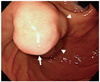
In cases with active bleeding, securing the endoscopic view to the fundus is difficult due to the large amount of blood in the gastric lumen. In such cases, changing the position of the patient to an upright or prone position is sometimes dramatically helpful. In a case with a large fundal varix, the dome area of the varix has the highest pressure with a high speed of blood flow (Fig. 2). Thus, it is better to inject at the side of the varix first, moving the injection to the dome in a stepwise manner.
Potential Complications of EVO
Cyanoacrylate injection may cause some serious complications, including embolization into the renal vein, IVC, pulmonary or systemic vessels, fever, paravariceal injection with mucosal necrosis and bleeding, intraperitoneal injection inducing severe pain, needle sticking in the varix, and adherence of the glue to the endoscope. However, most of these complications can be prevented by keeping a standardized injection technique, and the overall incidence of complications is low (25, 29). Antibiotic prophylaxis should be administered to all cirrhotic patients with GI bleeding, whether EVO is performed or not.
Summary
Early endoscopic evaluation is essential for the diagnosis and treatment of variceal bleeding. EVO with cyanoacrylate glue injection is the recommendation of choice for acute GV bleeding, with the option of TIPS where endoscopic therapy is not available. By keeping a standardized technique, EVO can be performed safely and effectively.
 XML Download