

 PDF
PDF ePub
ePub Citation
 Print
Print


The human heart can contain various amount of fat. There have been many reports regarding fat deposition or infiltration after pathologic conditions such as old myocardial infarction (MI), arrhythmogenic right ventricular dysplasia (ARVD) and cardiomyopathy (1-3). Additionally, recent reports have demonstrated that fatty tissue intermingled with myocardial tissue is present within the right ventricle (RV) in over 50% of healthy elderly people (1). However, little is currently known regarding a fat streak in the middle layer of the left ventricular (LV) myocardium in patients without a clinical history of MI or ARVD. In this study, we report a case of LV middle layer fat deposition that was detected on cardiac computed tomography (CT).
CASE REPORT
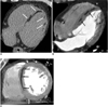
A 51-year-old man was admitted for chest discomfort and hematemesis. Her previous medical history included hypertension, type 2 diabetes mellitus, duodenal ulcer and chronic pancreatitis with pseudocyst. He had been a heavy smoker for 45-pack years, and he had a history of heavy alcohol consumption (although the patient was not currently drinking). The echocardiography (ECG) revealed normal sinus rhythm without Epsilon waves or a prolonged QRS complex. On abdomen CT, an incidental finding of regional hypoattenuated lesions involving the myocardium of the LV was noted (not shown). The patient denied any cardiac problems. Contrast-enhanced ECG-gated 64-slice multidetector CT coronary angiography (CA) was conducted in order to rule out old MI or ARVD. CTCA revealed a curvilinear middle layer fat deposition in the LV septum and a patchy middle layer fat deposition in the LV lateral wall without any associated wall thinning (Fig. 1). No stenotic lesions were noted in the coronary arteries, nor was fat deposition detected in the RV free wall. The end-diastolic volume (135 ml), the end-systolic volume (45 ml) and the ejection fraction (67%) were measured on the LV functional analysis with multiphase reconstruction of the image data set using cardiac CT. There were no regional wall motion abnormalities. Echocardiography and cardiac MRI were not conducted because the patient did not desire any further workup.
DISCUSSION
The differential diagnosis for the presence of LV myocardial fat includes old MI, ARVD, cardiac lipoma, lipomatous infiltration, hypertrophic cardiomyopathy, dilated cardiomyopathy and the sequelae of myocarditis or toxicity (2-6). Although fat deposition may be encountered under the benign condition of fatty replacement of the RV or as a normal variant, fat is generally not observed in the LV wall without a history of MI (7). The presence of myocardial fat was observed in up to 6% of the CT examinations conducted on ischemic heart disease patients and the typical CT findings of LV myocardial fat in old MI included linear or curvilinear foci of fat attenuation within the subendocardial portion of the LV myocardium, LV wall thinning and/or calcification (7). Another study reported that fat in the LV myocardium was detected at a frequency of 22% on the CT imaging of old MIs and it is more frequently associated with a longer postinfarct period, milder coronary artery stenosis and more regional wall motion abnormalities (6). ARVD is clinically characterized by ventricular arrhythmias with left bundle branch blockage, and the relevant CT findings are as follows: (i) dilation of the RV, (ii) fatty tissue in conspicuous trabeculae of the RV, and particularly in the anterior wall, apex and inferior wall, and (iii) a scalloped appearance of the RV wall (8). Hypertrophic cardiomyopathy may be associated with the presence of myocardial fat in a thickened LV wall, with this fat deposition being observed on CT in up to 11% of the cases (9). Dilated cardiomyopathy may also be associated with myocardial fat deposition, as seen on CT, in up to 18-24% of the cases (5, 9). Cardiac lipoma is a true neoplasm that is composed of an encapsulated mass of mature adipose tissue, and cardiac lipomatous infiltration is an unencapsulated mass of fatty tissue in the myocardium (10).
In our case, multiple curvilinear fat and nodular fat were distributed in the middle layer of the LV myocardium without coronary stenoses and without relation to the vascular territory or LV wall thinning. We detected no evidence of ARVD, such as Epsilon waves on ECG or the above-mentioned CT findings. Hypertrophic cardiomyopathy was excluded based on the CT findings. Capenter (10) previously insisted that the term 'fat infiltration' is a misnomer, implying invasion of the myocardium by epicardial adipose tissue, whereas the fat probably arises from metaplasia of the connective tissue. In our case, fat infiltration might be a misnomer, but we cannot prove that this is the result of metaplasia or that it is a consequence of any other causes. We have no satisfactory explanation for this finding. We have no idea regarding its incidence, the manner in which such patients should be managed or its prognosis. We assume that myocardial fat that is detectable by CT might be involved anywhere within the normal myocardial wall.
In conclusion, we report herein an unusual case of a curvilinear streak of fat deposition in the middle layer of the LV myocardium without any identified causes on CTCA. This report indicates that LV middle layer fat deposition should be further investigated in order to determine its etiology, pathogenesis and prognosis.
 XML Download