

 PDF
PDF ePub
ePub Citation
 Print
Print


Afibroxanthoma is a descriptive term for a benign fibrohistiocytic tumor that is characterized by a prominent xanthomatous change of the histiocytic component in the fibrocollagenous background (1). A primary fibroxanthoma in the central nervous system (CNS) is very rare (2). In the clinical literature, no case report has been identified for a primary fibroxanthoma that arose in the CNS in a patient younger than the age of one year. We present a case of an infantile fibroxanthoma that arose from the cranial dura mater in a six-month-old girl with US, MRI and PET/CT features.
CASE REPORT
A 6-month-old girl, who had a history of 37 weeks of gestational age, 2.6 kg birth weight and normal vaginal delivery, was admitted for the evaluation of macrocephaly (head circumference 46.7 cm, 97 percentile).
The infant showed motor developmental delay of approximately two months based on the Korean Child Development Review (K-CDR), but no significant abnormality was detected on neurological examinations.
The hemoglobin level (10.1 g/dL) was slightly decreased, the levels of liver parenchymal enzymes (alanine aminotransferase, 71 U/L; aspartate aminotransferase, 66 U/L) were increased, activated partial thromboplastin time (53.6 seconds) was prolonged, the level of α-fetoprotein (27.570 IU/mL) was increased and the level of β-HCG (< 2.00 mIU/mL) was normal.
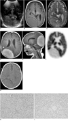
Initial cranial US performed through the anterior fontanel showed the presence of a large echogenic mass in the right parieto-occipital area, which was identified by the use of a 2-5 MHz convex probe and was not seen with the use of a 5-8 MHz sector probe (Fig. 1A).
Subsequent MR imaging was performed on a 1.5 Tesla unit with the patient under sedation. An approximately 8.3 × 8.1 × 5.0 cm sized extra-axial tumor that arose from the dura matter was seen with iso-signal intensity on spin-echo T1-weighted image (Fig. 1B). The mass was seen with heterogeneous signal intensity with a 'tree ring' like appearance on turbo spin-echo T2-weighted image that characterized the concentric patterns of high and low signal intensities (Fig. 1C). The mass was seen with homogeneous enhancement on contrast enhanced T1-weighted image. In addition, a focal leptomeningeal enhancement, anterior to the tumor, was detected (Fig. 1D, E).
Under the radiological impression of a meningioma with possible malignant change, PET/CT was performed with the patient under sedation. As seen on PET/CT image, the mass was hypometabolic, and the focal lesion of leptomeningeal enhancement seen on contrast-enhanced T1-weighted image was not hypermetabolic (Fig. 1F). Precontrast CT scan obtained on PET/CT demonstrated the presence of a large, isoattenuated to slightly hypoattenuated mass to the gray matter without calcification or bony change (Fig. 1G).
The infant underwent tumor excision on the seventh hospital day. Dural attachment was severe but bony involvement adjacent to the tumor was not seen. The mass was yellowish in color and was difficult to remove in a piecemeal manner due to the severe fibrotic nature of the mass.
On a gross examination, the excised tumor showed a grayish yellow, solid, relatively homogeneous rubbery cut surface. Microscopically, the tumor cells had round to spindled cytoplasm and small round to ovoid or occasionally elongated bland-looking nuclei. The cytoplasm varied in granularity, showing an eosinophilic granular, clear to foamy appearance. Foamy cytoplasms were frequently found in the grouped round to polygonal cells. There was a fine delicate collagenous stroma intervening between the tumor cells (Fig. 1H). Immunohistochemically, the tumor cell cytoplasms were diffusely immunoreactive for histiocytic markers including CD68, A-1-AT (alpha-1-antitrypsin), A-1-ACT (alpha-1-antichymotrypsin) and were non-reactive for meningothelial or Langerhans cell markers including EMA, VM and CD1a (Fig. 1I). The tumor was morphologically diagnosed as a fibroxanthoma.
Just after surgery, bradycardia and cardiac arrest developed in the patient. Although placed in intensive care, the infant expired.
DISCUSSION
A fibroxanthoma (fibrous histiocytoma) is a neoplastic or quasi-neoplastic lesion composed of a mixture of fibroblastic and histiocytic cells that are arranged in sheets or short fascicles and are accompanied with a varying number of inflammatory cells, foam cells and siderophages (1).
Xanthomatous tumors of the CNS are very rare and have been classified into two types, the ventricular type and dural type, according to the location. The ventricular type is commonly located in the choroids plexus of the lateral ventricle, as has been reported as an incidental finding after an autopsy. The dural type is very rare and may be a complication of metabolic disease or a manifestation of systemic disease, such as Hand-Schuller-Christian disease, Weber-Christian disease, histiocytosis X and familial hyperlipidemia (3). The tumor in this case was a dural type.
In previous reports, intracranial fibroxanthomas have appeared slightly hypointense with central hyperintense area on T1-weighted image, completely hypointense on T2-weighted image, irregularly enhanced on contrast-enhanced T1-weighted image, hyperintense on proton density MR image and hypodense with central hyperdense area on CT scan (2, 4). These imaging appearances of intracranial fibroxanthomas on MR and CT scan may mimic meningiomas. In our case, the preoperative diagnosis was a meningioma, as was the situation for other cases reported in the literature (2, 4). A possible malignant change was considered, as a focal leptomeningeal enhancement that was anterior to the tumor was detected. In children, 7-16% of meningiomas are malignant. The prevalence of malignancy decreases with increasing age in children (5).
A meningioma typically appears as a sharply circumscribed round or smoothly lobulated mass that abuts a dural surface. As seen on precontrast CT scan, approximately 70-80% of all meningiomas are homogeneously hyperdense relative to adjacent brain; 25% of the lesions appear isodense. Hyperdense tumors are seen in 1% to 5% of cases. Regardless of the histological type, most meningiomas are isointense or slightly hypointense relative to the cortex on T1-weighted image, although the signal on T2-weighted image is variable, and nearly all meningiomas enhance rapidly and intensely following contrast administration (6).
In this case, however, the tumor appeared isointense on T1-weighted image. The tumor appeared heterogeneously intense with a 'tree ring' like appearance on T2-weighted image, the tumor appeared homogeneously strongly enhanced on contrast-enhanced T1-weighted image and CT scan demonstrated the presence of a large, isoattenuated to slightly hypoattenuated mass as compared to the gray matter. In addition, the lesion appeared as an echogenic mass on US and as a hypometabolic mass on PET/CT. These image findings for a fibroxanthoma on US and PET/CT have not reported previously. The image finding of a 'tree ring' like appearance as seen on T2-weighted image for the mass could not be correlated with histopathology as this appearance was found at a later date after the surgery. We suppose that this appearance can be associated with an uneven distribution of fibrotic and xanthomatic components of the tumor.
The differential diagnosis of an intracranial fibroxanthoma in childhood includes meningeal-based tumors in addition to meningiomas, such as infantile myofibromatosis, intracranial plasma cell granuloma, meningeal solitary fibrous tumor and meningeal sarcoma. Radiologically, it is difficult to differentiate a fibroxanthoma from these rare tumors that arise from the dura mater.
Infantile myofibromatosis is the most common fibrous tumor of infancy and early childhood. Intracranial involvement is caused by cephalad extension of an extracranial process. The lesions are typically seen in the vicinity of the dura and infiltrate or erode the adjacent calvarium (7). A plasma cell granuloma is an uncommon form of inflammatory pseudotumor of obscure pathogenesis. Contrast-enhanced T1-weighted images demonstrate intense enhancement and reveal interdigitation with the adjacent cortex, confirming that these lesions may be locally infiltrative (8). A solitary fibrous tumor is an uncommon spindle cell tumor. Radiologically and macroscopically, this tumor of the meninges is non-distinguishable from a meningioma, but histologically, the difference between a meningioma and a solitary fibrous tumor can be easily shown by the use of immunohistochemistry (9). A primary meningeal sarcoma is a highly aggressive mesenchymal tumor that shows poorly defined margins, heterogeneous contrast enhancement, cystic structures within the masses and a visible connection to the leptomeninges on CT or MR image. These imaging features are characteristic but not specific, as many intracerebral tumors (e.g. astrocytomas, medulloblastomas, meningiomas, neuroblastomas, lymphomas, and malignant melanomas) show similar imaging features (10).
To the best of our knowledge, this is the first case report of a primary fibroxanthoma of the CNS in an infant younger than one-year-old. A 19-month-old boy had been the youngest previous case for a primary fibroxanthoma of the CNS described in the clinical literature (2).
In conclusion, fibroxanthomas should be considered in the differential diagnosis for an intracranial dural mass with radiologic features that mimic meningiomas and other meningeal-based tumors in infants. Further studies are needed to elucidate the imaging features.
 XML Download