

 PDF
PDF ePub
ePub Citation
 Print
Print


INTRODUCTION
Flavonoids, which are secondary metabolites in plants, are considered relatively non-toxic bioactive substances and have diverse biological effects, such as anti-inflammatory, anti-oxidant, anti-allergic, hepatoprotective, anti-thrombotic, anti-viral and anti-carcinogenic activities [1,2]. Among the flavonoids, quercetin has been given special attention since it is an antioxidant which efficiently scavenges highly reactive biological species such as peroxynitrite and the hydroxyl radical [3]. Rumecis folium is a family of polygonaceae and this plant was used for disinfestations, treating diarrhea, fever, edema, jaundice and constipation in traditionally oriental medicine. In the previous study performed. QGC (Quercetin-3-O-β-D-Glucuronopyranoside) was isolated from a Rumecis folium through several steps, it QGC had a more potent effect than quercetin on the inhibition of experimental acute and chronic gastritis and ethanol-induced gastritis in SD rats in vivo [4].
The QGC EXT was extracted from Rumecis folium by ethanol. In previous studies, QGC EXT revealed protective effects on indomethacin induced gastric damage [5]. In the present study, to perform part of a preclinical evaluation of QGC EXT, the acute toxicity and general pharmacological effects of QGC EXT on general behavior, central nervous system, digestive system, smooth muscles, cardiovascular and respiratory systems were investigated.
METHODS
Animals
Male Sprague-Dawley rats (200 g), guinea pigs (300 g), mice (30 g), or cats (3.5 kg) were used for the experiments. Animals were housed in a controlled room (temperature of 22±20℃, relative humidity of 50±5%), were maintained in a light controlled room and were given solid diet and tap water ad libitum. All animals were fasted over night before the experiment. This experiment was approved by the Institutional Animal Care and Use Committee of the Chung-Ang University Medical School.
Drugs
QGC EXT was separated and extracted from Rumex aquaticus, phenylephrine, chlorpromazine, acetic acid, naproxen, zolazepam, strychnine, valproic acid, BaSO4, acetylcholine, urethane and other materials were purchased from Sigma Chemical Co. (St. Louis, Mo, USA).
Single dose toxicity in mice and rats
Drugs were administered orally at a dose rate of 0.5, 1, 2, 3, 5 g/kg in both sexes, respectively. Over the course of 2 weeks, observations regarding the variation of physical states, weights, intake of feed & water, once a day or more by naked eyes were taken. The above parameters were measured before and after treatments. Dead animals were displayed on 4th, 7th, 10th, 14th day of observation. To evaluate the maximum tolerated dose, consecutive doses for 4 days (1, 2, and 3 g/kg) were administrated orally in rats. The number of death animal was recorded. Autopsies were performed and checked hematological findings, main organs weights and other phenomena (diarrhea, constipation, stool state, stomach mucosa, etc).
General behavior
The methods used were based on the procedures described by Irwin [6] and Lee [7]. The effect of QGC EXT was observed on locomotor activity, writhing response, fighting, convulsion, tremor, exophthalmos, ptosis, piloerection, tail elevation, traction, motor incoordination, muscle tone, catalepsy, righting reflex, pain response, pinna reflex, skin color, respiration, lacrimation, salivation, diarrhea, vocalization and death were observed. One night-fasted animals were administrated with the drugs, General actions about 24 items were indicated as "+" or "-" after specific times. There were no changes on the 24 parameter of the behavior.
Muscle cooperation
Mice were put on the Rotarod (Ugo Basile, ITALY) to discipline them. Rota rod rotation stick was upside down and rotated 5 times per minute described by Dunham et al. [8]. The time it took to fall down was measured and if the animal fell down within 2 minutes, we judged that motion hindrance occurred. Chlorpromazine 5 mg/kg was used as a positive control.
Writhing reaction test
We put mice into the cage and beaker and adjusted them for 30 minutes Koster et al. [9]. Normal saline-diluted with 0.6% acetic acid was injected into the abdominal cavity by 0.1 ml/10 g for writhing reaction (repetition of stretching, squirining, abdominal constriction response). The number of writhing reactions was recorded 10 minutes and 20 minutes after the test. The drug was administrated before acetic acid in mice administration. Considering the effective maintenance time from the preliminary experiment, administration route, and methods, we observed the writhing reaction at the maximum-effect time. 500 mg/kg naproxen was administered as a positive control group.
Randall-Selitto test
In each group 10 mice were used. According to the Randall and Selitto [10] method, 20% yeast suspension was injected into the sole and the sample was administrated orally 2 hours later. Using the Randall-Selitto (UGO Basile, Italy) machine, Analgesy meter's needle was put on the foot of mice, and then we started a motor. When we increased the pressure of the needle gradually, the weight that made mice pull out their foot became the threshold for pain. QGC EXT 10, 30, 50 mg/kg was administrated orally to each group, and then the values after 1 hr, 2 hrs were measured. 500 mg/kg naproxen was used as a control.
Sleeping time
Zolazepam at 12.5 mg/kg was injected into the abdominal cavity after the administration of QGC EXT in animals through the oral route. The index losing more than 5 seconds of righting reflex, estimated the start time of sleeping and duration of sleeping. Chlorpromazine 20 mg/kg was injected into the abdominal cavity as the positive control group.
Strychnine induced convulsion
Strychnine (1.5 mg/kg) was injected in abdominal cavity 30 min after the drug was administered [11,12]. We watched for symptoms and compared them with the control group. The incidence of Tonic Flexion (TF), Tonic Extension (TE), Clonic convulsion (CL) and mortality were measured. Valproic acid 50 mg/kg was used as the positive control.
Body temperature
Temperature was measured through the rectal route. Temperature was measured 0.5, 1.0, 2.0 and 3.0 hrs after the drug was administered. Chlorpromazine (20 mg/kg) was used as the positive control group.
Large intestine movement
1 hour before the experiment, we chose animals which did not have diarrhea. 30 minutes after oral administration of the drug, 25% BaSO4 suspension (0.1 ml/10 g) was administered. Then we estimated the time of BaSO4 to come out and compared the control group with the test group.
Intestine motility
We used cat or guinea pig low esophageal smooth muscle, ileum and relaxed it in a 1 ml organ bath with Krebs solution. The muscle strips were stimulated with a pulse train of 80 V in amplitude and 10 s in duration with a pulse duration of 1 ms through platinum wire electrodes placed longitudinally on either side of the strip. Acetylcholine was treated with various concentrations, and applied to 4 Hz electronic stimulation and measured the contraction using a polygraph chamber (Grass Instrument Co, Quincy, Mass, USA). Single muscle cells were isolated according to Sohn et al. [13] and Biancani et al. [14]. Muscle strips were incubated overnight in a normal potassium-HEPES buffer containing collagenase. The next day we incubated the tissue in a water bath at 31℃ for 30 min. After incubation, the digested tissue was poured out over a 360-µm Nitex filter mesh, rinsed in collagenase-free HEPES buffer to remove any trace of collagenase, and then incubated in this solution at 31℃. The cells were allowed to dissociate freely for 10 to 20 min. Suspensions of single muscle cells were harvested by filtration through a 500 µm Nitex filter mesh.
Respiratory and cardiovascular effects
After anesthesia with a urethane polyethylene (PE-10) tube was inserted to the carotid artery and femoral vein, blood pressure and heart rates were tested. We applied the physiological recording system (polygraph, Grass Instrument Co., Quincy, Mass, USA) using a pneumograph to measure respiratory rate. Blood pressure and heart rate was carried out using the tachograph. As a positive control, phenylephrine 10 µg/kg was administered.
Statistical analysis
Data are expressed as means±SEM. Statistical significance was determined using a two-tailed Student's t-test for paired observations or Chi-square test. A difference was considered as significant when the p value was lower than 0.05. Traces were representative of at least three experiments on three or more muscle preparations.
RESULTS
Single dose toxicity in mice and rats
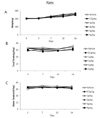
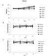
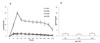
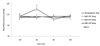
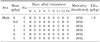
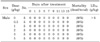
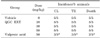
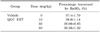
Drug at a dose rate of 0.5, 1, 2, 3, or 5 g/kg was applied both in male and female experimental animals, respectively. There were no changes in physical states, weights, intake of feed & water, or the recordings about the variation performed once per 3~4 days (Fig. 1 of rats, Fig. 2 of mice). We found that LD50 was larger than 5 g/kg in both mice and rats. Dead animals and these data were recorded on the 1st, 3rd, 5th, 7th, 9th, 11th, 13th, 15th of the observation. We found no dead animals with regards to every dose in rats and mice (Table 1 of rats and Table 2 of mice). In addition, changes of hematological findings, main organ weights and other phenomena (diarrhea, constipation, stool state, stomach mucosa) were not found. To evaluate the maximum tolerated dose, consecutive doses for 4 days (1, 2, and 3 g/kg) were administered orally in rats. The number of dead animal was also recorded. We did not find any differences in hematological findings, main organs weights or other phenomena (data not shown).
General behavior
The chronic oral administration of QGC EXT (10 mg, 30 mg, 50 mg/kg) caused no observable change in general behavior responses (stereotyped behavior, convulsion, exophthalmos, ptosis, piloerection, tail elevation, traction, motor incoordination, muscle tone, analgesia, abnormal tone, catalepy, righting reflex, pinna reflex, pupil reflex, skin color, respiration, lacrimation, salivation, urination, diarrhea, death during 4 hr periods) of the rats and, compared to the control group, no significant changes in body weight, food intake and utilization of food in treated mice. Both the control and treated rats appeared uniformly healthy at the end of study.
Muscle cooperation
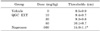
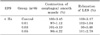
Administration of QGC EXT at doses of 10 mg, 30 mg, 50 mg/kg showed no observable changes in muscle cooperation in mice at 30, 60, 90, 120, 180, 240 minutes. In contrast, chlorpromazine, which was a positive control, showed muscle cooperation disturbance (Table 3).
Writhing reaction test
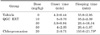
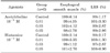
Administration of QGC EXT at doses of 10 mg, 30 mg, 50 mg/kg showed no observable change in No. of Writhings in comparison with the vehicle group. However, when naproxen (positive control) was treated, the No. of writhing reactions decreased significantly (p<0.05) (Table 4).
Randall-Selitto test
Administration of QGC EXT at doses of 10 mg, 30 mg, 50 mg/kg showed no observable change in thresholds in comparison with vehicle. Naproxen (positive control) increased pain thresholds significantly (p<0.05) (Table 5).
Sleeping time
Zolazepam increased sleeping time remarkably. Administration of QGC EXT at doses of 10 mg, 30 mg, 50 mg/kg showed no observable change in onset time and sleeping time. Chlorpromazine (positive control) decreased onset time and increased sleeping time significantly (p<0.05) (Table 6).
Strychnine induced convulsion
Strychnine induced CL and TE, and death in all cases. Administration of QGC EXT at doses of 10 mg, 30 mg, 50 mg/kg showed no observable change (Table 7). Valproic acid prevented its convulsion. QGC EXT itself has no convulsive effect (data not shown).
Body temperature
Administration of QGC EXT at doses of 10 mg, 30 mg, 50 mg/kg showed no significant changes in body temperature in mice during 240 minutes periods (Table 8).
Large intestine movement
Administration of QGC EXT at doses of 10 mg, 30 mg, 50 mg/kg showed no significant changes in large intestine movement in mice during 240 minutes periods (Table 9), it was the same as the vehicle treatment.
Intestine motility
Both in cat esophageal smooth muscle and cat LES, the dose of QGC EXT 0.01, 0.03, 0.05 mg/ml did not cause a contraction (Fig. 3). When we treated with 10-7, 10-6, 10-5 M Acetylcholine to esophageal smooth muscle and LES, the concentration-dependent contraction was exhibited. There was no change by treatment with 0.01, 0.03, 0.05 mg/ml QGC EXT (Fig. 4) Electrical stimulation induced contractions of esophageal smooth muscle and relaxation of LES of cats. When QGC EXT was added, there was no significant variation (Table 10). When we used acetylcholine and histamine as agonists, the same result was obtained in guinea pigs (Table 11). The muscle strips were stretched 2.5 gm. for optimal force development and equilibrated. When we treated 10-7 M acetylcholine, the maximum contraction occurred at 30 second, and then the contraction maintained during 20 minutes. When QGC EXT was also added, the contraction was inhibited (Fig. 5) By electrical stimulation, esophageal smooth muscle exhibited the contraction. In contrast, LES showed the relaxation effect. EXT had no effect.
We examined the effect of extracts when the maximum contraction was 100%. There was no effect on its own self like esophagus and LES. So, the response of acetylcholine on the contraction and relaxation was expressed as a percentage.
Mean blood pressure, heart rates and respiration rates
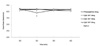
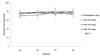
We confirmed statistically significant vasoconstriction and reflective heart rate lowering effects using adrenergic alpha-1 agonist phenylephrine. When phenylephrine (10 µg/kg i.v.) was administered, the mean blood pressure was increased in 5~10 min and recovered and sustained for several hours (Fig. 6). With regards to heart rate, after administering the same amount of phenylephrine, it decreased and after 5~10 min, it recovered and sustained itself for several hours (Fig. 7). There were no changes in respiration rates after administration of phenylephrine (Fig. 8).
DISCUSSION
Although medicinal plants may produce several biological activities in humans, generally very little is known about their toxicity and the same applies for QGC EXT. Safety should be the overriding criterion in the selection of medicinal plants for use in healthcare systems [15]. One should, in addition to the use of historical documentation on QGC EXT, also have formal toxicological evaluation of this plant to optimize its safe use as a medicine.
QGC showed potent efficacy on the development of reflux esophagitis and indomethacin-induced gastritis, by the inhibition of gastric secretion and the prevention of oxidative stress [16]. Flavonoids also have gastric antisecretory activity [17,18]. It has been reported that flavonoid compound inhibits gastric H+, K+-ATPase where the inhibition was competitive with respect to ATP [19,20] described the gastric antisecretory activity is as effective as cimetidine in reducing gastric acid secretion. Glucuronide flavonoids have antiulcer and gastroprotective activity. It appeared to have antiulcerogenic properties in rats and guinea pigs; such properties appeared to be of interest with respect to the adverse effect of gastric ulceration, which develops commonly in subjects taking anti-inflammatory drugs [21]. It has been shown that apigenin blocked cytokine-induced expression of intercellular adhesion molecule-1 [22,23] vascular cell adhesion molecule-1, and E-selectin on human endothelial cells.
Quercetin is a natural flavone with various bioactivities. Quercetin was found to be highly efficient at scavenging free radicals in cell-free systems [24] and to be more active in this respect than the traditional antioxidants vitamins C and E [25,26] found that flavonoids, especially an apigenin, blocked the cytokine-induced expressions of intercellular adhesion molecule-1 (ICAM-1), vascular cell adhesion molecule-1 (VCAM-1), and E-selectin on human endothelial cells [27]. Quercetin was also found to be an active anti-inflammatory agent in a rat paw carrageenan model and to reduce contact sensitivity in mice. In a study on gastric secretion, the oral administration of QGC reduced gastric content significantly and dose-dependently, and when QGC inhibited gastric acid output, the flavonoids extensively prevented the development of reflux esophagitis. These results suggest that QGC has inhibitory effects on reflux esophagitis and gastritis in rats, and our findings support the antiulcer, gastroprotective, and gastric anti-secretory activities of QGC. Furthermore, in feline esophageal epithelial cells, QGC was found to have a protective effect on ethanol induced cell damage by inhibiting ROS generation, activation downstream of ERK and downstream signal transduction induced by interleukin-1 [28].
In this study, the extract of QGC was found to be non-toxic in mice and rats when administered orally in doses up to 5 gm/kg. Based on the classification of Loomis and Hayes [29] viz. that substances with LD50 between 500 and 5,000 and between 5,000 and 15,000 mg/kg bodyweight are regarded as being slightly toxic and practically non-toxic, respectively, the present results suggested that GQC EET safety falls between these 2 categories.
There were no effects on the central nervous system, cardiovascular system, gastrointestinal system, and respiratory system. From the data found in this experiment we conclude that QGC EXT did not induce any adverse effects in experimental animals.


 XML Download