

 PDF
PDF ePub
ePub Citation
 Print
Print


INTRODUCTION
Hop (Humulus lupulus L.), native to Europe, North America and parts of Asia, have been used in brewing since Roman times [1]. Hop has been used in brewing for centuries, with the original purpose of this bitter herb being to preserve the beer [2]. The flowers, which are typically called "cones", contain several prenylated phloroglucinol derivatives that have antimicrobial properties. These compounds are typically divided into the heat-labile a-acids (humulone, cohumulone, adhumulone) and the more stable b-acids (lupulone, colupulone, adlupulone) [3]. The hop has a long history as a medicinal plant useful to treat sleep disturbances, restlessness and excitability besides to promote healthy digestion [4]. Moreover hop has been used as a folk remedy to treat a wide range of complaints, including spasms, cough, fever, inflammation, earache and toothache [5]. In addition, hop extracts are traditionally used for their sedative properties, but few studies have been performed on specific components of hop. Humulone, an alpha acid of hop, has been reported to inhibit induction of COX-2 and subsequent PGE2 production in a murine cell model, but clinical studies have not been reported [6,7].
Although the several therapeutic potentials of hop have been suggested, the role of hop in the regulation of antinociception has not been well characterized. Thus, we, in the current study, attempted to characterize the antinociceptive profiles and pharmacological mechanisms of hop extract in several pain animal models.
METHODS
These experiments were approved by the University of Hallym Animal Care and Use Committee (Registration Number: Hallym 2009-05-01). All procedures were conducted in accordance with the 'Guide for Care and Use of Laboratory Animals' published by the National Institutes of Health and the ethical guidelines of the International Association for the Study of Pain.
Experimental animals
Male ICR mice (MJ Co., Seoul, Korea) weighing 20~25 g were used for all the experiments. Animals were housed 5 per cage in a room maintained at 22±0.5℃ with an alternating 12 hr light-dark cycle. Food and water were available ad libitum. The animals were allowed to adapt to the laboratory for at least 2 hr before testing and were only used once. Experiments were performed during the light phase of the cycle (10:00~17:00).
Oral administration, and intraperitoneal (i.p.) and intrathecal (i.t.) injections
Oral administration was performed with gage in a volume of 500 µl/25 g body weight. I.p. injection was conducted to an anesthesized mice with volume of 250 µl. The i.t. administration was performed following the method of Hylden and Wilcox [8,9] using a 30-gauge needle connected to a 25 µl Hamilton syringe with polyethylene tubing. The i.t. injection volume was 5 µl and the injection site was verified by injecting a similar volume of 1% methylene blue solution and determining the distribution of the injected dye in the spinal cord. The dye injected i.t. was distributed both rostrally and caudally but with short distance (about 0.5 cm from the injection site) and no dye was found visually in the brain. The success rate for the injections was consistently found to be over 95%, before the experiments were done.
Acetic acid-induced writhing and intraplantar formalin tests
For the writhing test [10], 1% acetic acid was injection i.p. and then, the animals were immediately placed in an acrylic observation chamber (20 cm high, 20 cm diameter). The number of writhes was counted during 30 min after the injection of acetic acid. A writhe was defined as a contraction of the abdominal muscles accompanied by an extension of the forelimbs and elongation of the body. For the formalin test [11], 10 µl of 5% formalin was injected subcutaneously under the plantar surface of the left hindpaw. Following injection of formalin, the animals were immediately placed in an acrylic observation chamber, and the time spent licking, shaking and biting the injected paw was measured with a stop-watch timer and considered as indication of nociception. The early phase of the nociceptive response normally peaked 0 to 5 min, and the last phase 20 to 40 min after formalin injection, representing the direct effect on nociceptors and inflammatory nociceptive responses, respectively [12]. Animals were pretreated orally once with vehicle (control) or hop extract at various doses (from 25 to 100 mg/kg) 30 min prior to performing the acetic acid-induced writhing and formalin tests.
Substance P- or glutamate induced nociceptive behavioral test
Vehicle (control) or 100 mg/kg of hop extract was pretreated orally 30 min prior to performing i.t. injection of substance P (0.7 µg/5 µl) or glutamate (20 µg/5 µl). Immediately after i.t. injection with substance P or glutamate the mice were placed in an observation chamber (20 cm high, 20 cm diameter) and their nociceptive behavioral responses were recorded during 30 min. The cumulative response time of licking, scratching and biting episodes directed toward the lumbar and caudal region of spinal cord were measured with a stop-watch timer [9].
Pretreatment of antagonists
At first, mice were pretreated i.p. with either saline, naloxone (5 mg/kg), methysergide (5 mg/kg) or yohimbine (5 mg/kg) 10 min before oral administration of vehicle as a control or a fixed dose of hop extract (100 mg/kg). And then, the writhing response was tested 30 min after the treatment with either vehicle or hop extract [13-19].
Drugs
All drugs were purchased from Sigma Chemical Co. (St. Louis, MO, USA). Naloxone, yohimbine, methysergide were dissolved in saline. Hop extract was prepared following steps: (step A) 1 g of hop extract was dissolved in 0.5 ml of ethanol plus 0.5 ml of polyethylene glycol 400. (step B) Separately, 100 mg of sodium carboxymethylcellulose was dissolved in 9 ml of distilled water. (step C) Finally, step (A) Solution and step (B) Solution were vigorously mixed. This solution excluding hop extract was used as vehicle control. All drugs were prepared just before use.
Statistical analysis
Data were presented as the mean±SEM. The statistical significance of differences between groups was assessed with one-way ANOVA with Bonferroni's post-hoc test using GraphPad Prism version 4.0 for Windows XP (GraphPad Software, San Diego, CA, USA); p<0.05 was considered significant.
RESULTS
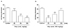
Effect of hop extract on the nociceptive behavior induced by acetic acid administered i.p.
Hop extract attenuated the acetic acid-induced writhing numbers in a dose-dependent manner (Fig. 1A). Treatment with hop extract at the dose of 100 mg/kg led to 72% decrease in the acetic acid-induced writhing response compare to the control group of mice. In addition, the time-course study showed that pretreatment with hop extract for 30 and 60 min attenuated the acetic acid-induced writhing response compare to the control group of mice (Fig. 1B). However, pretreatment with hop extract for 120 min did not affect acetic acid-induced writhing response (Fig. 1B).
Effect of hop extract on the nociceptive behavior induced by formalin injected into the plantar of the hindpaw
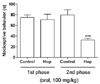
In vehicle-treated control group, injection of 5% formalin caused acute, immediate nociceptive formalin responses (i.e., licking/flinching and biting the injected paw) that lasted for 5 min (1st phase response). The 2nd phase nociceptive responses began about 20 min after formalin administration and lasted for about 20 min (20~40 min after formalin injection). In hop extract-treated mice, the nociceptive behaviors induced by intraplantar injection of formalin were decreased as compared with control group of mice during the only 2nd phases (Fig. 2). Treatment with hop extract at the dose of 100 mg/kg did not affect in the 1st phase of formalin test over the control group of mice. However, the effect of hop extract led to 66% decrease in the 2nd phase of formalin test over the control group of mice.
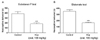
Effect of hop extract on the nociceptive behavior induced by substance P and glutamate administered i.t.
In vehicle-treated control mice, i.t. injection of substance P (0.7 µg) or glutamate (20 µg) caused acute, immediate behavioral responses, i.e., licking, scratching and biting the lumbar or caudal region, which lasted about 30 min. As shown in Fig. 3A, hop extract attenuated the cumulative nociceptive response times induced by substance P administered i.t. In addition, as shown in Fig. 3B, hop extract significantly attenuated the cumulative nociceptive response induced by glutamate administered i.t.
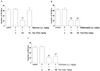
Effect of opioidergic-, serotonergic- or adrenergic-receptor antagonist on the antinociception induced by hop extract
The possible involvement of opioidergic, serotonergic or adrenergic receptors in the regulation of antinociception induced by hop extract was investigated. The treatment of naloxone, methysergide or yohimbine at the given dose did not affect the writhing response (Fig. 4). The blockade of opioidergic receptor with systemic pre-administration of naloxone significantly reversed the inhibition of the writhing response induced by hop extract (Fig. 4A). However, the pretreatment with methysergide (a serotonergic receptor antagonist, Fig. 4B) or yohimbine (an α2-adrenergic receptor antagonist, Fig. 4C) did not affect the inhibition of the writhing response induced by hop extract.
DISCUSSION
In the present study, we found that hop extract administered orally produces antinociception in various pain models. We examined the effect of hop extract on the acetic acid-induced writing response. I.p. injection of acetic acid can produce the peritoneal inflammation (acute peritonitis), which causes a response characterized by contraction of the abdominal muscles accompanying an extension of the forelimbs and elongation of the body. This writhing response is considered as a visceral inflammatory pain model [10; for review, see 20]. This behavior is considered to be evidence of peritoneovisceral pain, since acetic-acid directly activates visceral and somatic nociceptors innervating the peritoneum and induces inflammation not only in subdiaphragmatic visceral organs, but also in subcutaneous muscle walls [21]. There is evidence that polymodal C fibers and A fibers are present in the gut [21,22]. Acetic-acid causes tissue damage and releases pain-producing substances that activate nociceptors on the sensory nerve fibers [23]. In the present study, we clearly showed the antinociceptive effect of hop extract in an acetic acid-induced writhing test. The results of the present study also show that the duration of antinociceptive action was maintained at least 60 min as demonstrated in the writhing test.
Moreover, in the formalin test, we found that hop extract has an antinociceptive effect at the dose of 100 mg/kg during the only 2nd phase. It is widely agreed that the nociceptive behaviors manifested during the acute 1st phase in the formalin test may be caused by the direct effect on peripheral nociceptors activating primary afferent fiber. It is followed by the tonic 2nd phase, which may be resulted from the tonic inflammatory nociceptive response [11,12,24-26]. Shibata et al. have reported that peripherally acting drugs such as aspirin and glucocorticoid only inhibit the 2nd phase in the formalin test. In contrast, aminopyrine and mefenamic acid, which act on both central and peripheral sites, inhibit nociceptive behaviors manifested during the both phases [27]. Therefore, it is speculated that hop extract may be, at least, a peripherally acting compound, because oral treatment with hop extract inhibited the only 2nd phase in the formalin test. Furthermore, it has been reported that i.t. injection of substance P or glutamate in mice can also elicit nociceptive responses, consisting of biting, scratching and licking the caudal parts of the body [12,28]. We found in the present study that hop extract was also effective in attenuating substance P- or glutamate-induced nociceptive responses. These results suggest furthermore that hop extract may exert their antinociceptive effect via the central sites, possibly spinally mediated mechanisms.
The roles of opioid, serotonergic and adrenergic receptors in the regulation of modulation of nociceptive processing have been demonstrated in many previous studies. For example, it is well known that opioid receptors are involved in the antinociception [29-31]. Also, it has been reported that blockade of the spinal serotonergic or noradrenergic receptors by spinal injection of methysergide or yohimbine antagonize the antinociception induced by morphine administered supraspinally [30,32,33]. We observed in the present study that blockade of opioidergic receptors by naloxone, but not serotonergic and α2-adrenergic receptors, attenuated pain behaviors manifested in the writhing pain model, suggesting that opioid receptors appear to be involved in orally administered hop extract-induced antinociception. Since opioids produce the antinociception by acting on central nervous system [17], it is also suggested that hop extract appear to produce antinociception by, at least, acting on the central nervous system.
In conclusion, our results suggest that hop extract shows an antinociceptive property as manifested in various pain models related to inflammation, peripheral and central nerves pains. Furthermore, this antinociceptive effect induced by hop extract appears to be mediated by opioidergic receptors, but not serotonergic and α2-adrenergic receptors.
 XML Download