

 PDF
PDF ePub
ePub Citation
 Print
Print


INTRODUCTION
Alcohol consumption is one of the major causes of liver disease, which is a spectrum including simple alcoholic steatosis, alcoholic hepatitis and cirrhosis. Severe alcoholic hepatitis can be life-threatening and a common reason for hospitalization after heavy alcohol drinking. The progression of alcoholic liver diseases is regulated by various factors and the increased levels of pro-inflammatory factors are known to be important contributors. Heavy alcohol intake causes increase of endotoxin/lipopolysaccharide (LPS) in the blood, which in turn activates Kupffer cells, the resident hepatic macrophage, leading to the increased production of several proinflammatory cytokines, such as TNFα and interleukin-6. In particular, TNFα has been shown to play a critical role in the progression of alcoholic liver disease [1,2]. Previous animal studies have shown that rats treated with TNFα antibody and TNFα receptor I knock-out mice are resistant to alcohol-induced liver injury [3,4]. In addition, the treatment of antibiotics which reduce the level of blood endotoxin prevents the hepatic steatosis and inflammation in rats after exposure to alcohol [5].
A significant amount of studies, therefore, have focused on the regulation of TNFα production in liver exposed to alcohol to develop effective therapies for alcoholic hepatitis. The expression of TNFα in LPS-stimulated Kupffer cell is regulated by a number of signal molecules, including reactive oxygen species (ROS) and cAMP [6,7]. Among the regulators of TNFα expression, it has been clearly demonstrated that the elevation of cyclic AMP (cAMP) suppresses the expression of TNFα in macrophage [8,9]. In consistent with these observations, phosphodiesterase (PDE) inhibitors suppressed TNFα production in macrophage through elevating intracellular cAMP [10-12].
In recent years, pentoxifylline, a non-selective PDE inhibitor has attracted interest in clinical trials and has been pointed as a preferred first-line agent for treatment of alcoholic hepatitis due to its safety compared to glucocorticoids [13]. However, the studies on its efficacy are limited and its effects are unclear in some clinical situations [14]. At this time, no other therapeutic modalities for severe alcoholic hepatitis are available. Therefore, more studies on pentoxifylline are needed as well as new pharmacological therapy is urgent.
Cilostazol is a selective PDE3 inhibitor, which is known to inhibit platelet aggregation by increase of intracellular cAMP. Thus, cilostazol is widely used for treatment of peripheral vascular diseases [15,16]. In addition to its antiplatelet effect, recent studies have suggested its various pharmacologic effects including anti-inflammatory, antioxidant and anti-apoptotic effects via cAMP-dependent and -independent pathways [17-19]. Cilostazol has also shown a beneficial effect on liver steatosis in non-alcoholic fatty liver disease animal model [20].
In the present study, we have examined the effects of cilostazol on ethanol-mediated TNFα expression in in vitro and in vivo model using RAW264.7 murine macrophage and binge drinking mice and the effect of cilostazol was compared to that of pentoxifylline.
METHODS
Materials
Cilostazol was donated by Otsuka Pharmaceuticals (Tokushima, Japan). Dulbecco's modified eagle's medium (DMEM), fetal bovine serum (FBS) and penicillin/streptomycin, and 5-(and-6)-carboxy-2',7'-dichlorodihydrofluorescein diacetate (carboxy-H2DCFDA) were purchased from Invitrogen (Carlsbad, CA, USA). LPS (Escherichia coli 0111:B4), db-cAMP, 5-Aminoimidazole-4-carboxamide 1-β-D-ribofuranoside (AICAR), pentoxifylline, ethanol, carboxymethylcellulose, 3-(4,5-dimethylthiazole-2yl)-2,5-diphenyl-2H-tetrazolium bromide (MTT) and protease inhibitors (aprotinin, leupentin, pepstatin A) were obtained from Sigma-Aldrich (St. Louis, MO, USA). The phospho-AMPK antibody was from Cell Signaling Technology (Beverly, MA) and GAPDH antibody was from Santa Cruz Biotechnology (Santa Cruz, CA, USA).
Cell culture and treatment
The RAW264.7 murine macrophage was obtained from the Korean Cell Line bank (Seoul, Korea) and cultured in DMEM containing 10% FBS, 100 U/ml penicillin, 100 µg/ml streptomycin and maintained at 37℃ in a humidified incubator with 5% CO2 atmosphere. For experiments, cells were plated at a density of 1×10⁵/cm2 and treated with 25 mM ethanol for 24 h in the presence or absence of cilostazol and pentoxifylline [21,22]. Then, cells were stimulated with 50 ng/ml LPS.
Ethanol binge
Seven-week old male C57BL/6 (18~22 g) mice were obtained from Central Lab. Animal Inc. (Seoul, Korea). Mice were housed in a specific pathogen-free animal care facility under a 12 h light/dark cycle and were allowed to free access to standard laboratory chow and tab water. Ethanol binge model developed by Carson and Pruett [23] was used. After one week acclimatization, mice were divided into seven groups: control, ethanol (6 g/kg body weight, p.o.), cilostazol (100 mg/kg/day, i.p.), cilostazol (50 and 100 mg/kg/day, i.p.)+ethanol, pentoxifylline (50 and 100 mg/kg/day, i.p.)+ethanol. Mice were administered cilostazol, pentoxifylline, or vehicle (0.5% carboxyl methylcellulose) for 4 days before ethanol administration. Ethanol was diluted with sterile water (32% w/v) and was given orally 1 h after the last treatment with cilostazol or pentoxifylline. Mice were sacrificed 6 h after ethanol administration and liver was collected. The doses of cilostazol or pentoxifylline used in this study were selected by following previous studies reported by others [20,24-26]. The protocol for animal care and use was approved by Animal Care and Use Committee of Yeungnam University.
Cell viability
Cell viability was measured based on the conversion of water soluble tetrazolium MTT to water-insoluble blue formazan by viable cells. Cells were treated with various concentrations of cilostazol or pentoxifylline in the presence or absence of 25 mM ethanol for 24 and 48 h. Then, cells were treated with MTT (5 mg/ml in PBS) and incubated for 4 h. The formation of formazan was dissolved in DMSO and the optical density was measured at 570/620 nm.
Protein extracts
Cells were lysed on ice in lysis buffer (20 mM HEPES (pH 7.5), 1 mM EDTA, 1 mM EGTA, 50 mM NaF, 2 mM MgCl2, 150 mM NaCl, 10 mM KCl, 1% NP-40, 1 mM Na3VO4, 1 mM DTT, 1 mM benzamide, 1 mM PMSF, and protease inhibitors). Liver was homogenized in lysis buffer supplemented with 1% glycerol. After centrifugation at 13,000 g, the supernatant was taken and protein concentration was determined by Bradford reagent (Sigma, St. Louis, MO).
Enzyme-linked immunosorbent assay (ELISA) of TNFα
The levels of TNFalpha in cell and liver were determined using mouse TNFα ELISA kit (R&D Systems, Inc., Minneapolis, MN). One hundred microliter of cell culture media or liver extract was used and the assay was performed according to protocol provided by manufacturer. The amount of TNFα production was expressed as pg/mg protein.
Semiquantative- and real time RT-PCR
Total RNA was extracted from RAW264.7 macrophage or liver using Tri reagent (Sigma, St. Louis, MO). RNA was reverse transcribed to cDNA from 1 µg of total RNA using a High-Capacity cDNA Reverse Transcription Kit (Applied Biosystems, Foster City, CA, USA). For semiquantitative PCR, the following primers were used: TNFα (307 bp: forward, 5'-GGCAGG-TCTACTTTGGAGTCATTGC-3'; reverse, 5'-ACATTCGAGGCTCCAGTGAATTCGG-3'); 18s rRNA (209 bp: forward, 5'-CCCGGGGAGGTAGTGACGAAAAAT-3'; reverse 5'-CGCCCGCTCCCAAGATCCAACTAC-3'). Quantitative real-time PCR was performed using the Real-Time PCR 7500 system and Power SYBR Green PCR master mix (Applied Biosystems) according to the manufacturer's instructions. The thermal cycling conditions were initial incubation at 95℃ for 10 minutes, followed by 45 cycles of 95℃ for 15 seconds, 55℃ for 20 seconds, and 72℃ for 35 seconds. Primers for mouse TNFα (71 bp: forward, 5'-CTATCTCCAGGTTCTCTTCAA-3'; reverse, 5'-GCAGAGAGGAGGTTGACTTTC) and β-actin (121 bp: forward, 5'-TGGACAG-TGAGGCAAGGATAG-3'; reverse, 5'-TACTGCCCTGGCTCCTAGCA-3') were designed using the Primer Express program (Applied Biosystem). The expression level of β-actin was used as an internal control.
Reactive oxygen species (ROS) measurement
To determine ROS generation, FACS analysis was performed. Cells were incubated with 50 µM carboxy-H2DCFDA (Invitrogen, Carlasbad, CA) for 40 min. Cells were washed with PBS and subjected to flow cytometry using a Becton-Dickinson FACS Caliber and analyzed by Cell Quest software (Becton-Dickinson, San Jose, CA).
Intracellular cAMP measurement
The concentration of cAMP was measured using cAMP EIA kit (Cayman, Ann Arbor, MI) according to the manufacturer's protocols. Briefly, cells were lysed in 1 ml of lysis buffer and 50 µl of supernatant after centrifugation was used for assay and then the absorbance was measured at 405 nm.
Western blotting
Equal amounts of proteins from cell lysates were separated by 10% SDS-PAGE gel and transferred to nitrocellulose membrane (Bio-Rad, Hercules, CA). The membrane was blocked with 5% non-fat dry milk in Tris-buffered saline. The blots were incubated with primary antibody for phospho-AMPK (Cell signaling, Beverly, MA) and then, reacted with a peroxidase-conjugated secondary antibody. The protein bands were detected using an enhanced chemiluminescence detection system (Millipore, Billerica, MA). The density of respective bands was analyzed by the LAS-3000 imaging system (Fuji film, Tokyo, Japan). The membrane was reprobed with anti-GAPDH antibody, which was used as loading control.
RESULTS
The effects of cilostazol on LPS-stimulated TNFα production in RAW264.7 macrophage exposed to ethanol
We first examined the cytotoxic effects of cilostazol and pentoxifylline on RAW264.7 cells after ethanol exposure. The treatment of cells with cilostazol (0~100 µM) or pentoxifylline (100 µM) for 24 h did not cause cytotoxicity at any concentration used regardless of the presence of ethanol. At 48 h, pentoxifylline reduced cell viability ~8% and ethanol did not have any effects on cell viability (Fig. 1). In the subsequent experiments, cells were treated with cilostazol or pentixifylline for 24 h.
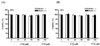
Next, the effect of cilostazol on LPS-stimulated TNFα expression in the presence of 25 mM ethanol was determined. LPS increased about 260 fold (~29 ng/mg protein) of TNFα production in culture media, which was significantly reduced to 56%, 65% and 76% by pretreatment with 100 µM cilostazol, 50 µM cilostazol and 100 µM pentoxifylline, respectively, compared to DMSO, a vehicle control (Fig. 2A). The level of TNFα mRNA was also substantially reduced by cilostazol and pentoxifylline, showing similar results to that of ELISA (Fig. 2B). These results indicate that cilostazol reduces LPS-stimulated TNFα production in RAW 264.7 cells exposed to ethanol in dose-dependent manner and the degree of its inhibition is comparable to that by pentoxifylline.
The role of ROS, cAMP and AMPK in the inhibition of TNFα production by cilostazol
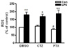
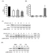
Since ROS has been shown to be an important contributor to LPS-stimulated TNFα production in macrophage [7,27], the effect of cilostazol on ROS production in RAW264.7 macrophage was examined to elucidate the underlying mechanisms. The production of ROS was increased about 2 fold by LPS treatment for 4 h (Fig. 3). However, the increase was not ameliorated by pretreatment with cilostazol or pentoxylline. This result indicates that suppression of ROS production is not involved in the action of cilostazol or pentoxifylline. Next, the role of cAMP in the action of cilostazol was examined. As shown in Fig. 4A, pretreatment with db-cAMP, a cell-permeable cAMP analog significantly reduced LPS-stimulated TNFα production by 39%. However, the intracellular cAMP was not changed by cilostazol or pentoxifylline (Fig. 4B), indicating that the inhibition of TNFα by cilostazol occurs via cAMP-independent pathway.
Recent studies have shown the regulation of TNFα expression by AMP-activated protein kinase (AMPK) [28,29]. We therefore examined whether the inhibition of LPS-stimulated TNFα production by cilostazol in macrophage exposed to ethanol was attributable to activation of AMPK. The cells were treated with LPS (50 ng/ml) for different time (0~4 h) and the activation of AMPK was measured by Western blotting. The treatment with LPS decreased AMPK activation about 60% at 30 min (Fig. 4C). In contrast, cilostazol treatment (0~4 h) increased AMPK activation about 2 fold within 5 min and then gradually returned to basal. A pharmacological activator of AMPK, AICAR increased AMPK activation about 30%. The pretreatment with cilostazol increased basal AMPK activity about 2-fold and normalized the level of AMPK activation in LPS-stimulated RAW264.7 cells, whereas pentoxifyllin did not affect the activation of AMPK (Fig. 4D). Consistent with this result, AICAR reduced LPS-stimulated TNFα production in RAW264.7 cells exposed to ethanol (Fig. 4A). These results indicate that AMPK but not cAMP is involved in the inhibition of TNFα production by cilostazol in RAW264.7 cells.
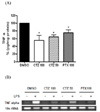
The inhibition of TNFα expression by cilostazol in binge drinking mice
The effects of cilostazol on TNFα production by ethanol was examined in in vivo animal model. Acute ethanol exposure to mice by gavage at a single dose 6 g/kg body weight has shown to significantly increase TNFα production with maximum increase at 6 h after ethanol administration [30,31]. The same protocol was followed in the present study for development of binge drinking model. The levels of TNFα mRNA and protein in liver after ethanol administration were determined by real-time PCR and ELISA, respectively. Unexpectedly, the level of TNFα mRNA was decreased at 6 h after ethanol treatment by 25~30%. The pretreatment with cilostazol (50 and 100 mg/kg) and pentoxifylline (50 and 100 mg/kg) further reduced the TNFα mRNA, although cilostazol itself did not change the level of TNFα expression (Fig. 5A). The ELISA assay also shows similar results (Fig. 5B). These results indicate that cilostazol decreases the expression of TNFα mRNA and protein in liver after ethanol exposure in vivo with similar extent to pentoxifylline, although ethanol treatment reduced the level of TNFα in this model.
DISCUSSION
The results from this study have shown that cilostazol significantly suppressed LPS-stimulated TNFα production in RAW 264.7 murine macrophage exposed to ethanol and in liver from binge drinking mice.
Numerous clinical and experimental studies have shown that the increased level of hepatic TNFα and monocyte TNFα production in alcoholic hepatitis are positively correlated with disease severity [2,32]. Chronic ethanol exposure enhances LPS-stimulated TNF production in RAW264.7 cells as well as Kupffer cells [21,22]. In accordance with this observation, inhibition of TNFα by TNFα antibody reduced liver injury in alcohol fed rats [3]. Recent clinical reports have provided beneficial effects of pentoxifylline, a TNFα suppressor for treatment of alcoholic hepatitis and have suggested this treatment as an alternative to glucocorticoids [13]. At this time, however, its efficacy on survival benefit is controversial [14,33]. It is necessary to conduct more studies on the effects of pentoxifylline while the development of new pharmacological therapy for alcoholic hepatitis is needed. Our results show that cilostazol significantly suppresses LPS-stimulated TNFα production in ethanol-primed RAW264.7 as well as binge drinking mice and the extent of inhibition by cilostazol is comparable to that of pentoxifylline. The overproduction of TNFα by ethanol has been shown to be associated with increased TNF mRNA expression [22,34]. In agreement with these results, the level of TNFα mRNA was increased by ethanol and LPS in RAW264.7 cells, which was decreased by cilostazol and pentoxifylline. These data suggest that both cilostazol and pentoxifylline suppress the production of TNFalpha at transcription level.
The underlying mechanisms by which ethanol increases TNFα expression in macrophage have been extensively studied. LPS increases ROS production in Kupffer cells and ethanol exposure further increase LPS-induced ROS production, which leads to increase of TNFα production [7]. We have also observed that LPS simulation increases ROS production in RAW264.7 cells exposed to ethanol. However, neither cilostazol nor pentoxifylline reduced LPS-stimulated ROS production. This indicates that the inhibition of TNFα production by cilostazol occurs via ROS-independent pathway. Although previous study has shown that cilostazol attenuates NADPH oxidase-derived ROS production in macrophage [35], intracellular ROS can be generated from various sources including NADPH oxidase and mitochondrial electron transport chain [36]. A lot of studies have shown that mitochondria plays a critical role in ethanol-induced ROS production and liver injury [37,38]. Furthermore, Chandel et al. [27], have shown that antioxidants had no effect on LPS-induced TNFα expression in murine macrophage.
Enhanced intracellular cAMP has been well known to suppress LPS-stimulated TNFα production in macrophage in vivo and in vitro [39,40]. Moreover, chronic ethanol exposure results in decrease of cAMP, which is one of mechanisms responsible for increased TNFα production from Kupffer cells and macrophages [21,22]. Concomitant with these observations, previous studies have documented that various cAMP elevating agents including adenylyl cyclase activator and PDE inhibitors invariably suppress TNFα production in macrophages. Our data also shows that db-cAMP significantly reduced TNFα production in RAW264.7 cells, supporting previous reports by others. However, cilostazol and pentoxifylline did not increase intracellular cAMP levels in the present study, indicating that cilostazol inhibition of TNFα production in ethanol treated macrophage is cAMP-independent. Similarly, previous studies have also shown that cilostazol did not change intracellular cAMP in RAW264.7 macrophage [19,24].
The activation of AMPK has been shown to repress inflammatory responses stimulated with LPS in vitro and in vivo [28,29]. Recent studies have shown that the anti-inflammatory effects of cilostazol in vascular smooth muscle cells and endothelial cells occur by activation of AMPK pathway which is independent of cAMP [28,29]. Our data support this findings demonstrating that AICAR, an activator of AMPK suppressed ethanol-mediated TNFα production and cilostazol enhanced LPS decreased AMPK activation in RAW264.7 cells exposed to ethanol. These results suggest that cilostazol exerts the inhibitory effects on TNFα production in ethanol-primed RAW264.7 cells through activation of AMPK.
Various alcohol binge animal models have been employed to study alcoholic hepatitis [41,42]. In preliminary experiments, we observed significant increase in serum ALT/AST, makers of liver injury and hepatic TNFα mRNA expression 6 h after gavage of a single dose of 6 g/kg or three doses of 5 g/kg ethanol, similar results to previous reports by others [31,42]. In subsequent repeated experiments, however, we obtained the opposite results that TNFα expression was rather decreased in mice gavaged with 6 g/kg ethanol compared with that in control mice. Recent study has shown similar finding that the administration of either a single dose or three dose of ethanol (5 g/kg) to rats suppressed TNFα expression in liver collected 4 h after ethanol gavage [41]. Several previous studies have demonstrated that binge ethanol causes dual effects, pro- and anti-inflammatory responses; mostly anti-inflammatory effects at early after ethanol treatment (0~6 hr) and pro-inflammatory effects later after ethanol treatment (after 24 hr) [43,44]. In addition, the differential effects of binge alcohol drinking on hepatic TNFα production in animal model may be due to different animal species and different animal batches used in experiments. Although we failed to present the increase of hepatic TNFα expression by ethanol binge itself, our data clearly show that cilostazol does decrease the expression of TNFα in liver exposed to ethanol in vivo and the degree of inhibition by cilostazol is similar to that by pentoxifylline. However, additional studies to examine the effect of cilostazol on the TNFα production in ethanol-fed animal model are needed to make concrete conclusion.
Taken together, these data demonstrate that cilostazol suppresses ethanol-mediated TNFα expression in RAW264.7 macrophage and in liver from binge drinking mice and the effects of cilostazol involves AMPK activation. Furthermore, the effects of cilostazol are comparable to that of pentoxifylline.
 XML Download