Abstract
Calcium (Ca2+) is an intracellular second messenger associated with neuronal plasticity of the central nervous system. The calcium-binding proteins regulate the Ca2+-mediated signals in the cytoplasm and buffer the calcium concentration. This study examined temporal changes of three calcium-binding proteins (calretinin, calbindin and parvalbumin) in the medial vestibular nucleus (MVN) during vestibular compensation after unilateral labyrinthectomy (UL) in rats. Rats underwent UL, and the changes in the expression of these proteins at 2, 6, 12, 24, 48, and 72 h were examined by immuno-fluorescence staining. The expression levels of all three proteins increased immediately after UL and returned to the control level by 48 h. However, the level of calretinin showed changes different from the other two proteins, being expressed at significantly higher level in the contralateral MVN than in the ipsilateral MVN 2 h after UL, whereas the other two proteins showed similar expression levels in both the ipsilateral and contralateral MVN. These results suggest that the calcium binding proteins have some protective activity against the increased Ca2+ levels in the MVN. In particular, calretinin might be more responsive to neuronal activity than calbindin or parvalbumin.
REFERENCES
Arai R., Winsky L., Arai M., Jacobowitz DM. Immunohistochemical localization of calretinin in the rat hindbrain. J Comp Neurol. 310:21–44. 1991.

Baizer JS., Baker JF. Immunoreactivity for calcium-binding proteins defines subregions of the vestibular nuclear complex of the cat. Exp Brain Res. 164:78–91. 2005.

Billing-Marczak K., Kuanicki J. Calretinin – sensor or buffer – function still unclear. Pol J Pharmacol. 51:173–178. 1999.
Celio MR. Calbindin D-28k and parvalbumin in the rat nervous system. Neuroscience. 35:375–475. 1990.

Caicedo A., d'Aldin C., Eybalin M., Puel JL. Temporary sensory deprivation changes calcium-binding proteins levels in the auditory brainstem. J Comp Neurol. 378:1–15. 1997.

Chard PS., Bleakman D., Christakos S., Fullmer CS., Miller RJ. Calcium buffering properties of calbindin D-28k and parval-bumin in rat sensory neurons. J Physiol. 472:341–357. 1993.
Forster CR., Illing RB. Plasticity of the auditory brainstem: cochleotomy-induced changes of calbindin-D28k expression in the rat. J Comp Neurol. 416:173–187. 2000.
Fortin M., Marchand R., Parent A. Calcium-binding proteins in primate cerebellum. Neurosci Res. 30:155–168. 1998.

Ghosh A., Greenberg ME. Calcium signaling in neurons: molecular mechanisms and cellular consequences. Science. 268:239–247. 1995.

Jeon MH., Jeon CJ. Immunocytochemical localization of calretinin containing neurons in retina from rabbit, cat, and dog. Neurosci Res. 32:75–84. 1998.

Kevetter GA., Neonard RB. Use of calcium-binding proteins to map inputs in vestibular nuclei of the gerbil. J Comp Neurol. 386:317–327. 1997.

Kim MS., Choi MA., Kim YK., Rhee JK., Jin YZ., Park BR. Asymmetric activation of extracellular signal-regulated kinase 1/2 in rat vestibular nuclei by unilateral labyrinthectomy. Brain Res. 1011:238–242. 2004.

Kim MS., Jin BK., Chun SW., Lee MY., Kim JH., Park BR. Effect of MK801 on cFos-like protein expression in the medial vestibular nucleus at early stage of vestibular compensation in uvulonodullectomized rats. Neurosci Lett. 231:147–150. 1997.

Kim MS., Kim JH., Jin YZ., Kry D., Park BR. Temporal changes of cFos-like protein expression in medial vestibular nuclei following arsanilate-induced unilateral labyrinthectomy in rats. Neurosci Lett. 319:9–12. 2002.

Kitahara T., Takeda N., Saika T., Kubo T., Kiyama H. Role of the flocculus in the development of vestibular compensation: immunohistochemical studies with retrograde tracing and flocculectomy using fos expression as a marker in the rat brainstem. Neuroscience. 76:571–580. 1997.

Lacour M., Vidal PP., Xerri C. Visual influences on vestibulospinal reflexes during vertical linear motion in normal and hemilabyrinthectomized monkeys. Exp Brain Res. 43:383–394. 1981.

Lane RD., Allan DM., Bennett-Clarke CA., Rhoades RW. Differential age-dependent effects of retinal deafferentation upon calbindin- and parvalbumin-immunoreactive neurons in the superficial layers of the rat's superior colliculus. Brain Res. 740:208–214. 1996.
Murakawa R., Kosaka T. Diversity of the calretinin immunoreactivity in the dentate gyrus of gerbils, hamsters, guinea pigs, and laboratory shrews. J Comp Neurol. 411:413–430. 1999.

Orrenius S., Nicotera P. The calcium ion and cell death. J Neurol Transm. 43(Suppl.):1–11. 1994.
Sans N., Moniot B., Raymond J. Distribution of calretinin mRNA in the vestibular nuclei of rat and guinea pig and the effects of unilateral labyrinthectomy: a non-radioactive in situ hybridization study. Brain Res Mol Brain Res. 28:1–11. 1995.

Fig. 1.
Immunofluorescence findings of calretinin in the medial vestibular nucleus after unilateral labyrinthectomy (UL). A & B, left (Lt) & right (Rt) MVN, respectively, in control rat; C & D, ipsilateral (left) & contralateral (right) MVN to the lesion, respectively, 2 h after UL; E & F, ipsilateral & contralateral MVN, respectively, 6 h after UL; G & H, ipsilateral & contralateral MVN, respectively, 24 h after UL; I & J, ipsilateral & contralateral MVN, respectively, 48 h after UL; MVNmc, magnocellular portion MVN; 4thV, 4th ventricle.
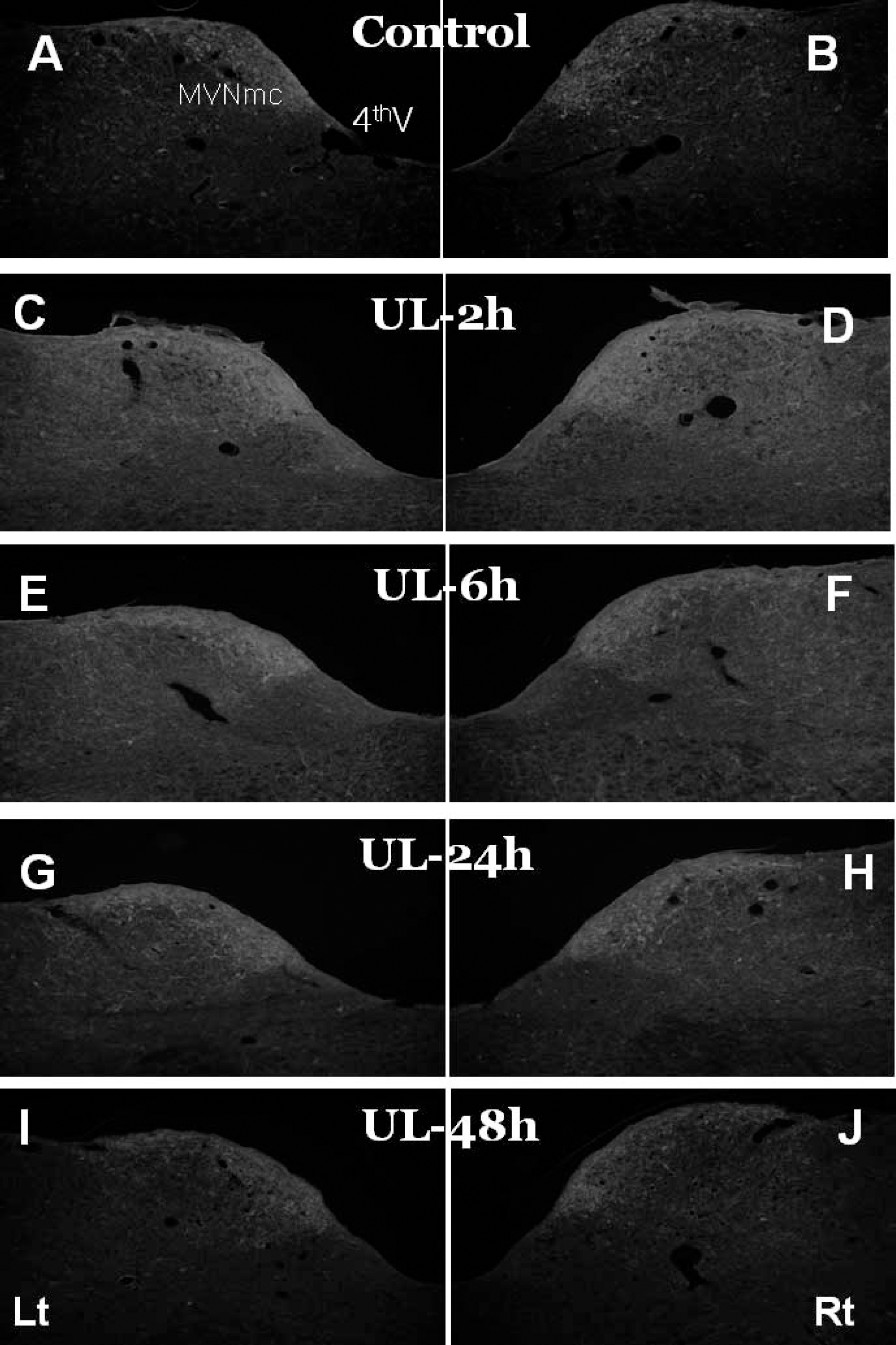
Fig. 2.
Changes in the area of calretinin staining in the medial vestibular nucleus after UL. 0 hour represents control; ipsilateral, ipsilateral MVN to the lesion; contralateral, contralateral MVN to the lesion. Relative ratio was calculated from the ratio of the areas in UL animals to those in control animals. Values are mean±SE. ∗p<0.05, ∗∗p<0.01, significantly different from control; †p<0.05, significantly different from the opposite side.
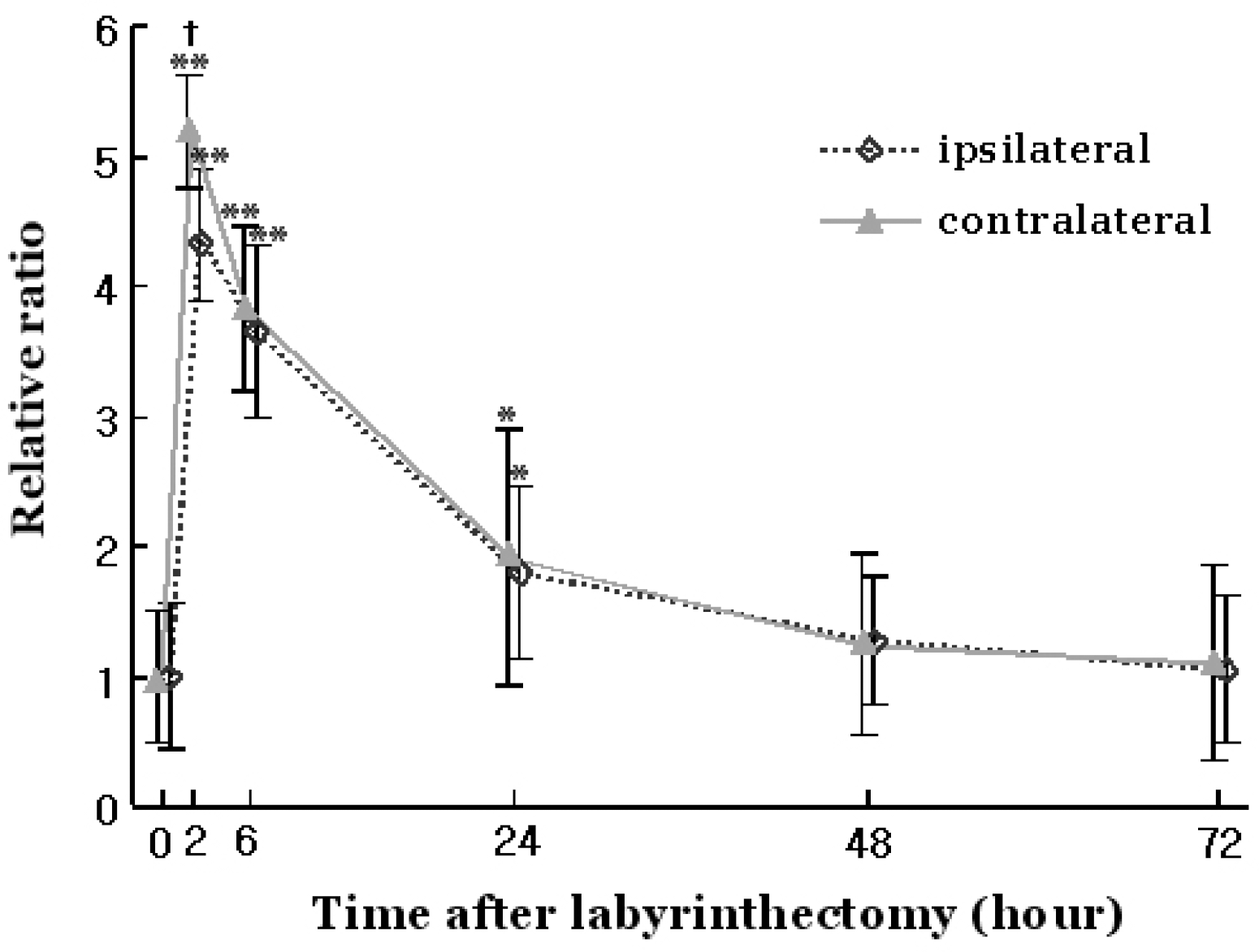
Fig. 3.
Immunofluorescence findings of calbindin in the medial vestibular nucleus after UL. A & B, left (Lt) & right (Rt) MVN, respectively, in control rat; C & D, ipsilateral (left) & contralateral (right) MVN to the lesion, respectively, 2 h after UL; E & F, ipsilateral & contralateral MVN, respectively, 6 h after UL; G & H, ipsilateral & contralateral MVN, respectively, 24 h after UL; I & J, ipsilateral & contralateral MVN, respectively, 48 h after UL. In E & F, the large square in the lower panel is a magnification of the small square in the upper panel. Triangle indicates the nerve terminals and arrow indicates the nerve fibers.
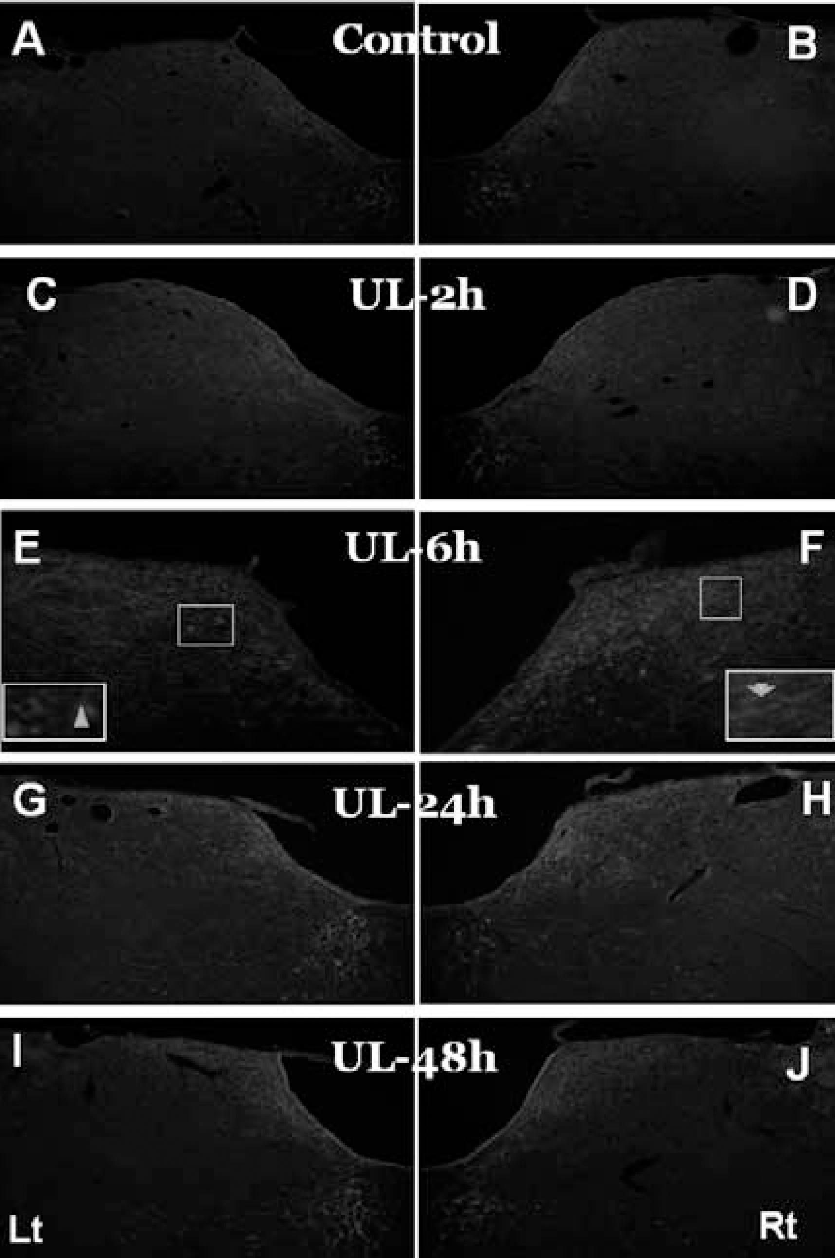
Fig. 4.
Changes in the area of calbindin staining in the medial vestibular nucleus after UL. 0 hour represents control; ipsilateral, ipsilateral MVN to the lesion; contralateral, contralateral MVN to the lesion. Relative ratio was calculated from the ratio of the areas in UL animals to those in control animals. Values are mean±SE. ∗p<0.05, ∗∗p<0.01, significantly different from control.
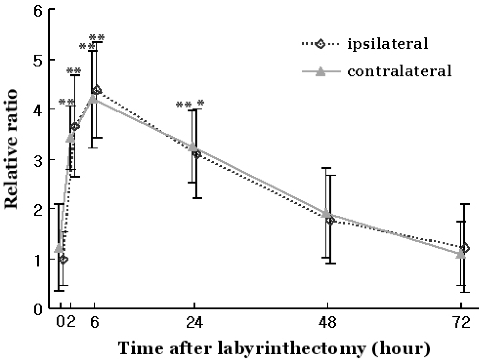
Fig. 5.
Immunofluorescence findings of calbindin in the medial vestibular nucleus after UL. A & B, left (Lt) & right (Rt) MVN, respectively, in control rat; C & D, ipsilateral (left) & contralateral (right) MVN to the lesion, respectively, 2 h after UL; E & F, ipsilateral & contralateral MVN, respectively, 6 h after UL; G & H, ipsilateral & contralateral MVN, respectively, 24 h after UL; I & J, ipsilateral & contralateral MVN, respectively, 48 h after UL.
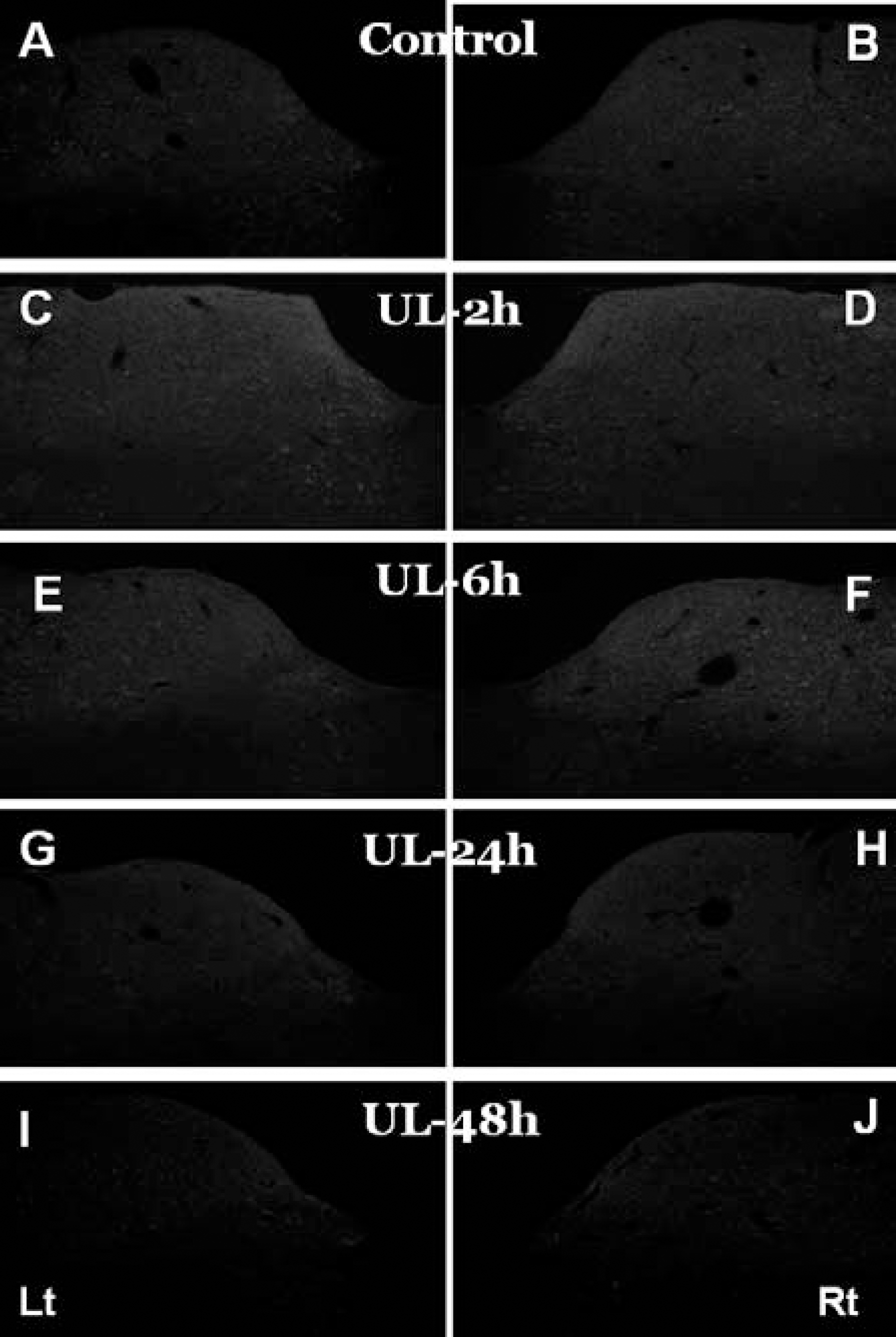
Fig. 6.
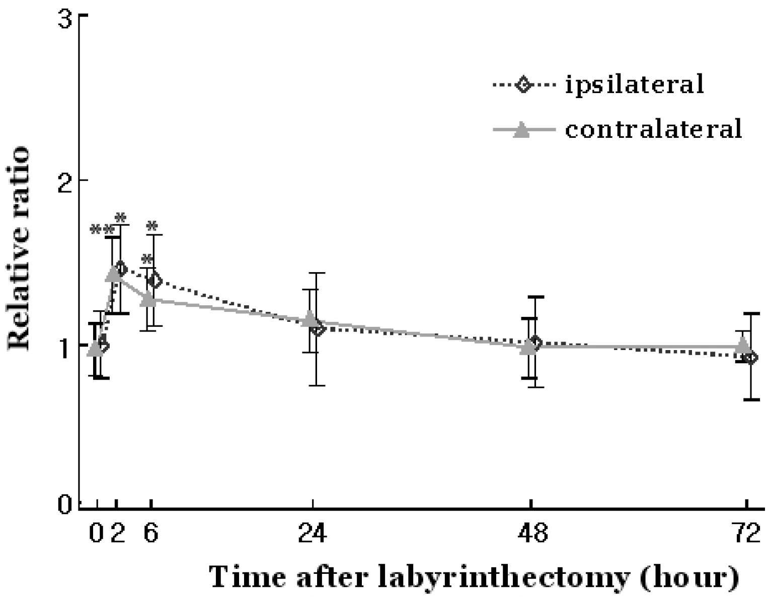
Changes in the area of parvalbumin staining in the medial vestibular nucleus after UL. 0 hour represents control; ipsilateral, ipsilateral MVN to the lesion; contralateral, contralateral MVN to the lesion. Relative ratio was calculated from the ratio of the areas in UL animals to those in control animals. Values are mean±SE. ∗p<0.05, ∗∗p<0.01, significantly different from control.


 PDF
PDF ePub
ePub Citation
 Print
Print


 XML Download