Journal List > J Korean Med Sci > v.31(7) > 1023008

Moon, Park, Kim, Choi, Song, Lee, Jo, Kim, Yang, Kim, Kim, and Yun: Two Cases of Intravascular Lymphoma Presenting with Pulmonary Involvement
Intravascular lymphoma (IVL) is a rare type of extranodal non-Hodgkin's lymphoma. It is characterized by the proliferation of malignant lymphocytes within the lumen of small vessels and sparing of surrounding tissue (1235). Pulmonary presentation of IVL is rare in Asian population and shows poor prognosis (45). It can be also a cause of unexplained interstitial lung disease. Because the prognosis is poor, the need of more intensive treatment should be considered.
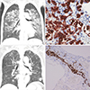
Two women, 56 years old (patient 1) and 59 years old (patient 2), commonly had a one month history of fever, weight loss, and upper respiratory symptoms. They did not show skin lesions and hepatosplenomegaly. Chest computed tomography (CT) scans also commonly showed ground-glass opacities and consolidations in both lungs (Fig. 1). In the bone marrow study, secondary hemophagocytosis (patient 1) and lymphoma involvement (patient 2) were observed, respectively. A positron emission tomography (PET) scan with 2 patients revealed diffuse increased glucose uptake in both of the lungs. These findings of CT scan and PET scan considered atypical bacterial or viral pneumonia, and acute hypersensitivity pneumonitis as possible diagnosis. An open lung biopsy for a confirmative diagnosis showed that CD20, CD79a, bcl-2, and MUM-1 were positive in the intravascular atypical cells (Fig. 1). Patient 1 was given best supportive care because of poor general condition due to delayed diagnosis. It was much delayed (2 months [patient 1], 1 month [patient 2], respectively) to diagnose finally from the initial hospital visit. On several reports, the prognosis of IVL is very poor, early introduction of systemic chemotherapy may be important to improve prognosis (46). The previous reports recommended an anthracycline-based chemotherapy regimen including CHOP. Rituximab is added in cases of B-cell lineage (236). Therefore we decided to use intensive chemotherapy for patient 2, who was given 3 cycles of R-hyper CVAD regimen. After 2 cycles of R-hyper CVAD, partial remission was observed. However, disease rapidly progressed and the patients eventually expired at three (patient 1) and four months (patient 2) after initial presentation, respectively. For early diagnosis of IVL with lung involvement, the early suspect based on symptoms, chest CT scan and PET scan are important because of no specific findings for IVL of lung involvement, and early aggressive diagnostic procedures (such as trans-bronchial biopsy, open lung biopsy) are required. Because the prognosis of IVL is poor, the more intensive treatment including front-line autologous transplantation should be explored. Here, we reported two cases of intravascular large B-cell lymphoma involving the lungs with their images and histopathology findings.

Figures and Tables

Fig. 1
Findings of 56-year-old (A, B) and 59-year-old (C, D) women with pulmonary intravascular lymphoma. (A) Chest CT scan shows patchy GGOs and consolidations in both lungs, mainly located in the central portion. (B) Open lung biopsy specimen shows that many atypical lymphoid cells occupy the capillary vessels and widening of alveolar septae filled with atypical tumor cells. The atypical lymphocytes are positive for CD20 (× 400). (C) Chest CT scan shows widespread GGOs mixed with multifocal non-calcified nodules in both lungs. (D) Lung specimen by wedge resection shows that many atypical lymphoid cells occupy the capillary vessels. The atypical lymphocytes are positive for CD20 (× 200).
jkms-31-1011-g001

References

1. Kawamura T, Sando Y, Tajima S, Hosono T, Sato M, Maeno Y, Maeno T, Suga T, Kurabayashi M, Nagai R. Pulmonary intravascular lymphoma complicated with Pneumocystis carinii pneumonia: a case report. Jpn J Clin Oncol. 2001; 31:333–336.
2. Zuckerman D, Seliem R, Hochberg E. Intravascular lymphoma: the oncologist's "great imitator". Oncologist. 2006; 11:496–502.
3. Martusewicz-Boros M, Wiatr E, Radzikowska E, Roszkowski-Sliz K, Langfort R. Pulmonary intravascular large B-cell lymphoma as a cause of severe hypoxemia. J Clin Oncol. 2007; 25:2137–2139.
4. Niida T, Isoda K, Miyazaki K, Kanoh S, Kobayashi H, Kobayashi A, Kimura F, Hayashi K, Kusuhara M, Ohsuzu F. Pulmonary intravascular lymphoma diagnosed by 18-fluorodeoxyglucose positron emission tomography-guided transbronchial lung biopsy in a man with long-term survival: a case report. J Med Case Reports. 2011; 5:295.
5. Ferreri AJ, Campo E, Seymour JF, Willemze R, Ilariucci F, Ambrosetti A, Zucca E, Rossi G, López-Guillermo A, Pavlovsky MA, et al. Intravascular lymphoma: clinical presentation, natural history, management and prognostic factors in a series of 38 cases, with special emphasis on the ‘cutaneous variant'. Br J Haematol. 2004; 127:173–183.
6. Souza CA, Quan K, Seely J, Kravcik S, Burns B. Pulmonary intravascular lymphoma. J Thorac Imaging. 2009; 24:231–233.
TOOLS
ORCID iDs

Ji Young Moon
https://orcid.org/http://orcid.org/0000-0003-2768-7810

Won Hong Park
https://orcid.org/http://orcid.org/0000-0003-0716-7914

Jin Man Kim
https://orcid.org/http://orcid.org/0000-0003-3129-3355

Yoon Seok Choi
https://orcid.org/http://orcid.org/0000-0002-0054-3641

Ik-Chan Song
https://orcid.org/http://orcid.org/0000-0002-6938-970X

Hyo Jin Lee
https://orcid.org/http://orcid.org/0000-0001-8378-3001

Deog-Yeon Jo
https://orcid.org/http://orcid.org/0000-0002-8267-4214

Samyong Kim
https://orcid.org/http://orcid.org/0000-0001-5993-1430

Young Jun Yang
https://orcid.org/http://orcid.org/0000-0003-4394-7993

Song Soo Kim
https://orcid.org/http://orcid.org/0000-0002-3078-2184

Jin Hwan Kim
https://orcid.org/http://orcid.org/0000-0002-1632-2421

Hwan-Jung Yun
https://orcid.org/http://orcid.org/0000-0003-0696-1235

Similar articles