

 PDF
PDF ePub
ePub Citation
 Print
Print


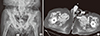
An 83-year-old man presented with acute onset of consciousness disturbance and dyspnea. He had a history of diabetes mellitus and congestive heart failure. However, there was no fever, chills, cough, sputum, vomiting, diarrhea, dysuria, or urinary frequency. Physical examination revealed swelling with erythematous changes over the perineal region. Laboratory data revealed white blood cells of 28,200/mm3, and a C-reactive protein of 226.8 mg/L (normal reference < 5 mg/L). Plain radiography of the pelvis showed subcutaneous emphysema over the left perineal region (Fig. 1A, arrow). Computed tomography (CT) of the abdomen showed the presence of air over left perineum, inguinal, and lower abdominal wall region (Fig. 1B, arrows). He received empirical antibiotic treatment with ertapenem and underwent an emergent fasciectomy. Two days later, extended-spectrum beta-lactamase (ESBL) producing E. coli was isolated from the specimen of blood and wound. The post-operation course went smoothly and he was discharged uneventfully one month later.
Fournier's gangrene (FG) is defined as a rapidly progressive necrotizing infection of the perineal, perianal, and genital regions and, most important of all, it is a life-threatening disease associated with high mortality (12). Although early diagnosis is critical, the initial signs and symptoms of FG may be nonspecific. The formation of gas is the characteristic finding in patients with FG; however, it is not always detected on physical examination. Besides physical examination, several imaging studies can be used to evaluate necrotizing infections. Despite that CT is usually the confirmation image (3), its use may be limited by the concern about contrast-related adverse effects such as acute renal failure. In contrast to CT, plain radiography has the advantage of being time-saving. Therefore, it may be another option, which may disclose typical findings - subcutaneous gas in the involved tissue planes in some occasions.
 XML Download