

 PDF
PDF ePub
ePub Citation
 Print
Print


INTRODUCTION
Angiogenesis, the process of generating new microvascular networks, plays an important role in a wide range of physiological and pathological conditions, including embryonic development, wound healing, tissue regeneration, and tumor growth (1, 2, 3). Vascular endothelial growth factor (VEGF) is one of the most potent angiogenic factors known to date (4, 5); it is secreted by a variety of cell types for functions such as regulating angiogenesis and tumor metastasis (6, 7, 8). Clinical studies of colorectal cancer have shown that the VEGF monoclonal antibody, bevacizumab, in combination with cytotoxic therapy positively affects patient survival rates (9). The VEGF-receptor (VEGF-R) tyrosine kinase inhibitor, vatalanib, has also shown to have an antitumorigenic effect in colorectal cancer as a result of antiangiogenic activity (9).
Many herbs and their natural products are traditionally used in anticancer treatments and are known to exhibit antiangiogenic properties through various interdependent processes (10). Grape seed proanthocyanidins inhibit angiogenesis (11), and Gleditsia sinensis thorn extract has been shown to prevent colon cancer and angiogenesis both in vitro and vivo (12, 13).
Acer tegmentosum (Acereaceae) has been used in Korean traditional medicine for the treatment of hepatic disorders (14). Diarylheptanoids (15), rhododendrol glycoside (16), and tannins (17) have been found in and isolated from the genus Acer. Compounds from the Acer tegmentosum Maxim methanol extract has been shown to be cytotoxic to cancer cell lines (18); however, no studies have examined its effect on angiogenesis or the underlying mechanisms. In this study, we investigated the effects of the A. tegmentosum maxim water extract (ATME) on angiogenesis and its underlying signal mechanism and found that the extract exhibits antiangiogenic potential both in vitro and in vivo.
MATERIALS AND METHODS
Preparation of the plant extract
A. tegmentosum Maxim twigs were collected from the Taebaeg area of Kangwondo, Korea. The dried and chopped twigs (170 g) were extracted twice with hot water (1.5 L) for 4 hr. This extract was filtered and lyophilized with a freezing dryer. The dry weight of the extract was 4 g. The dried extract was reconstituted in distilled water for the subsequent in vitro, ex vivo, and in vivo studies.
Cell culture and animal maintenance
Human umbilical vein endothelial cells (HUVECs) were prepared from human umbilical cords by collagenase digestion as previously described (19). They were maintained in M199 medium (Invitrogen, Carlsbad, CA, USA) supplemented with 20% fetal bovine serum (FBS), 100 U/mL penicillin, 100 µg/mL streptomycin, 3 ng/mL basic fibroblast growth factor (Upstate Biotechnology, Lake Placid, NY, USA), and 5 U/mL heparin at 37℃ and 5% CO2 with humidity. The HUVECs used were from between 4-6 passages for all experiments. The human pancreatic tumor cell line MIAPaCa-2, murine colon adenocarcinomas CT-26 cell line, and human hepatoblastoma HepG2 cell line were maintained in Dulbecco's modified Eagle's medium (DMEM) containing 10% heat-inactivated FBS (Life Technologies, Gaithersburg, MD, USA) at 37℃ and 5% CO2 with humidity. Sprague-Dawley rats (age, 7 weeks) were obtained from Orient Co. and were maintained on standard chow and water ad libitum.
Cell viability assay
Cell viability was evaluated with using the 3-(4,5-dimethylthiazol-2-yl)-2,5-diphenyltetrazolium bromide (MTT) assay. HUVECs (1×10⁵ cells) were plated in 24-well plates and cultured overnight. Cells were treated with 50 µg-1 mg ATME for 48 hr. The medium was then replaced with fresh medium containing 0.5 mg/mL MTT (USB Corporation, Cleveland, OH) for 4 hr. After incubation, the medium was carefully removed from the plate, and dimethyl sulfoxide (DMSO) was added to solubilize the formazan produced from MTT by viable cells. Absorbance was measured at 560 nm using a microplate reader (Molecular Devices, Sunnyvale, CA, USA).
Proliferation assay
The MTT assay was used to determine the effects of ATME on the proliferation of HUVECs in response to VEGF (R&D Systems, Minneapolis, MN, USA). Cells were seeded at a density of 1×10⁵ cells per well in 12-well plates. Proliferation and normal growth were determined at 48 hr after ATME treatment by using a colorimetric assay based on the uptake of MTT by viable cells. In brief, MTT solution was added to the cells and incubated at 37℃ to allow cleavage of the tetrazolium ring by mitochondrial dehydrogenases and formation of blue formazan crystals. After 4 hr, the residual MTT was carefully removed, and the crystals were dissolved by incubation with DMSO for 30 min. The plates were shaken for 5 min, and the absorbance at 560 nm was measured using a microplate reader. The optical density of untreated cells represented 100% viable cells, and the background color formation of MTT with DMSO added to an empty plate represented 0% viable cells.
Endothelial cell migration assay
The chemotactic motility of HUVECs was assayed using a Transwell apparatus (Corning Costar, Cambridge, MA, USA) with 6.5-mm diameter polycarbonate filters that had a pore size of 8 µm pore size, as described previously (20). Briefly, the lower surface of the filter was coated with 10 µg gelatin. The fresh M199 medium (1% FBS) containing VEGF was placed in the lower wells. Cells were trypsinized and suspended to a final concentration of 1×10⁵ cells/mL in M199 containing 1% FBS. ATME was added to the cells for 30 min at room temperature before seeding. One hundred microliters of the cell suspension was loaded into each of the upper wells. The chamber was incubated at 37℃ for 4 hr. Cells were fixed and stained with hematoxylin and eosin (H&E) stain. Non-migrating cells on the upper surface of the filter were removed by wiping with a cotton swab, and chemotaxis was measured with an optical microscope (200×) by examining the number of cells that migrated to the lower side of the filter. Ten fields of view were counted for each assay.
Endothelial cell invasion assay
The effect of ATME on HUVEC invasion was measured using a Cultrex® Cell Invasion Assay Kit (Trevigen, Gaithersburg, MD, USA). The membrane of the upper invasion chamber was coated with basement membrane extract (BME). HUVECs (5×10⁴ cells) were resuspended in 100 µL of low-serum medium (0.5% FBS) and seeded onto the culture inserts. They were then deposited into the 24-well companion plate with 600 µL of low-serum medium containing VEGF (20 ng/mL) and various concentrations of ATME. Wells containing VEGF alone served as a positive control. After 16 h of incubation, the media from the wells was withdrawn and the non-invasive cells on the upper surface of the membrane were removed by wiping with cotton swabs. The cells that had penetrated the BME-coated membrane and migrated onto the lower surface of the membrane were stained with H&E staining and mounted onto microscope slides. Images of the invasive cells were captured at 100× magnification using an optical inverted microscope. Following this, HUVEC invasion was quantified by counting the number of cells per insert.
In vitro capillary-like tube formation assay
Tube formation assays were performed as previously described (20). Briefly, 250 µL of growth factor-reduced Matrigel (Collaborative Biomedical Products, Bedford, MA, USA) was pipetted into a 16-mm diameter tissue culture well and polymerized for 30 min at 37℃. HUVECs incubated for 6 hr in M199 medium containing 1% FBS were harvested by trypsin treatment and suspended in M199 medium containing 1% FBS. ATME was incubated with the cells for 30 min at room temperature prior to plating the cells onto a layer of Matrigel at a density of 2×10⁵ cells per well, at which point 10 ng/mL of VEGF was added. After 20 hr, the cultures were imaged (40×). The area covered by the tube network was determined using an optical imaging technique in which pictures of the tubes were scanned in Adobe Photoshop and quantified using Image-Pro Plus (Media Cybermetics, Bethesda, MD, USA).
Rat aortic ring sprouting assay
The assay was performed as previously described (21). Briefly, aortic rings were obtained by cross-sectioning the thoracic aorta of 7-week-old Sprague-Dawley rats at 1-mm intervals. Plates (48 wells) were coated with 120 µL Matrigel; subsequently, rings were placed individually on the bottom of the plates with the luminal axis lying parallel to the bottom of the plate and sealed in place with an overlay of 50 µL Matrigel. VEGF with or without ATME was added to the wells in a final volume of 600 µL of human endothelial serum-free medium (Invitrogen). On day 7, the cells were fixed and stained with Diff-Quick, neovessels were imaged at 100× magnification using an Olympus CKX41 inverted microscope, and the angiogenic response was measured by quantifying the number of neovessels sprouting out of the rings over time. Sprouting was measured using the following scale: 0=no sprouting; 1=migrated cells without sprouting; 2=isolated sprouting; 3=sprouting in 25%-50% of the arterial ring circumference; 4=sprouting in 50%-75% of the circumference; and 5=sprouting in 75%-100% of the circumference. The assay was scored from 0 to 5 in a double-blinded manner. Each data point was quantified in sextuplet.
Western blot analysis
Cells were harvested and lysed with RIPA buffer containing 2 mM EDTA, 137 mM NaCl, 20 mM Tris-HCl (pH 8.0), 1 mM sodium vanadate, 10 mM NaF, 1 mM PMSF, 1% Triton X-100, 10% glycerol, and a protease-inhibitor cocktail. The protein concentration of each sample was determined using a BCA protein assay kit (Pierce, Rockford, IL, USA). Proteins were electrophoresed on sodium dodecyl sulfate-polyacrylamide electrophoresis gels and transferred to polyvinylidene fluoride membranes (Pall Corporation, East Hills, NY, USA). The blocked membranes were then incubated with the indicated antibodies, and immunoreactive bands were visualized using a chemiluminescent substrate. Antibodies for phospho-p44/42 MAP kinase (Thr 202/Tyr 204), p44/42 MAP kinase, phospho-p38 MAP kinase (Thr 180/Tyr 182), p38 MAP kinase, and Akt were obtained from Cell Signaling (Beverly, MA, USA).
In vivo matrigel plug assay
C57BL/6 mice (7 weeks of age) were given s.c. injections of 600 µL of Matrigel (Collaborative Biomedical Products, Bedford, MA) at 4℃, containing the indicated amount (25 µg) of ATME, 100 ng VEGF, and 10 units heparin. After injection, the Matrigel rapidly formed a plug. After 7 days, the skin of the mouse was pulled back to expose the Matrigel plug, which remained intact. After quantitative differences were noted and imaged, hemoglobin was measured using the Drabkin method and Drabkin reagent kit 525 (Sigma) to quantify blood vessel formation. The hemoglobin concentration was calculated using a known amount of hemoglobin assayed in parallel.
RESULTS
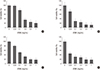
Effect of ATME on human endothelial cell viability
To rule out any toxicity from ATME, we tested endothelial cell viability using the MTT assay. As shown in Fig. 1A, the exposure to ATME induced cytotoxicity in a dose-dependent manner. We also investigated the cytotoxic effect of ATME on tumor cells. Similarly, ATME treatment exhibited cytotoxic effects in a dose-dependent manner in tumor cell lines as well. At 50% cytotoxic concentration, the order of cytotoxicity for each cell line was CT-26>MIAPaCa-2>HepG2>HUVECs. Addition of 50 µg/mL ATME was not toxic for HUVECs, indicating that the cytotoxic effect of ATME is specific to cancer cells.
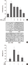
Inhibitory effects of ATME on VEGF-induced human endothelial cell proliferation and migration
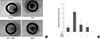
To assess the antiangiogenic activity of ATME, the effect of ATME on VEGF-induced proliferation of endothelial cells was evaluated. HUVECs were pretreated for 30 min with a given concentration of ATME prior to treatment with 20 ng/mL VEGF. ATME inhibited VEGF-induced proliferation, with half-maximal inhibition at approximately 20 µg/mL (Fig. 2A). Migration of endothelial cells is essential for tumor angiogenesis. The effect of ATME on the chemotactic motility of HUVECs was measured using a Transwell assay. After stimulating HUVECs with 20 ng/mL VEGF for 4 hr, a large number of cells migrated to the lower side of the filter in the Transwell Chamber. This VEGF-induced migration effect was significantly downregulated by ATME treatment (Fig. 2B, C). Treatment of ATME alone slightly inhibited the migration of endothelial cells, although this effect was not significant. These results indicated that ATME can block VEGF-induced angiogenesis in vitro.
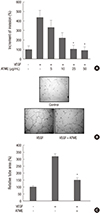
Effect of ATME on VEGF-induced endothelial cell invasion and tube formation
We subsequently studied the effect of ATME on the invasion of human endothelial cells by using the Transwell culture plate. As shown in Fig. 3A, VEGF-treated cells serving as positive controls exhibited increased invasion; however, the number of cells invaded in response to VEGF significantly reduced in a dose-dependent manner with ATME treatment. Next, we examined the effect of ATME on tube formation. When HUVECs were placed on growth factor-reduced Matrigel in the presence of VEGF, we observed the formation of elongated and robust tube-like structures that were found in greater frequency in the VEGF-treated cells than the control cells. ATME effectively abrogated the width and the length of the VEGF-induced endothelial tubes (Fig. 3B, C). These results indicate that ATME can block VEGF-induced angiogenesis in vitro.
Inhibition of VEGF-induced vessel sprouting ex vivo by ATME
The sprouting of vessels from aortic rings was investigated to determine whether ATME inhibited VEGF-induced angiogenesis ex vivo. Treatment with VEGF (20 ng/mL) significantly stimulated vessel sprouting when compared to the results with medium alone. However, the enhanced vessel sprouting induced by VEGF significantly reduced with ATME treatment (Fig. 4A, B).
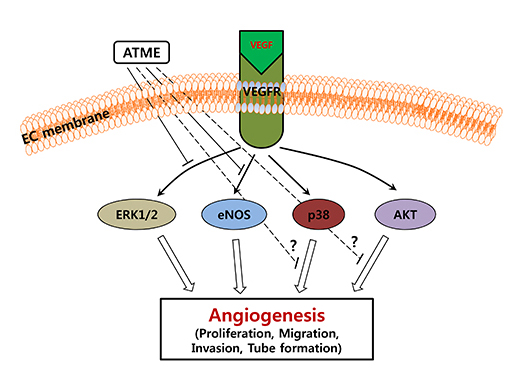
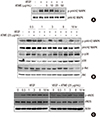
Effects of ATME on ERK and eNOS phosphorylation
To investigate the effects of ATME on cell signaling, we first examined the ERK pathway because it is a well-known pathway wherein VEGFs stimulate the phosphorylation of ERK. When we added VEGF to the human endothelial cells, we could induce ERK phosphorylation (Fig. 5A). However, as expected, ATME inhibited VEGF-induced phosphorylation of p44/42 in a dose- and time-dependent manner (Fig. 5A, B). In addition, phosphorylation of p44/42 MAP kinase, p38, Akt, and eNOS are important signaling events for proliferation, migration, and morphogenesis of endothelial cells induced by various angiogenic factors. Next, we performed western blot analysis to analyze p38, Akt, and eNOS phosphorylation and found that eNOS phosphorylation was inhibited by ATME but that VEGF-induced Akt and p38 phosphorylation was not affected by ATME treatment (Fig. 5B, C).
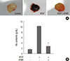
ATME downregulates angiogenesis in vivo
Next we evaluated the effect of ATME on the ongoing angiogenesis process by using an in vivo mouse Matrigel plug assay (Fig. 6). Matrigel and heparin (10 units/600 µL) with or without ATME (25 µg) were s.c. injected into C57BL/6 mice, and 7 days later, the Matrigel plug formed in mice was examined. Plugs with Matrigel alone were pale in color, indicating no or little blood vessel formation. In contrast, plugs mixed with VEGF appeared dark red (Fig. 6A). The new vessels were abundantly filled with intact RBCs, which indicate the formation of a functional vasculature inside the Matrigel and blood circulation in the newly formed vessels by angiogenesis induced by VEGF. However, when we cotreated ATME with VEGF, VEGF-induced angiogenesis significantly reduced (Fig. 6A, B). We also measured the hemoglobin content inside the Matrigel plugs to quantify the angiogenesis induced by ATME. The hemoglobin level in control cells was nearly 1.8 g/dL, and VEGF markedly enhanced the hemoglobin level to approximately 10 g/dL (Fig. 6B). The increase in the hemoglobin level by VEGF was potently decreased by ATME (Fig. 6B). These results indicate that ATME might be a powerful, natural antiangiogenic compound.
DISCUSSION
Angiogenesis is one of the most important processes for tumor growth and metastasis. VEGF, the most potent of the angiogenic growth factors, plays a crucial role during tumor angiogenesis. Suppression of angiogenesis using natural plant extracts has been shown to effectively block tumor growth (10). However, the antiangiogenic effects of A. tegmentosum Maxim, which is used as a traditional Korean medicine, have not been reported. To our knowledge, this study is the first to demonstrate that VEGF-induced angiogenesis is inhibited by ATME. ATME was tested for its ability to inhibit cell viability, cell proliferation, migration, invasion, and tube formation in vitro by using HUVECs; aortic ring sprouting ex vivo by using rat aortae; and new vessel formation in vivo by using the matrigel plug method. We found that ATME significantly reduces cell proliferation, migration, and extracellular matrix degradation and therefore inhibits tube formation, highlighting the role of A. tegmentosum Maxim as an antiangiogenic therapy for cancer prevention.
Although cell viability was not affected by ATME in this study, ATME significantly suppressed the stimulatory effect of VEGF on endothelial cell proliferation and migration (Fig. 2). As shown in Fig. 1, ATME had no significant effect on the normal growth of endothelial cells with a dose of up to 50 µg/mL; its antiproliferative affects at low concentrations seem to reflect a specific response of endothelial cells stimulated by the angiogenic growth factor VEGF. We also found that the extract has the capacity to increase cell death in a dose-dependent manner and that the cancer cells (human pancreatic tumor cell line MIAPaCa-2, human hepatoblastoma cell line HepG2, and murine colon adenocarcinoma cells CT-26) are more sensitive to ATME than healthy control cells, specifically MDCK (canine kidney epithelial) cells (data not shown) and HUVECs (human umbilical vein endothelial cells). These results suggest that ATME can specifically induce cancer cell death at a concentration at which it does not influence normal endothelial cells.
Although the VEGF signaling pathway in endothelial cells is not fully understood, signaling molecules such as those involved in the Src, PI3K/Akt, MAPK, and eNOS/NO signal pathways have been reported to be involved in the VEGF signaling cascades (22, 23, 24). MAPK pathways constitute a large modular network that regulates a variety of physiological processes such as cell growth, migration, differentiation, and apoptotic cell death (25, 26, 27, 28). Currently, three subfamilies of MAPK have been characterized: extracellular signal regulated kinase 1/2 (ERK 1/2, p44/42 MAP kinase), c-Jun N-terminal kinase 1/2 (JNK 1/2), and p38 MAP kinase (p38). They are activated by growth factors, inflammatory cytokines, and stress stimuli and play a critical role in the angiogenesis process (29). The activation of p44/42 and p38 MAP kinase pathway plays an important role in endothelial cell proliferation and migration (30, 31). In these experiments, we found that ATME downregulated the phosphorylation of p44/42 MAP kinase in response to VEGF but not phosphorylation of p38 MAP kinase (Fig. 5). The current results demonstrated that ATME inhibits VEGF-induced angiogenesis by inhibiting p44/42 MAP kinase activation.
Nitric oxide (NO) has recently been shown to be an important signaling molecule and regulator of angiogenesis. NO donors, such as nitroprusside, promote endothelial cell proliferation and migration; in contrast, inhibitors of nitric oxide synthase (NOS) suppress this response (32). In addition, several lines of evidence have indicated that NO is an angiogenic downstream mediator in the angiogenic response of a variety of growth factors (33). NO lies downstream from and mediates the effects of VEGF (34, 35). VEGF has also been shown to upregulate eNOS massage, NO production, and eNOS protein (36, 37). The activation of the PI3K/Akt/eNOS/NO signaling pathway is an important event in angiogenesis (24, 38). Angiogenesis is significantly reduced in response to growth factors in eNOS-deficient mice (39). In this study, we examined the effect of ATME on Akt and eNOS phosphorylation in response to VEGF. ATME prevented VEGF-induced phosphorylation of eNOS but had no effect on Akt phosphorylation (Fig. 5). These results indicate that the inhibitory effect of ATME on eNOS phosphorylation is not due to the PI3K/Akt pathway and that the antiangiogenic activity of ATME may be due to the blockage of p44/42 and eNOS activation. The mechanism underlying the inhibitory effect of ATME on VEGF-induced angiogenesis is yet to be determined.
In addition to cell proliferation and migration, invasion and tube formation are essential to the angiogenic process. Vessel formation requires degradation of both the laminin-rich basement membrane surrounding the endothelial cells and proteolysis of the collagen-rich extracellular matrix of the connective tissue and requires the assembly of endothelial cells into vessel tubes. In the current study, we found that ATME significantly reduces the number of invaded cells and the mean tube length of endothelial tubes induced by VEGF. Thus, ATME inhibits the angiogenic factor-induced invasion and tube formation effects of HUVECs in vitro. Similarly, when added to rat aorta rings that had been maintained in a three-dimensional matrigel to allow the sprouting of newly formed vessels, ATME remarkably suppressed sprouting of endothelial cells in the rat aorta in response to VEGF (Fig. 4). This ex vivo antiangiogenic activity may be explained by the inhibitory effect of ATME on the proliferation, migration, invasion, and differentiation of endothelial cells in response to angiogenic growth factors such as VEGF. Moreover, ATME strongly inhibited angiogenesis in an in vivo matrigel plug assay (Fig. 6).
In conclusion, our present data demonstrate that ATME inhibits angiogenesis in vitro and in vivo. ATME may be useful for preventing or treating angiogenesis-dependent human disease conditions, including tumors.
 XML Download