

 PDF
PDF ePub
ePub Citation
 Print
Print


INTRODUCTION
Orientia tsutsugamushi is a rickettsial organism and a causative pathogen for Scrub typhus (1). It causes acute and chronic infection, leading to symptoms of fever, rash, pneumonitis, encephalitis, and ultimately death (2). It is found endemically mainly in Northeast Asian regions, China, India, and Australia. Scrub typhus is one of the most common vector borne diseases occurring in Korea and the Asia pacific regions (3). More than 5,000 cases are reported annually in Korea, with incidents currently on the rise (4).
Gram-negative bacteria produce OMVs (outer membrane vesicles) of 50-250 nm in diameter from the outer membrane (5). To date, OMVs originating from many bacteria, including Escherichia coli, Neisseria meningitides, and Pseudomonas aeruginosa have been documented (4). OMVs are secreted from the bacterial surface membrane, and therefore consist of outer membrane proteins (OMP), lipopolysaccharides (LPS), phospholipids and other periplasmic components (6). OMVs have been reported to play various roles associated with the functions of secretion and delivery, supporting the survival and pathogenesis of bacteria (7). OMVs have also been observed in intracellular gram-negative bacteria of Salmonella spp., Franciella spp. and Chlamydia spp. (8, 9, 10). However, no report has yet confirmed whether OMVs are produced by O. tsutsugamushi, an obligate intracellular bacterium. In this regard, we intend to investigate whether O. tsutsugamushi produces OMVs and purifies microvesicles by immunoprecipitation.
MATERIALS AND METHODS
Preparation of O. tsutsugamushi
O. tsutsugamushi Boryong strain was propagated in ECV-304 cells (CLS, Germany) cultivated in M199 (WelGENE, Korea) with 10% (v/v) fetal bovine serum (Corning Cellgro, USA). Confluency of bacteria in ECV304 was confirmed by immunofluorescence assay (IFA). When ECV-304 cells were heavily infected, they were gathered and used for electron microscopic observation of O. tsutsugamushi in cytosol of host cells. Heavily infected cells were disrupted with glass beads (diameter, 1.0 mm) to release bacteria from the cells and bacteria were purified with 40% percoll density solution utilizing the same method of Tamura et al. (11). Purified bacteria were also observed by electron microscope.
Purification of OMVs
ECV304 cells, heavily infected with O. tsutsugamushi, were centrifuged at 13,000 rpm for 30 min at 4℃, after which the centrifugal media was passed through a 0.22 µm pore size filter system (Corning, MA, USA). The cell free medium was concentrated to 50 mL by ultra-filtration using a QuixStand Benchtop system (GE Healthcare Bio-Sciences, Uppsala, Sweden) with a 100 kDa hollow fiber membrane and harvested by ultracentrifugation at 150,000 g for 3 hr at 4℃. The pelleted OMVs were resuspended in 1 mL of PBS and then purified as described previously with modifications (12). After ultracentrifugation in a sucrose gradient solution, each fraction was analyzed by SDS-PAGE, Coomassie Brilliant Blue staining, and western blotting. The fractions of sucrose density gradient showing protein profiles corresponding to O. tsutsugamushi in immunoblot bands were collected and centrifuged at 150,000 g for 3 hr at 4℃. The resulting pellets of purified OMVs were resuspended in PBS containing protease inhibitor cocktail (Sigma-Aldrich Co., MO, USA). The suspended OMVs were observed using an electron microscope. The purified OMVs were quantified using DC protein assay reagents (Bio-Rad Laboratories Inc., Hercules, CA, USA) and aliquots of the OMVs were stored at -70℃. Purified OMVs were taken for immunoenrichment and immunoblot analysis.
Immunoenrichment of O. tsutsugamushi derived OMVs
For enrichment of O. tsutsugamushi derived OMVs from a mixed population of vesicles, FS15 mouse monoclonal antibody reacting against 56 kDa protein of O. tsutsugamushi Boryong strain was combined with 10 µL of protein G magnetic beads (NEW ENGLAND BioLabs., MA, USA) and incubated at room temperature for 1 hr while rotating (25). The resultant was washed three times with IP buffer (25 mM Tris pH 7.5, 150 mM NaCl, 2.5 mM EDTA, 0.05% Triton X-100) and then combined with appropriate concentrations of purified OMVs overnight while rotating at 4℃. The mixture was washed four times with IP buffer and the final wash was performed with PBS. Pellets in reducing sample buffer (50 mM Tris-Cl pH 6.8, 100 mM dithiothreitol (DTT), 2% SDS, 0.1% bromophenol blue, 10% glycerol) were solubilized by boiling for 10 min at 100℃. The solubilized samples were loaded on a 10% polyacryl amide gel. The proteins from the OMVs were transferred to a PVDF (Millipore, Darmstadt, Germany) membrane. The membrane was blocked with 5% nonfat dry milk in PBST (0.1% tween20 in PBS) for 1 hr at room temperature and then incubated overnight at 4℃ with O. tsutsugamushi polyclonal antibody. The membrane was washed three times with PBST and incubated with HRP-conjugated secondary antibody (Jackson Immunoresearch Laboratories, PA, USA) for 1 hr at room temperature. The membrane was washed again three times with PBST and developed with enhanced chemiluminescence (ECL) solution (GE Healthcare Life-Sciences, Uppsala, Sweden). Antibody used for the western blot assay, which was purified from the serum of a patient infected with O. tsutsugamushi Boryong, was confirmed by nested PCR amplifying the 56 kDa region. The two pairs of primers used were as follows: outer primers, 1F (5'-ATAATTAATGTATTTTCGAACG-3') and 2R (5'-CCTKCA AAGGACTTTTAGCT-3'), and inner primers, 1Fn (5'-AACACAGTGTTTTATAGATTGTTTA-3'), and 2Rn (5'-RCATTAATTGCTACACCAAGT-3'). The amplified length was 1,562 bp. The PCR product was purified and sequenced by GenoTech Corp. (Daejeon, Korea). The resulting sequence was identified as 56-kDa TSA gene using BLAST (http://ncbi.nlm.nih.gov/blastn).
Transmission electron microscopy (TEM)
To observe the blebbing of O. tsutsugamushi in infected ECV304 cells and purified bacteria, each pellet was fixed with 2.5% glutaraldehyde in 0.1 M phosphate (pH 7.4) for 2 hr. The samples were washed and placed in 1% osmium tetroxide for 30 min, dehydrated in ethanol and embedded in epoxy resin (Epon 812, Electron Microscope Sciences, UK) in Beam capsules. Ultrathin sections were cut using an ultramicrotome (UltraE, Reichert-Jung, USA) and stained with uranyl acetate and citrate. For observation of purified OMVs structure, samples were placed on 200 mesh carbon-coated copper grids until settled on the film for 5 min. The grids were stained for 5 min with 1% uranyl acetate. After air drying, the samples were analyzed under a CM200 transmission electron microscope (Phillips, Netherlands).
RESULTS
Electron microscopic observation of OMVs derived from O. tsutsugamushi
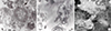
Electron microscopic examination showed that OMVs were secreted from the bacterial surface membrane in cytosol of host cells (Fig. 1A). Several microvesicles that might have been derived from O. tsutsugamushi were observed near the bacteria. The budding OMVs had a monolayer membrane and were approximately 130 nm in diameter. Microvesicles were observed in the vicinity of purified O. tsutsugamushi (Fig. 1B). Purified OMVs were observed as round or spherical vesicles of relatively diverse size ranging from 50-150 nm in diameter (Fig. 1C).
Identification of O. tsutsugamushi derived OMVs
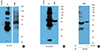
Western blot analysis of purified OMVs with FS15 monoclonal antibody, specific to O. tsutsugamushi 56-kDa protein showed the presence of proteins corresponding to O. tsutsugamushi (Fig. 2A). In control experiments, ECV 304 cell lysates used for cell culture did not react with either FS-15 monoclonal antibody or polyclonal antibody. This result validates that microvesicles observed in the electron microscopic examination were derived from O. tsutsugamushi. Western blot analysis with O. tsutsugamushi polyclonal antibody showed that a 56-kDa protein band was present as a prominent band (Fig. 2B).
Immunoenrichment of O. tsutsugamushi derived OMVs
To further purify O. tsutsugamushi derived OMVs from mixed populations with host derived microvesicles, we enriched bacterial OMVs by immunoprecipitation with FS15 monoclonal antibody. Western blot analysis of enriched OMVs showed 42, 46, and 56-kDa protein bands, corresponding to the surface antigens of O. tsutsugamushi (Fig. 2C). As expected, the 56-kDa protein was the most abundant protein band. ECV304 cell lysates used for cell cultures did not have a responding band in the western blot analysis using either FS15 monoclonal or polyclonal antibody. This result shows that OMVs from O. tsutsugamushi could be enriched with immunoprecipitation.
DISCUSSION
Scrub typhus is an important cause of febrile diseases in endemic regions. In Korea scrub typhus occurs mainly in the autumn and the incidence is increasing (4). While most patients respond favorably to antibiotic treatment some may experience severe complications and others may suffer chronic infection even after recovery from the disease (2). In this regard, a good understanding of host-pathogen interaction in this disease is needed for development of effective treatment and prevention.
In the current study, we were able to demonstrate that OMVs are derived from Orientia tsutsugamushi, an intracellular pathogen. OMVs derived from the outer membrane layer of bacteria are related to the survival of pathogens inside host cells (7, 13). The secretion of OMVs is dependent on growth conditions or environmental factors. OMVs produced from O. tsutsugamushi might play roles for their survival. Many of the components constituting OMVs might be attributed to those found abundantly in the bacteria (13). The protein profile of OMVs may be similar, but not entirely identical, to that of bacterial outer membrane. The major antigens for O. tsutsugamushi are 110, 80, 70, 56, 46, and 25-kDa in size (14). The most abundant and important surface antigen of O. tsutsugamushi is the 56-kDa protein, which is also the major antigen isolated from OMVs of the current study. Being one of the most important antigens of O. tsutsugamushi, the 56-kDa protein has a role in the attachment on and penetration into the host cell (15). The immune response to 56-kDa protein has an important role in prevention of the disease. The 56-kDa protein has been shown to induce neutralizing antibodies in animals immunized using the 56-kDa protein (16).
O. tsutsugamushi requires host cells for their growth and proliferation. Hence, OMVs obtained from such cell culture may contain vesicles not only derived from the pathogen but also from the host cell used for survival. As a result, demonstration or isolation of vesicles derived from intracellular pathogens is not easy. In our experiment, we isolated OMVs from the culture media of heavily infected cells. Frohlich et al. (17) successfully demonstrated that vesicles were derived from bacteria using an immunoenrichment method in Chlamydia, which is an intracellular organism. In our study, we also isolated bacterial microvesicles using antibody against O. tsutsugamushi. OMVs with a high purity could contribute towards the understanding of host-pathogen interactions or further studies to delineate the function of microvesicles.
An OMV derived from the outer membrane layer measures 20-200 nm in diameter, and it characteristically represents the serotype of the pathogen as it contains the pathogen's outer membrane proteins and surface antigens (7). Consequently, the potential of OMVs to function as a vaccine or adjuvant has been suggested since OMVs have similar properties as found in various pathogens and can be recognized effectively by antigen presenting cells to induce immune reactions (18). It is also expected that OMVs could be modified by combining with heterogeneous antigens (19). The OMV-conjugate vaccine created by the unified genetic fusion method proved to have desirable effects (20). In fact, a Meningococcus group B vaccine using OMVs has been used successfully to control an epidemic (21). Vaccine research for O. tsutsugamushi had been limited by the fact that vaccines induced only strain specific immune responses and that a durable protection period was not provided (3, 22). OMVs obtained in this study contain a 56-kDa, which is the most important antigen for O. tsutsugamushi and other surface antigens. Likewise, OMVs derived from O. tsutsugamushi could be utilized to induce immune reaction against various antigens or reinforce immunogenicity as an adjuvant.
Since the outer membrane of most Gram negative strains consists of LPS, OMVs, having been derived from that source, also consist of LPS (7). However, despite being a gram-negative pathogen, the outer membrane of O. tsutsugamushi is known not to consist of LPS or peptidoglycans (23). If OMVs obtained from other gram-negative pathogens are used as immune adjuvants, the safety of LPS comes into question since it works precisely due to endotoxin contained in OMVs. Some safety measures are based on alteration of the OMVs structure or use of non-pathogens to lower LPS endotoxicity (24). OMVs derived from O. tsutsugamushi, conversely, would be clinically safer since they do not contain LPS.
To summarize, we demonstrated that OMVs were produced from Orientia tsutsugamushi, which were purified with ultracentrifugation and immunoprecipitation. We also found that OMVs contain a 56-kDa protein which is a neutralizing antigen. Further studies are required to examine the function of OMVs and methods for development of vaccines or adjuvants based on such findings.
 XML Download