

 PDF
PDF ePub
ePub Citation
 Print
Print


INTRODUCTION
We attempted virtual endoscopic and laparoscopic exploration of the ascending colon based on sectioned images from a cadaver. The realistic sectioned images and their derived outlined images were the source of a high resolution volume model with actual body color that could be piled or peeled. This innovative trial has efficiently demonstrated tissue layers of the ascending colon, which are not revealed by clinical and microscopic images (1). We decided to apply the established procedures to the stomach, because endoscopy and laparoscopy of the stomach have become more important in hospitals.
The aim of this study was to ascertain virtual endoscopy and laparoscopy based on sectioned images to provide anatomical detail of the stomach. A raw sectioned image including the stomach was examined in relation to the corresponding endoscopic and laparoscopic views, where the luminal outlines were continuously expanded at appropriate thicknesses.
MATERIALS AND METHODS
The subject was switched from a male cadaver to a new female cadaver, who was 26 yr old and had a standard body size (length, 1.69 m; weight, 52 kg), when she died of stomach cancer and pneumonia. But she did not undergo surgery of the stomach or lung. Using the methods upgraded by our Visible Korean team (2), the cadaver was serially sectioned to yield improved sectioned images (resolution, 5,616×3,744; pixel size, 0.1 mm; color depth, 24 bits color) in 2010.
We previously formulated and detailed the methods to produce endoscopic and laparoscopic views. According to these methods, luminal outlines of the stomach were traced in the serial sectioned images and employed to acquire a volume model. While the outlines were expanded, the stomach was observed endoscopically and laparoscopically in comparison with a chosen sectioned image (1).
The luminal outlines between the lumen and mucosa were prominent, because the gastric lumen was filled with blue gelatin and food. In the previous study with the ascending colon, the boundary between the muscular layer and adventitia was demarcated. Consequently, the present study only tried to expand the outlines, while the previous one attempted to expand and shrink the outlines (1, 3, 4, 5, 6, 7).
Another new trial was the interpolation of luminal outlines (intervals, 1 mm) of the stomach. Consequently, the original sectioned images (intervals, 0.2 mm) could be utilized to enhance the quality of volume model.
RESULTS
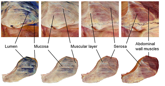
The sectioned image showed that the posterior wall of the stomach was invaded by the gastric cancer, but the anterior wall was intact. We focused on the normal anterior wall in this study (Fig. 1).
The gastric rugae were sporadic. The rugae involved the mucosa and submucosa but not the muscular layer as it is known (Fig. 1). The rugae are flattened when the stomach is filled with food (8). In the Visible Korean experiment, blue gelatin was placed in the gastric lumen during serial sectioning; the gelatin was not injected to swell the stomach before serial sectioning.
The four layers of the stomach were discernible by their proper colors in sections of the non-embalmed cadaver. The mucosa, submucosa, muscular layer, and serosa were light brown, dark brown, white, and light red, respectively. The genuine serosa could not be distinguished from the parietal peritoneum and extraperitoneal fat at the anterior side of the stomach. We designated them as just serosa for convenience. Outside the serosa were abdominal wall muscles such as the transversus abdominis and rectus abdominis (Fig. 1).
Smooth muscles differed in color from the dark red color of the skeletal muscles (Fig. 1). This color difference between the two muscle types was found in the sectioned images of other cadaver (1), and even in dissected cadavers.
In this study, one volume model consisted of about 370 million voxels, as 510 images (resolution, 880×830) were stacked after interpolation. Once the outline-associated numbers were marked on the voxels of the volume model, laparoscopic and endoscopic visualization was not retarded (1, 9, 10).
All layers in the endoscopic and laparoscopic views, except the submucosa, were identified by use of their own colors. The colors of the mucosa, muscular layer, serosa, and abdominal wall muscles in the volume model were consistent with those in the sectioned images. The volume models of the original stomach, not expanded, showed the lumen (blue) and mucosa (light brown) simultaneously. This heterogeneous composition resulted from imperfect demarcation; actually, the semiautomatic or manual outlining was not free from errors despite our efforts (Fig. 1, 2).
Four sets of endoscopic and laparoscopic views were made by serial expansion of the outlines at 2 mm thickness. Interestingly, the mucosa, muscular layer, and serosa were seen simultaneously in the first and second views, the second and third views, and the third and fourth views, in that order (Fig. 2). This was caused by the sectioned images where each layer had an approximate 2 mm thickness (Fig. 1). The three muscular layers, which are internal circular, external longitudinal, and oblique were not discriminated in either the endoscopic and laparoscopic views or the sectioned images (Fig. 1, 2). Laparoscopy of the most expanded (6 mm thickness) part of the stomach showed the abdominal wall muscles and the serosa. Their topographic distribution seemed to result from the anatomical configuration of the stomach: the fundus-side body, pressed by the anterior abdominal wall, had a flattened wall, while the pylorus-side body had a thick wall (Fig. 2).
DISCUSSION
Even if the virtual endoscopy and laparoscopy using cadaver images differ from the real practices for patients, our trial has the definite merit. These computer-processed images are not reproducible during actual endoscopy or laparoscopy in clinics. The computed tomography (CT) scans and magnetic resonance images (MRIs), showing stereoscopic contours of organs by means of the structures' density difference, cannot yield the curved sectional surfaces without outlining process (1, 11, 12). Therefore, this morphological investigation will help clinicians comprehend the stomach anatomy. For example, they will recognize that the abdominal wall is close to the gastric lumen on the fundus side (Fig. 2).
We demonstrated that a volume model can be piled or peeled at the intended intervals as if the structure's surface were expanded or shrunken. The original sectioned images, virtual endoscopic and laparoscopic images, all of which are corresponding, compensate for one another. This means that some layers of the stomach are more visible in an image set than in others (Fig. 1, 2).
The lifelike images of non-embalmed cadaver are to be broadened. By use of our raw data and algorithm, more endoscopic and laparoscopic images can be obtained to demonstrate neighboring organs such as the liver, gallbladder, pancreas, spleen, and kidney (13). Further, the serial enlargement of the outlines would disclose each organ's distance for the baseline. The images are available for clinical practice (e.g., endoscopic ultrasonography).
Several decades ago, sectional anatomy was just the perspective for anatomy education (14, 15, 16, 17). However, after various tomographies became current in hospitals, sectional anatomy has occupied a great deal in the anatomy field. Similarly, our new approach to pile or peel a volume model resulting in the curved sectional surfaces has the possibility to be widely applied in near future.
Any researcher can manufacture their own curved sectional surfaces of organs or regions. It requires two sets of sectioned images and outlined images besides the computer technique (1). The free software to browse sectioned and outlined images of cadaver is downloadable at the Visible Korean site (anatomy.co.kr); then the image data can be extracted (18). Moreover, investigators are supplied with a full high resolution data set after fulfilling an agreement with the authors. Further delineation is feasible after referring to the sectioned images (6, 7, 19, 20). Clinical images such as CTs and MRIs that are rapidly advancing in quality could be raw data for this experiment so as to disclose other aspect of the pathologic organ's shape. Serial histological images including confocal microscopic sections are alternative good data source for our suggested investigation to reveal additional characteristics of the stained tissues.
In summary, we have devised an expanding and shrinking stomach volume model, followed by two different visualization modes. This virtual endoscopic and laparoscopic exploration provides morphologic information for interested physicians and surgeons, respectively.
 XML Download