

 PDF
PDF ePub
ePub Citation
 Print
Print


INTRODUCTION
Supernumerary muscles have been reported in various regions throughout the human body, and some of them are well known by their relatively high frequency. The incidence of the accessory belly of the digastric muscle is relatively high at 5.1%-40.0% (1,2,3). In addition, the supernumerary head of the biceps brachii muscle has been found at an incidence of approximately 20% (4). In contrast, some of the muscular variations such as the palmaris profundus are only found in extremely rare cases (5). Moreover, unfamiliar variations such as an extra slip between the biceps femoris and semitendinosus have been rarely reported (6, 7).
Because these variations all have similar ectopic attachment sites between other normal muscles, they may have developed from a common factor during embryogenesis. Therefore, exploration into concurrent appearances may be helpful in understanding the causes of developmental aberration. Here, we report on the existence of multiple muscular variations in the neck, upper limb, and lower limb biased toward the left side of a single cadaver.
CASE DESCRIPTION
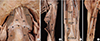
In a routine anatomical dissection in 2013, four kinds of muscular variations were found in an adult Korean male cadaver who died at his age of 76 yr. In the anterior neck, accessory bellies were found bilaterally in the digastric muscles (Fig. 1A). These bellies were triangular and located deeper than the anterior bellies were. They originated from a 57.4 mm long fibrous band between the intermediate tendons of the digastric muscles at both sides and inserted near the digastric fossa. The widths of the lower borders were 26.9 and 22.4 mm at the right and left, respectively, and the lengths from the origin to the insertion were 27.9 and 26.7 mm at the right and left, respectively. The branches from the nerves to mylohyoid muscles innervated these bellies.
In the left arm, a supernumerary head of the biceps brachii muscle was found with its origin at the humeral shaft inferior to the insertion of the coracobrachialis (Fig. 1B). This head joined deep to the main body of biceps brachii at 160.4 mm distal from its origin. At the widest part, the width was 11.1 mm. The musculocutaneous nerve innervated this muscle.
In the left forearm, an aberrant muscle named the palmaris profundus originated from the interosseous membrane near the middle part of the ulnar shaft and inserted onto the styloid processes of the radius and ulna. This aberrant muscle was deeper than the flexor pollicis longus and flexor digitorum profundus were (Fig. 1C). A 58.1 mm long muscular proximal part in the shape of spindle and a 52.6 mm flat tendinous distal part comprised it. The maximal width of the muscular part, the minimal width of tendinous part, and the width at the insertion were 7.9, 3.6, and 36.2 mm, respectively.
In the left thigh, an aberrant muscular slip between the biceps femoris and semitendinosus muscles was found (Fig. 1D). This muscular slip originated from a common origin with the long head of the biceps femoris, which was approximately 127.7 mm lower than was the ischial tuberosity and was united with the semitendinosus 110.5 mm above its insertion. The total length was 138.8 mm. A branch from the tibial part of sciatic nerve arose and innervated the aberrant muscle.
DISCUSSION
To the best of our knowledge, this is the first case with two or more muscular variations of these types coexisting in the same cadaver. Previous cases in the literature have only been found to have one of these muscular variations. Each type of variation does have the potential to cause clinical manifestations or abnormal presentations of various degrees of uncomfortness.
The dimensions and innervations of the digastric muscle accessory belly have been reported to vary (1, 2, 8, 9). It can be misinterpreted as a lymph node of a pathologic condition or a pseudomass requiring surgical intervention (10).
The presence of a third head in the biceps brachii muscle has been described in several reports. According to the classification by Rodriguez-Niedenfuhr et al. (4), supernumerary heads were classified as a superior, infero-medial, or infero-lateral humeral head depending on the origin and location. The third head in the present case can be classified as the infero-medial humeral head and is a rare variant (9% of all variations). This kind of variation has been known to be associated with the ulnar nerve entrapment (11). The size and attachments of the palmaris profundus have been found to be variable (12). It was reported to be related to ulnar nerve compression (13).
The existence of an accessory muscular slip between the long head of biceps femoris and semitendinosus muscles was reported previously by Sinav et al. (7). The aberrant muscular bundle in their report originated 12 cm below the ischial tuberosity and joined the long head of biceps femoris at 12.5 cm above the medial epicondyle, which is similar to that of present case. The innervations of the muscle by the branch from tibial part of the sciatic nerve are also identical between the two cases. Because this type of muscular slip runs across the sciatic nerve, it may compress the nerve and cause symptoms of sciatic nerve compression, which is also seen in the piriformis syndrome.
Therefore, the existence of multiple muscular variations in this cadaver could have induced multiple idiopathic presentation such as bilateral cervical pseudomasses and unexplained multiple nerve entrapment syndromes in the upper and lower extremities during his life.
The discovery of multiple muscular variations biased toward to one side of the body might suggest that a common, single cause has affected the symmetry of the musculoskeletal system. The muscular variations in this case do not seem to result from variations of the innervation because no remarkable nervous variations around the variant muscular slips were found in this cadaver, except in the small nervous branches innervating them. In developing mouse embryos, the elimination of sensory and motor innervation in muscle masses before segmentation was found to not affect the patterning of muscles (14). Moreover, the aberrant invagination of the epimysium or perimysium is not considered to cause muscular variations because connective tissue is scarce even after the perinatal period, and epimysium or perimysium seems to be too weak to affect the pattern of the muscles (15). Although the asymmetric arterial supply is related to severe bilateral musculoskeletal anomalies such as clubfoot disease (16, 17), we did not find any unilateral or bilateral arterial variations in the areas of concern in this case.
Rather, the multiple muscular variations may be related to the errors in establishment of attachments between the muscle precursors and tendon cell precursors or the errors in their migrations to the target regions. During craniofacial and limb myogenesis in vertebrates, muscle precursors and tendon precursors migrate to the proper sites and the elongated myotubes affect the maturation of the tendons before the attachments between them are formed (18). The ectopic origins and insertions of the muscular slips in the present case may have resulted from the migration or attachment of the muscle and tendon precursors prematurely at the midpoint of proper development.
The bias of the site of variation toward the left side of the body may have resulted from transient disturbances in the formation of embryonic somites. So far, an interruption to retinoid acid signaling in the developing embryo is known as the cause of the asymmetric development of the musculoskeletal system. In the developing embryos of chickens, mice, and zebrafish, the absence of retinoid acid signaling resulted in asynchronous somitogenesis between the left and right sides (19). Thus, in the present case, a transient interruption by an unknown environmental factor might have caused a slight disruption to the synchronization of somitogenesis in the arm, forearm, and thigh between both sides. Moreover, proper segmentation in the anterior neck might have also become disrupted by same factor only enough to development of mild muscular variations which are not critical or remarkable.
In conclusion, we found multiple variations biased toward the left side in the neck, upper arm, forearm, and thigh in a single cadaver without any variations to the neurovascular system. By applying knowledge of animal embryogenesis, we hypothesize that the simultaneous appearance of multiple muscular variations may have resulted from a single factor affecting the proper somitogenesis or myogenesis in this case.
 XML Download