

 PDF
PDF ePub
ePub Citation
 Print
Print


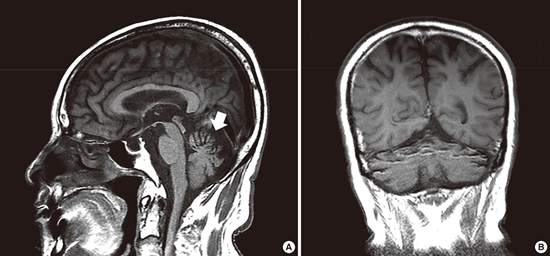
A 79-year-old man presented with progressive gait disturbance and repeated falls on 8 June 2015. He had consumed 1-1.5 L of Korean hard liquor daily for 30 years. He had no family history of neurological disorders. Neurological examinations revealed showed normal mental status and cognitive function. However, there were postural instability, ataxic gait, bilateral gaze-evoked nystagmus, and cerebellar tremor in both arms. Laboratory investigations, including liver function test, were normal. Abdominal sonography revealed chronic alcoholic liver disease.
Magnetic resonance imaging demonstrated significant atrophy of the cerebellum, especially in the anterior portion of the vermis (the lingula and central lobule - Panel A, white arrow) and the anterior portion of the hemisphere (the anterior lobe and simple lobule - Panel B), which appeared as more prominent folia than that in posterior portion of cerebellum. The results of other tests including nerve conduction studies were unremarkable.
Alcoholic cerebellar degeneration occurs prominently in anterior portions of the vermis in early stages of the disease, and lesions spread to posterior portions of the vermis as well as the adjacent portions of the lateral hemisphere of anterior lobe at later stages. This characteristic selective regional vulnerability can differentiate alcoholic cerebellar degeneration from other such as spinocerebellar ataxia, paraneoplastic cerebellar degeneration, and multiple systemic atrophy, which show more diffuse distribution of degeneration.
This is a typical case of early-stage alcoholic cerebellar degeneration, clinically and radiologically, showing prominent atrophy of anterior portion of the vermis and lateral hemisphere of anterior lobe. This finding can help diagnosing early stage of alcoholic cerebellar degeneration.
 XML Download