

 PDF
PDF ePub
ePub Citation
 Print
Print


INTRODUCTION
Adrenocortical oncocytomas are benign tumors consisting of oncocytes in which the cytoplasm becomes eosinophilic due to accumulation of abnormal mitochondria. Oncocytomas develop in various organs and are frequently found in the salivary gland, kidneys, thyroid gland, parathyroid gland, and hypophysis (1). Adrenocortical oncocytomas have been rarely reported; the first reported cases were documented by Kakimoto et al. (2) in 1986. Since then, only 110 cases of adrenocortical oncocytomas have been reported in the English-language literature, which has primarily focused on pathologic studies, especially of functioning adrenocortical oncocytomas. Adrenocortical oncocytomas are extremely rare; only 9 cases have been reported (3-8), of which there were 3 cases that displayed androgenic hormone being secreted in female patients. One case produced interleukin-6, 4 cases showed Cushing syndrome, and one man was found with gynecomastia and elevated serum prolactin and estradiol. These oncocytomas must be treated with great care to exclude metastasis from the extra-adrenal primary site or primary adrenal carcinoma, which is a more common phenomenon. In these present cases, one case of functioning oncocytoma presented with precocious puberty, and the other case was an incidentally detected nonfunctioning oncocytoma that was found via multi-detector computed tomography (CT) and magnetic resonance (MR) imaging.
CASE DESCRIPTION
Case 1
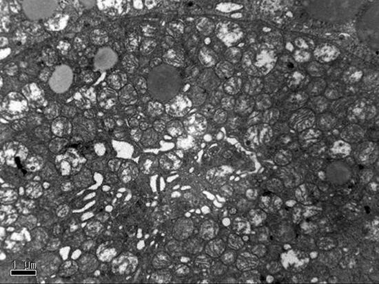
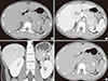
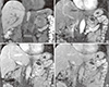
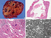
In May 2012, a 10-yr-old girl presented to our outpatient clinic for the evaluation of precocious puberty, which was diagnosed elsewhere. The patient reported no specific past history and no specific clinical symptoms, such as flushing, fever, or abdominal pain. A physical examination revealed elevated breast buds and the start of menstruation. The results of laboratory evaluations showed increased levels of androstenedione (23.1, normal range: 1.7-2.7 ng/dL) and estradiol (46.61, normal range: 0.2-3.0 ng/mL). Testosterone was mildly increased (1.64, normal range: 0.03-0.68 ng/mL), cortisol was slightly decreased (5.49, normal range: 6.2-19.4 µg/dL), and normal ranges of follicle stimulating hormone, luteinizing hormone, and adrenocorticotropic hormone were observed. There were no chromosomal anomalies. Abdominal CT scans detected a well-circumscribed, oval-shaped, 6-cm mass in the left adrenal gland, located superior to the left kidney, which demonstrated stippled calcifications on a precontrast scan. After the injection of a nonionic contrast medium (Omnipaque 350®, GE Healthcare, USA), volume of 100 mL of contrast medium was injected at a rate of 3 mL/s via an antecubital vein, heterogeneous well enhancement was visualized during the 1-min delayed scan, and enhancement washout was demonstrated on the 15-min delayed scan (Fig. 1). The CT Hounsfield unit (HU) was 40 HU on precontrast, 140 HU at 1 min, and 70 HU at 15 min. The absolute washout value ([Enhanced CT HU-Delayed CT HU]/[Enhanced CT HU-Unenhanced CT HU]×100) was approximately 70% (a value of more than 60% indicates adrenal adenoma), and the relative washout value ([Enhanced CT HU-Delayed CT HU]/Enhanced CT HU×100) was 50% (a value of more than 40% indicates adrenal adenoma) (9). Initially we thought that functional adrenal adenoma should be included in the differential diagnosis. A left adrenalectomy was performed. During the operation, there was no evidence of hypertension associated with palpation of the mass or direct invasion to the surrounding organs. The tumor was approximately 6×4 cm, well-encapsulated and dark brown in color (Fig. 2A). The tumor consisted of multiple small hemorrhagic vascular lakes with old blood. The microscopic examination revealed that the neoplasm consisted of polygonal cells with abundant eosinophilic cells and granular cytoplasm. Nuclear cellular atypia with enlarged nuclei were identified (×200, ×400 High Power Field [HPF], hematoxylin and eosin [H&E], Fig. 2B and C). An electron microscopic study was performed, and electron-dense inclusion and closely packed mitochondria with Golgi complex were found (×400 HPF, Fig. 2D). Thus, we concluded that the final diagnosis in this case was adrenocortical oncocytoma. The patient had an uneventful postoperative course and was doing well one year after surgery without new lesions.
Case 2
In June 2010, an incidentally detected left adrenal mass was found in a 54-yr-old man via CT performed at another hospital. Abdominal CT scans detected a lobulating contoured, well-demarcated, well-enhancing solid mass in the left adrenal gland, which contained the central necrotic portion.
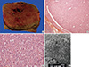
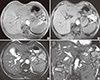
MR images were obtained with a 1.5 T unit using a contrast agent (Gadovist®, Bayer Healthcare, Germany). On a T2-weighted gradient echo image (TR/TE: 3.7/1.6), the mass was primarily of slightly high signal intensity with a hyperintense central portion. This mass primarily demonstrated hypointensity on T1-weighted gradient echo images (TR/TE: 175.0/5.0), without a definite signal drop (suggesting a fat component) on the opposed-phase correlated with the in phase. The previously highly hyperintense focus on T2-weighted images showed hypointensity on the T1-weighted images, suggesting a necrotic portion, and a central hemorrhagic component was revealed within the necrosis by demonstrating a hyperintense foci on a T1-weighted image and hypointense foci on a T2-weighted image (Fig. 3). Contrast-enhanced MR images heterogeneously demonstrated the enhancement of the solid tumor portion at the 1-min, peak enhancement at the 3-min, and slight washout at the 5-min delayed phases. Intratumoral hemorrhagic necrosis was not constantly enhanced (Fig. 4). The blood vanillylmandelic acid, total metanephrine, epinephrine, and norepinephrine were within the normal ranges.
Based on these observations, adrenal carcinoma and metastasis were included with the differential diagnosis. The patient underwent meticulous examination for the primary origin, including colonoscopy, gastroscopy, and chest CT; there was no evidence of a primary focus. The most probable preoperative diagnosis was adrenal carcinoma. The patient underwent laparoscopic adrenalectomy, and histology suggested adrenal oncocytoma with uncertain malignant potential.
The tumor was a soft oval mass measuring 9×6×5 cm in dimensions and 116 g in mass. The outer surface was well encapsulated by a thin fibrous capsule, and it was focally attached to a normal adrenal gland. Upon sectioning, the mass showed a brownish-yellow, fish-flesh-like cut surface with a multifocal brownish hemorrhagic necrosis and focal cystic changes (Fig. 5A and B).
The tumor was composed of epithelial cells with abundant acidophilic cytoplasm, with nuclear pleomorphism and a diffuse growth pattern, as shown using a light microscope (Fig. 5C), and the oncocytic cytoplasm was filled with a large number of mitochondria, as well as some lysosomes, Golgi bodies, small lipid particles, and glycogen particles (Fig. 5D), the histological features of which were consistent with adrenocortical oncocytoma. The tumor showed a low mitotic count (<5/50 HPFs), the absence of atypical mitosis, and venous invasion, but it also showed focal necrosis and inconspicuous capsule invasion, the histological features of which were consistent with uncertain malignant potential (borderline) behavior. Electron microscopy showed that microscopic and immunohistochemical findings were compatible with an adrenocortical oncocytoma. Because of this finding, we performed a careful follow-up study to exclude the possibility that the adrenal tumor was malignant over a three-year follow-up period. This workup included multiple abdominal ultrasounds examinations, as well as chest and abdominal CT scans. All of the imaging findings were unremarkable. We concluded that the final diagnosis in this case was primary adrenocortical oncocytoma.
DISCUSSION
Adrenocortical oncocytomas are classified as benign, borderline malignant potential, and malignant according to the Lin-Weiss-Bisceglia criteria (10). Proposed major criteria (high mitotic rate greater than 5 per 50 HPF, atypical mitoses, venous invasion) and minor criteria (large size and huge weight, necrosis, capsular invasion, sinusoidal invasion) in distinguishing malignant tumors are discussed. Their proposed working rules for diagnostic categorization of adrenocortical tumors are defined, with the presence of 1 major criterion indicating malignancy, 1 to 4 minor criteria indicating uncertain malignant potential (borderline), and the absence of all major and minor criteria indicative of benignancy (10). According to the proposed these criteria, our first case should be considered as benign, because there are no major or minor criteria, and our second case should be considered as uncertain malignant potential (borderline) due to focal capsular invasion and necrosis. Adrenocortical oncocytomas were reported as large neoplasms (>5 cm) that did not contain macroscopic evidence of necrosis or hemorrhage (1, 7, 11). One exception was the case reported by Nguyen et al. (1), which arose from heterotopic adrenocortical tissue separate from the adrenal glands within the retroperitoneum; that tumor measured only 3 cm and contained central necrosis. Our first case showed typical findings as a solid tumor without evidence of necrosis or hemorrhage and confirmed with benign oncocytoma, but our second case demonstrated atypical imaging findings as a large tumor with central hemorrhagic necrosis and surgically confirmed with borderline malignancy. Adrenocortical oncocytoma that arisen in the adrenal gland is very rare and usually benign and nonfunctioning, incidentally discovered by abdominal imaging performed to investigate an unrelated problem, and have arisen in patients ranging in age from 27 to 72 yr without a male or female predominance (12). Most to the best of our knowledge, only 3 cases of functioning adrenocortical oncocytomas have been reported in childhood.
The most widely studied oncocytoma from an imaging viewpoint has been the renal oncocytoma. Renal oncocytomas appear as solid masses on CT and characteristically have central radiating scars when large. The MR signal characteristics are nonspecific; currently, the tumor cannot be reliably differentiated from the more common renal cell carcinoma (13). The classic central radiating scar that has been described in renal oncocytomas is not inevitably present in adrenal oncocytomas. Recently segmental enhancement inversion during corticomedullary phase and early excretory phase was found to be a characteristic enhancement pattern of small (smaller than 4 cm) renal oncocytoma at biphasic multidetector CT (14), but there was no definite segmental enhancement inversion in our cases. We thought that the tumor mass of our two cases were larger in size than that of previously reported cases.
The imaging characteristics of our second case were very different from the previously reported cases but well correlated with the histopathology, heterogeneous signal intensity in MR imaging represent hemorrhagic necrosis detected on pathologic examination. Fibrous encapsulation is characteristic finding for adrenal oncocytomas and was found in both benign and malignant variants, and could be well detected by both CT and MR imaging. Given the large size (>6 cm), heterogeneous appearance and presence of necrosis or calcification in the tumor, adrenocortical carcinoma was obviously the most likely preoperative diagnosis. Non-functioning ones can be heterogeneous enhancement with central necrosis and hemorrhage, calcification may be present occasionally. Thus unfortunately, the imaging features of adrenocortical oncocytomas did not allow its differentiation from adrenocortical carcinomas (15). Cytopathology of adrenocortical oncocytoma by diagnosis of fine needle aspiration biopsy was reported (4). However, the morphologic features and ancillary studies of find-needle aspiration cytology material do not help in indicating the biological behavior of these tumors. Surgical resection remains the only tool for the definitive diagnosis.
Recently, MR imaging has become increasingly useful in characterizing adrenal masses, particularly using chemical shift imaging (in-phase and opposed-phase) to use the fat content of adenomas and lack of fat within metastases; opposed-phase imaging allows noninvasive distinction between these two entities with a high degree of accuracy (16). Adrenocortical carcinoma, which was a diagnostic consideration in our case, often can be differentiated from adenoma on CT or MR imaging based on its size (i.e., generally>5 cm), central necrosis, contrast enhancement, and frequent invasion of local organs and extension into the inferior vena cava (17). In the second case presented here, the tumor was heterogeneous hypointense with respect to the liver on T1-weighted and slightly hyperintense on T2-weighted imaging, and displayed mild homogeneous enhancement, this mass did not loss signal intensity on opposed-phase gradient echo imaging, a finding exhibited by most adrenal adenomas. These imaging characteristics correlated with the pathologic findings, which included a lack of significant amount of lipid. The lack of lipid is not typical of adrenocortical cells but may be secondary to the large amount of mitochondria, which instead pack the cytoplasm of oncocytic cells (18).
In summary, two rare primary adrenocortical oncocytomas with radiologic findings are presented with a focus on dynamic CT and contrast-enhanced MR imaging and histopathologic findings with immunohistochemical studies and electron microscopy.
Adrenocortical oncocytoma is rare disease entity, but can be included in the differential diagnosis of especially large, well-defined solid adrenal tumors. However, non-specific imaging features do not warrant a definitive preoperative diagnosis and unfortunately do not change the patient's management.
 XML Download