

 PDF
PDF ePub
ePub Citation
 Print
Print


INTRODUCTION
Fistula development between the kidney and gastrointestinal tract is a rare clinical event (1-3). It is mainly caused by various inflammatory diseases including reactions to foreign bodies, inflammatory bowel diseases, urolithiasis, benign and malignant neoplasms and pyogenic infections. Traumatic pyeloenteric fistulas can occur by trauma, surgery and interventional procedures. Many clinicians have believed the surgical nephrectomy and duodenal closure is the most successful treatment in this situation. To our knowledge, this is the first report of pyeloduodenal fistula (PDF) treated by endoscopic ligation without surgery.
CASE DESCRIPTION
A 74-yr-old woman was transferred to our hospital from a local clinic because of generalized weakness and high fever with chills on January 17, 2013. She had been complaining of abdominal discomfort. She had no underlying illness such as diabetes mellitus, hypertension, hepatitis, or tuberculosis. Her family history and social history were non-specific. For 15 days, prior to admission, she received conservative treatment for herpes zoster infection on the anterior chest without an anti-viral agent.
At the time of admission, her body temperature was 37.8℃ and blood pressure was 110/80 mmHg. Her heart rate was 112 beats per minute being faster than normal with sinus rhythm on ECG. Laboratory examination revealed pyuria, isomorphic hematuria, positive leukoesterase and bacteria on urinalysis. Systemic leukocytosis with a predominance of neutrophils (white blood cell count of 21,650/µL) and a high C-reactive protein level of 35.63 mg/dL were shown in the blood test. Blood urea nitrogen and serum creatinine level had risen to 69.4 mg/dL and 4.28 mg/dL, respectively, from normal values at baseline. Serum potassium was 6.0 mM/L, and coagulation test results were lengthened to a PT INR of 1.4 and an aPTT time of 70.2 sec.
Physical examination was otherwise unremarkable except diffuse abdominal tenderness. Through laboratory tests and physical examinations, the patient was suspicious of urinary tract infection combined with acute kidney injury. Next, a computed tomogram (CT) was checked and it revealed hydronephrosis with upper ureteral stones and multiple ill-defined hypodense lesions suggestive of acute pyelonephritis in both kidneys.
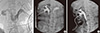
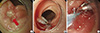
Percutaneous nephrostomy (PCN) was performed in both kidneys, and during this procedure, right PDF was visualized. Pyelography revealed a fistula to the descending part of the duodenum (Fig. 1). We prescribed total parenteral nutrition and intravenous antibiotics and planned surgery for PDF. However she was too old and was considered to be emaciated to tolerate nephrectomy and general anesthesia. Also, the surgeon was not able to find out the exact leakage site on CT. Therefore, gastroendoscopy was decided to avoid surgery. The gastroenterologist discovered PDF in the duodenal second portion and clipped the lesion 4 times (Fig. 2). After an appropriate course of parenteral antibiotics therapy with the clipping procedure, her temperature and white blood cell count returned to normal, and her urine culture grew Klebsiella pneumoniae which showed all sense to the antibiotics. The patient, however, had to undergo one more endoscopic procedure after 1 week because her follow-up fistulography revealed that there was still a communication between the pelvis and duodenum, but the amount of leakage was significantly reduced. The gastroenterologist ligated the fistula site with a detachable snare which is an elliptically shaped nylon loop used to tighten around the fistula site. This device has recently been used for closure of gastrointestinal fistula followed by application of a clip in the field of gastroenterology (4). Follow-up fistulography was performed after 1 week, and we were able to confirm there was no more leakage (Fig. 1). Her blood urea nitrogen and serum creatinine levels recovered to normal showing 10.7 mg/dL and 0.86 mg/dL, respectively. Her urine culture was returned to negative finding. She was discharged after confirmation of appropriate oral intake on admission day 30. The urologist removed PCN in the outpatient clinic, and then finally treated her stones by extracorporeal shockwave lithotripsy.
DISCUSSION
Pyeloduodenal fistula is a rare but serious condition, generally associated with chronic inflammatory renal diseases or trauma. It was first reported in 1893, and about 80 cases have been reported so far in the literature (5). In the early literature, tuberculosis was a frequently reported cause of renal inflammation associated with fistula formation. However, since the progression of effective anti-tuberculous treatment, few cases of pyeloduodenal fistula secondary to tuberculosis have been reported. Almost 65% of spontaneous renal fistulas occur secondary to nephrolithiasis which can cause chronic pyelonephritis and obstruction of urine flow and propagate to perinephric inflammation. It is believed that inflammation, urinary extravasation, abscess formation, and perinephric inflammation are main pathophysiologic mechanisms of non-traumatically caused PDF (1). The posterior aspect of the second portion of the duodenum lies in close proximity to the medial portion of the kidney and renal pelvis. When perirenal inflammation takes place, this portion of the duodenum is more easily involved because of relative immobility of the second part of the duodenum and its lack of a posterior peritoneal covering as well (6, 7). Our patient developed a fistula at the same site mentioned above. Patients with PDF complain of various symptoms such as persistent flank pain, epigastric pain, dyspepsia, malaise, weight loss etc. Common physical findings include fever, pyuria and flank tenderness (8). Since diverse and nonspecific symptoms and signs can be present, physicians should maintain a high index of suspicion regarding PDF as clinical impression, unless they miss the diagnosis. Diagnosis of PDF requires imaging studies of the urinary system. Retrograde pyelography is the method of choice which demonstrates the fistula in 64% of cases (8). Intravenous urography (IVU) can be used in only functioning kidneys, and CT often demonstrates fistula. Other reported imaging modalities include antegrade pyelography and upper GI studies (9). Nephrectomy and primary closure of the duodenum are traditional treatment methods for non traumatic PDF (8, 10-13). Either a transperitoneal or retroperitoneal approach may be used. The retroperitoneal approach in the face of an active suppurative process will avoid contamination of the abdominal cavity (8). In a review by Rodeny et al. (8), nephrectomy and duodenal closure were done in 21 cases of a total of 28 cases, and their prognosis was satisfactory. The rest of the patients were treated without surgery; one died of sepsis and others suffered long-term complications such as sinuses and recurrent infections. Because of poor prognosis of nonsurgical management, it was suggested that any attempt to spare the kidney should be avoided when the primary disease process is apparent in the kidney for a long time. There are few reports of PDF recently, and surgical management was the main therapeutic option in cases published since 2000 (14-16).
However, there have been a few previous reports of successful conservative management (9, 10, 17, 18). Conservative nonsurgical management of PDF primarily included appropriate intravenous antibiotics, relieving obstruction by employing nephrostomy or internal ureteric stents. Research in regard to which patients are suitable for surgical and non-surgical management is very scarce. The decision concerning whether to operate and/or possibly remove a kidney seems to largely depend on the function of the involved kidney. One researcher mentioned that if kidney function is adequate, nonsurgical therapy may be attempted (19). Such fistulas in minimally functioning kidneys, unfortunately, rarely respond to nonsurgical measure, and are associated with increased morbidity and mortality in some reports (20).
In our case, we were able to save the patient's kidney function and avoid a high risk of operation by endoscopic ligation which is the first case report to our knowledge. Although she had to suffer long-time fasting, her nutrition status was maintained in an appropriate level by total parenteral nutrition. A relatively small sized fistula and accessible location by gastroendoscopy may be favorable features for nonsurgical treatment of PDF. Treatment should be tailored to each patient, and every effort should be made to preserve kidney function as a nephrologist. To conclude, an endoscopic approach is a newly effective option for PDF in patients who cannot undergo surgery especially in elderly patients with concomitant diseases.
 XML Download