

 PDF
PDF ePub
ePub Citation
 Print
Print


INTRODUCTION
Papillary carcinoma is the most common type of thyroidal malignancy (1). Within variable morphologic variants of papillary thyroid carcinoma (PTC), follicular variant of PTC comprises 15%-20% of PTC, which is the second most common variant following the conventional PTC (2). Among the various subtypes of follicular variant PTC, diffuse follicular variant papillary thyroid carcinoma (DFVPTC) is very rarely found. As it is a variant of PTC, diagnostic nuclear features as nuclear clearing, grooves, and seldom nuclear pseudoinclusions are evident. Typically, it arises in young females and often has a poorer clinical course than other subtypes of follicular variant PTC. The tumor shows no encapsulation, diffuse permeating micro- and macrofollicular tumor cells devoid of papillary architecture. While usual follicular variant PTC has an excellent prognosis, DFVPTC is considered to have poor prognosis, based on the multicentricity of tumor, extensive extrathyroidal extension, vascular invasion and nodal metastasis, etc. We describe a case of DFVPTC arising in a 69-yr-old male rather than young female.
CASE DESCRIPTION
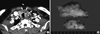
A 69-yr-old man was referred to our institution for the management of his neck mass. He has visited the previous hospital on February 17, 2011 with hoarseness for a few months. He had anti-reflux medication, but the hoarseness got worse. He was medicated for hypertension, and no other past history was found. The physical examination on him revealed firm, fixed mass on left anterior neck and limited motility of the vocal cord was observed. By further evaluation with computed tomography scan, a huge, ill-marginated mass was found, invading the left thyroid lobe and isthmus (Fig. 1A). Tracheal and esophageal invasion was suggested. Multiple enlarged lymph nodes were present in the left neck. At previous hospital, fine-needle aspiration was done to the mass in the left thyroid and left neck nodes, diagnosed as papillary thyroid carcinoma with lymph node metastases. In our institution, the aspiration cytology slides of thyroid and lymph nodes were reviewed, and also the diagnosis of papillary thyroid carcinoma with lymph node metastases was rendered.
After referred to our institution, he received bilateral total thyroidectomy with central compartment neck node dissection. After 17 days from the operation, the patient expired due to asphyxia caused by an unexplained bleeding in the gastrointestinal tract.
On examination of resected specimen, pathologic diagnosis of diffuse follicular variant papillary thyroid carcinoma (DFVPTC) was rendered. Multiple carcinomas were found in both thyroids, infiltrating into the esophagus, trachea, sternum, and mandible with multiple metastases in the regional lymph nodes.
The specimen was consisted of entire thyroid (total weight: 29.8 g, right lobe: 3.5 × 3.8 × 1 cm, left: 6 × 4 × 2.3 cm) with regional lymph nodes, and multiple biopsies from tracheal, esophageal, sternum, mandibular area. Left thyroid was firm and fibrotic in consistency. Sectioned surface showed poorly marginated yellowish and fibrotic tumor (3.7 cm), which was infiltrative and totally replacing the entire lobe (Fig. 1B). Right lobe seemed to be grossly normal, but a 0.9 cm-sized vague nodule was found in lower pole on sectioning.
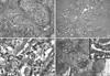
Hematoxylin-eosin (HE) stain revealed diffuse, vaguely nodular tumor in left thyroid (Fig. 2A). The tumor showed micro- and macrofollicular growth pattern, which was infiltrating into the thyroid parenchyma, extrathyroidal skeletal muscle and fibroadipose tissue. Dense collagenous fibrosis was accompanied by tumor cells (Fig. 2B). Inflammation was rarely found. Cytologically, each of tumor cells revealed vesicular nuclei with nuclear clearing and grooves, which are histologic findings of papillary thyroid carcinoma (PTC) (Fig. 2C). The other histologic features, such as intranuclear pseudoinclusion, psammomatous calcification, or discrete papillary structure were rarely seen.
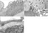
Six of 60 lymph nodes metastases were found from neck dissection, of which the largest size was 2.2 cm. Metastatic carcinoma in lymph nodes showed same histologic features as in the thyroid, like micro-and macrofollicular growth pattern of tumor cells harboring nuclear features of PTC (Fig. 2D). Permeating tumor cells were found in separately sent esophagus and tracheal cartilage, which also exhibited the same histologic features as in the thyroid (Fig. 3A-C).
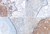
For ancillary interest, several immunohistochemical stainings - galectin-3, cytokeratin (CK) 19, CD56, thyroglobulin, Ki67, p53 - were performed on the left thyroid. Diffuse positive for galectin-3, CK19, and negative for CD56 were seen in tumor area, which was vice versa in normal thyroid tissue (Fig. 4A-C). Tumor showed decreased stain intensity for thyroglobulin than normal thyroid tissue (Fig. 4D). Ki67 labeled tumor cells were about 5% (found up to 37/high power field) and p53 nuclear expression was about 10% of tumor cells. Additionally, BRAF point mutation (V600E) was detected by sequencing anaylsis.
DISCUSSION
Follicular variant is common variant of papillary thyroid carcinoma, comprising 15%-20% of papillary carcinomas. Several subtypes of follicular variant of papillary thyroid carcinoma exist, such as encapsulated follicular, diffuse follicular, and macrofollicular variants (2). Encapsulated follicular and macrofollicular variants have an indolent course, and usually encapsulated. In contrast, diffuse follicular variant has aggressive biologic behavior. Diffuse follicular variant papillary thyroid carcinoma is first described by Sobrinho-Simões et al. (3). It usually occurs in young females. Histologically it typically lacks of encapsulation, reveals predominant microfollicular architecture, and vascular invasion and metastases to lymph nodes and distant area are common. In 1995, Mizukami et al. (4) reported one case of DFVPTC in 13-yr-old boy. Ivanova et al. (5) analysed clinicopathologic and immunohistochemical characteristics of 10 cases of DFVPTC in 2002. They compared DFVPTC with common FVPTC and common PTC, showed several distinguised properties of DFVPTC from others in clinical behaviour and histological features. The significant findings of DFVPTC were: female predominancy (male to female ratio was 1:9); young female (mean age of 26.8 yr); multicentricity; extrathyroidal extension; lymph node metastasis; venous invasion. Other morphologic feature of DFVPTC included no necrosis, no fibrosis, no lymphocytic infiltration, and rare psammoma bodies with low mitotic index (mean 1.3%).
We present a case of DFVPTC occurred in 69-yr-old male patient. The patient presented with refractory hoarseness and underwent bilateral total thyroidectomy with central compartment neck node dissection. Extensive tumor cell infiltration into left thyroid and surrounding structures, nodal metastases were found on pathologic examination. He was diagnosed as DFVPTC and expired on the 17 post operative day due to unexplained bleeding of surgical site. Diffuse infiltrative growth of tumor cells of micro- and macrofollicular growth pattern was distinctive. The nuclei of tumor cells demonstrated features of conventional PTC, such as nuclear clearing, grooves, and seldom pseudoinclusions. The tumor involved entire left lobe and part of right lobe, extended to the adjacent structure, including tracheal cartilage, esophagus, sternum, mandible, extrathyroidal skeletal muscle and fibroadipose tissue. On immunohistochemical staining, the tumor was positive for galectin-3, CK19, and lacked the CD56 expression compared to the juxtaposed normal thyroid tissue, which is consistent with conventional PTC. Thyroglobulin staining for tumor exhibited decreased intensity than normal thyroid. Some of tumor cells were labeled with Ki67 staining (about 5%, figure not shown) and expressed p53 (about 10%, figure not shown). Based on differentiated follicular architecture, cytologic and immunohistochemical features of PTC and biologic aggressiveness, present case is compatible with DFVPTC. However, unlike the study of Ivanova et al. (5), which showed the tendency of young female predominancy of occurence, present case is distinguished from the previously reported cases of DFVPTC in old age and male gender.
 XML Download