

 PDF
PDF ePub
ePub Citation
 Print
Print


INTRODUCTION
Many infectious agents and immunological mechanisms may play a role in pathogenesis of Guillain-Barre syndrome (GBS). It is possible to identify the four most common subtypes: the acute inflammatory demyelinating polyradiculoneuropathy (AIDP), the acute motor axonal neuropathy (AMAN), the acute motor and sensory axonal neuropathy (AMSAN) and Miller Fisher's syndrome (1). Above mentioned subtypes, AMSAN is considered to be a rare variant of GBS, and one that usually has a more serious clinical course and slower recovery than the classic demyelinating form of GBS (2). AMSAN has been associated with antecedent Campylobacter jejuni (C. jejuni) infection (2). Although viral hepatitis is common in Korea, acute viral hepatitis A (HA), as an antecedent infection of AMSAN, was extremely rare (3). We present a case of GBS-subtype AMSAN following acute HA infection and review the literature.
CASE DESCRIPTION
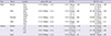
A 21-yr-old male was transferred to our hospital due to respiration difficulties and progressive weakness at 16th July 2010 from a local hospital where the patient had been admitted after a 7-day history of symptoms of acute viral hepatitis. In addition, the patient did not have a past history of respiratory or gastrointestinal infection within 1 month before onset of progressive weakness. Physical examination upon referral to our hospital showed the patient presented with anorexia, jaundice, and elevated transaminase levels (serum glutamic oxaloacetic transaminase, 385 IU/L; serum glutamic pyruvic transaminase, 376 IU/L). Immunoglobulin M (IgM) antibodies against HA were detected in blood and cerebrospinal fluid (CSF). Based on impaired liver function tests, clinical features, and the presence of anti-HA IgM antibody; the absence of markers for hepatitis B, C, D, E, diagnosis of acute HA was established. CSF analysis showed protein, 156 mg/dL; glucose, 61 mg/dL, with no pleocytosis. Results of serological and CSF tests for C. jejuni, cytomegalovirus (CMV), Epstein-Barr virus (EBV) and Haemophilus influenzae (H. influenzae) infection were negative. Anti-GQ1b and GM1 antibodies were not found in serum. An abdominal ultrasound showed altered echotexture of liver parenchyma. On neurological examination, the mental status of the patient was alert. By the Medical Research Council (MRC) grading, the patient had weakness with areflexia in both proximal upper limbs (MRC grading 4), both distal upper limbs (MRC grading 2), both proximal lower limbs (MRC grading 4), and both distal lower limbs (MRC grading 2). Pin-prick, vibratory, and joint position senses were reduced in glove and stocking distribution. Nerve conduction studies (NCSs) were performed at 2 weeks after the onset of weakness. The findings of motor NCSs showed markedly reduced amplitudes of CMAPs whereas distal latencies and motor conduction velocities were normal in bilateral peroneal, and posterior tibial nerves, without evidence of demyelination. The findings of sensory NCSs revealed decreased amplitudes of SNAPs and normal conduction velocities in bilateral median, ulnar, and sural nerves (Table 1). F-wave response was absent in the right median and both peroneal nerves. The needle electromyography examination demonstrated markedly abnormal reduction in recruitment, with positive sharp waves in the bilateral tibialis anterior and peroneus longus muscles. Based on clinical features with motor and sensory involvement, laboratory findings and electrophysiologic investigation, this patient was diagnosed the AMSAN subtype of GBS following acute HA viral infection according to proposed diagnostic criteria of GBS (4). The patient was treated with 0.4 mg/kg/day of intravenous immunoglobulin for 5 days and recovered slowly. Four months later, the patient continued to exhibit marked wasting and weakness of the distal upper and lower limb muscles.
DISCUSSION
AMSAN has been recently described as a subtype of GBS characterized by acute onset of distal weakness, loss of deep tendon reflexes, and sensory symptoms (2). AMAN and AMSAN, axonal subtypes of GBS, can be preceded by C. jejuni enteritis (2). However, in this case, we diagnosed the AMSAN subtype of GBS following acute HA infection using clinical evidence, electrophysiological studies, and HA virus-IgM antibodies in the serum and CSF. The exact pathogenesis by which the virus causes the disease is not clear. The involvement of the central nervous system in the viral disease could be due to direct invasion of the central nervous system by the virus, as evidenced by the presence of HA antibodies in the CSF. Therefore, it seems most likely that transport of antibodies occurs across a disrupted blood-nerve barrier during inflammatory reaction of nerve roots.
In AMAN and AMSAN, the pathological features differ from the features of AIDP in that macrophages invade the space between the Schwann cell and axon, leaving the myelin sheath intact (2). Griffin et al. (5) proposed the attractive hypothesis that AMAN and AMSAN are part of the spectrum of a single type of immune attack on the axon, but the relationship between AMAN and AMSAN has yet to be clarified. The AMSAN cases differ from the AMAN pattern of GBS in terms of slow recovery, in addition to sensory fiber involvement, but the pathologies are very similar (6). The number of GBS-subtype AMSAN cases is very small (< 10% of AMAN cases) (7). In this case, clinical, serological, and electrophysiological studies suggested a diagnosis of GBS-subtype AMSAN following acute HA infection.
Nervous system complications of HA viral infection appear to be very rare. A variety of neurological syndromes including GBS have been reported in serologically confirmed hepatitis A (8,9). Clinical features of GBS following HA can be summarized as follows: 1) Most of the patients are men. 2) The interval between the onset of hepatitis and the development of neuropathic symptoms is less than 14 days. 3) There is a frequent association with facial nerve palsy. 4) Joint position and vibratory sense are frequently impaired, in addition to superficial sensation. 5) A uniformly good outcome of the neuropathic symptoms is independent of the level of ALT, which corresponds to the severity of liver dysfunction (10). Most electrodiagnostic studies were compatible with an acquired demyelinating polyradiculoneuropathy, but a few cases showed dominant axonal involvement i.e., acute motor axonal neuropathy (AMAN) (11,12). The prognosis of HA virus-associated GBS was favorable, both in earlier reports (management with supportive care only) and in more recent reports (treatment with either intravenous immunoglobulin [IVIG] or plasmapheresis) (9). Therefore, HA virus-associated GBS appeared to run a course similar to "typical" GBS, and the outcome of GBS did not correlate with the severity of acute hepatitis (9).
The pathogenesis of HA virus-associated GBS is unknown. A cross-reaction between Schwann cells, myelin or other peripheral nerve antigens remains a possibility, but any molecular resemblance between hepatotropic viruses and structural components of peripheral nerves has not been explored (9). In our case, there was no recent infection of usual agents that have been reported to be associated with axonal degeneration, such as CMV, EBV, H. influenzae and C. jejuni (13). Taking into account these facts, we presume that the epitope of HA virus and the axonal component of peripheral neural tissue might have molecular mimicry. Further studies are needed to ascertain this exact mechanism.
Our patient had the AMSAN subtype of GBS following acute HA viral infection. AMSAN subtype has a fulminant course with slow recovery. Determination of the well-known agents related with GBS, such as C. jejuni and CMV, is clinically and immunologically important to diagnosis and prognosis of GBS subtypes. Also, unusual agents associated with GBS subtypes including AMSAN may be identified in order to clarify the pathogenic mechanism of GBS subtypes. Therefore, we suggest that a careful and broad view is needed to determine the causative relationship between an infection and the development of GBS subtypes.
 XML Download