

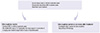
 PDF
PDF ePub
ePub Citation
 Print
Print


INTRODUCTION
Regular red blood cell (RBC) transfusions are the major supportive care for many Korean patients with aplastic anemia (AA), myelodysplastic syndromes (MDS), or other rare anemias. Physicians most commonly use the hemoglobin (Hgb) level to determine whether or not to transfuse (1). However, most guidelines recommend that transfusions should be given for symptoms of anemia and that the decision to transfuse should not be based on the Hgb level alone (2).
An optimal RBC transfusion regimen involves administering sufficient RBCs to maximize clinical benefits while avoiding unnecessary transfusions that increase costs and expose patients to potential risks, including infection and iron overload. Hence, the Korean Society of Blood Transfusion (KSBT) recommends transfusion to be considered at Hgb concentrations of 7 g/dL or less in chronically anemic patients. KSBT does not recommend routine RBC transfusion when Hgb>7 g/dL. Furthermore, KSBT only recommends RBC transfusion in those patients with symptoms such as dyspnea, palpitation on exertion, or edema (3).
Chronic transfusion therapy inevitably leads to secondary iron overload, which can cause significant damage to many organs such as the liver, heart, and endocrine system. Recently, deferasirox (DFX), an oral iron chelator, has been introduced and has been shown to improve the treatment of iron overload (4, 5). To summarize the evidence and provide practical guidelines for iron chelation in Korea, the Korean Society of Hematology Aplastic Anemia Working Party (KSHAAWP) describes here general considerations regarding iron overload in transfusion dependent patients and proposes guidelines for the treatment of iron overload based on published clinical evidence and the experience of the expert panel.
PATHOPHYSIOLOGY OF IRON OVERLOAD
Iron metabolism and the mechanism of organ damage
Iron is involved in many critical steps of cellular metabolism such as cellular respiration, heme synthesis, production of oxygen radicals, antioxidation, and DNA synthesis and repair, as well as in cellular proliferation and inflammation. Normal body iron stores are 3-4 g; an excess of iron of 20 g or more can lead to organ damage (6). Most additional iron can be stored within the reticuloendothelial system (RES). However, if the iron level exceeds the capacity of the RES to retain it, the excess iron will be released into the plasma. Normally, transferrin binds this released iron but, if the plasma iron concentration continues to rise, transferrin saturation may result. This scenario may occur with sequential transfusions: when the RES cannot retain all of the iron, it then diffuses into the plasma in amounts that exceed the capacity of transferrin to bind it. As a result, non-transferrin-bound iron, which seems to be the major mediator of extrahepatic tissue damage in transfusional iron overload, appears in the plasma (7). The harmful effects of excess iron may result from deposition into tissues and organs, but also from oxidative stress. Non-transferrin-bound plasma iron is deposited specifically in tissues with high level of transferrin receptors (eg, liver, heart, anterior pituitary, and pancreas). As the human body has no mechanism for excreting excess iron, and each unit of transfused RBCs contains 200-250 mg of iron, iron overload can readily occur in patients given multiple transfusions, typically after 10 to 20 transfusions (6). In the absence of treatment to reduce iron overload, progressive cardiomyopathy, cirrhosis, endocrinopathy and diabetes can have a serious impact on morbidity and mortality. Iron chelation therapy in patients given multiple transfusions aims to prevent or reverse some of these consequences and has been associated with a reduced morbidity and mortality (8, 9).
Measuring and monitoring body iron
Serum ferritin is the most common indirect parameter used to assess body iron stores in the clinical setting. Measurement of serum ferritin is easy and inexpensive but individual values may be affected by infection, inflammation, liver disease, vitamin C deficiency, and hemolysis (10). Although ferritin is not the most accurate marker of iron overload, ferritin levels do correlate with transfusion burden (11) and liver iron concentration (LIC) (10), and trends in ferritin levels are useful for following iron load.
Liver biopsy is considered the gold standard for direct measurement of total body iron, but the use of this invasive technique is excluded in many patients because of thrombocytopenia and neutropenia, which may predispose to bleeding and infectious complications (12).
Non-invasive measurement techniques are being developed, including T2* MRI and magnetic susceptometry using a superconducting quantum interference device (SQUID). These techniques have been shown in some studies to provide results that correlate well with liver biopsy-determined iron concentrations (13-15), but they are not widely available worldwide.
The data reported in a recent study indicate that serial measurements of serum ferritin are useful for monitoring chelation therapy with DFX so that doses may be adjusted according to the ongoing iron load due to transfusion (5). The DFX prescribing information recommends monitoring serum ferritin monthly to assess the patient's response to therapy and adjusting the DFX dose, if necessary, every 3 to 6 months (16).
CLINICALLY AVAILABLE IRON CHELATORS
Currently, three chelating agents are available: deferoxamine, deferiprone, and DFX.
Deferoxamine (Desferal™)
Deferoxamine (DFO) has been in widespread clinical use since the late 1970s and has provided unequivocal evidence that effective chelation therapy can arrest the progression of, and prevent early death from, iron overload (17). DFO is a trihydroxamic acid sideraphore secreted by Streptomyces pilosus, a fungus. DFO is a hexadentate chelator with a very high and selective affinity for iron. One molecule of DFO binds one atom of iron. DFO is administered as long parenteral infusions because the plasma half-life is short (min) and it is not active orally. It is usually given as an overnight subcutaneous infusion 5 to 7 nights per week. It is mainly distributed extracellularly and the protein binding in plasma is low (<10%). After complexing with iron it is excreted rapidly as ferrioxamine, mainly through the kidney and one-third into bile through the feces. Thus, its efficacy is also dependent upon adequate urine output and may be facilitated by dialysis in the case of kidney dysfunction. This requirement is extremely demanding and can result in poor adherence, thereby compromising efficacy and outcomes (18).
Deferiprone (Ferriprox™)
Deferiprone (L1; CP20; 1,2-dimethyl-3-hydroxypyrid-4-one) is an orally active hydroxypyridineone first used in humans in 1987. Deferiprone is a bidentate chelator. Three molecules of deferiprone are needed to bind one atom of iron. An advantage of this compound is that the iron chelate of deferiprone carries no net charge and, therefore, can penetrate membranes easily, allowing removal of potentially toxic iron from tissues (19). Other major advantages include oral administration and rapid absorption through the gastrointestinal tract. Deferiprone is mainly metabolized as glucuronide conjugates and is excreted via the renal route. It has a half-life of 2-3 hr. The typical dosage of deferiprone is 75 mg/kg/day in 3 divided doses, up to 100 mg/kg daily (20, 21). Deferiprone is rapidly absorbed mainly from the stomach and reaches the circulation quickly. However, there may possibly be food-drug interactions or other gastric factors that delay the appearance of the drug in the blood following oral administration. Wide variations in the metabolism and clearance of deferiprone among patients have been observed, mainly depending on the iron overload and availability of chelatable iron (22). This agent is approved in some Asian and European countries for second-line treatment of iron overload, but not in the USA and Canada due to its narrow therapeutic index and safety concerns including a risk of agranulocytosis (23, 24).
Deferasirox (Exjade™)
DFX (ICL670) is a new oral tridentate iron (Fe3+) chelator developed specifically for the treatment of chronic iron overload. It is an N-substituted bis-hydroxyphenyl-triazole selected from more than 700 compounds screened as part of a national drug development program (25). Two molecules of DFX are needed to bind one atom of iron. With a plasma half-life of 8 to 16 hr, once-daily dosing permits circulating drug to continuously scavenge non-transferrin-bound "labile plasma iron," which is the chemical species responsible for generating toxic oxygen intermediaries that can cause tissue damage in iron-overloaded subjects (26). Hepatocytes readily take up DFX, which chelates hepatocellular iron. The DFX-iron complex is then excreted in the bile (27). Within cells, DFX chelates cytosolic iron, leading to ferritin degradation by the proteasome (28).
CLINICAL BENEFITS
Iron chelation therapy has been shown to be beneficial in patients with transfusion-dependent anemia, especially β-thalassemia major. One small, randomized trial and many other observational studies have demonstrated that maintenance of low serum ferritin levels using iron chelators was associated with a reduction in end-organ toxicity as well as prolongation of survival in patients with transfusion-dependent anemia (29, 30). In Korea, thalassemia is very rare and major indication for iron chelation therapy includes AA and MDS (31). Guidelines for the management of AA or MDS suggest that iron chelation therapy should be considered for the patients with iron overload, although the guidelines often differ in specific details (32, 33).
Few studies have assessed the efficacy of iron chelation therapy in patients with AA. The prospective 1-yr Evaluation of Patients' Iron Chelation with DFX (EPIC) study enrolled the largest number (n=116) of AA patients and showed the effectiveness of DFX in reducing body iron (4). However, no trial has established the long-term effectiveness of iron chelation therapy in preventing organ toxicity or improving survival in patients with AA.
Clinical effects of iron chelation therapy in iron overloaded patients with MDS have been studied in several aspects: survival prolongation, improvement of organ function, improved outcomes after allogeneic hematopoietic stem cell transplantation (HSCT), and cytopenia improvement in lower risk MDS. However, these potential benefits of iron chelation therapy in MDS patients remain controversial (34). For example, iron chelation therapy may not be beneficial in patients with MDS whose expected survival is less than one year (35). In individual patients receiving long-term transfusion, the benefits of iron chelation therapy may vary according to the morbidity associated with the therapy, the estimated prognosis of MDS, and the latency period between the onset of transfusion and the development of clinical manifestations of iron overload (30).
Recent retrospective data suggest that efficient iron chelation therapy may prolong survival in patients with MDS (36, 37). The Groupe Francophone des Myelodysplasies reported a retrospective study in which 97 patients with low or intermediate-1 IPSS risk presenting for RBC transfusion over a one-month period were analyzed, and 53 patients (55%) received iron chelation therapy for at least 6 months. Median survival was 124 months in the group of patients receiving standard or adequate chelation (n=41, deferoxamine by infusion at least 3 days per week, or deferiprone, deferasirox, or a combination of agents) compared to 85 months in those receiving low or weak chelation (n=12; deferoxamine by intermittent bolus) and 53 months in non-chelated patients (n=44) (P<0.01) (36). In a retrospective study from Vancouver, the four year survival was 80% for chelated patients with low or intermediate-1 IPSS risk MDS and it was 44% for non-chelated patients (P<0.03) (37). So far, there have been no randomized trials examining whether morbidity or mortality would be improved with iron chelation therapy in patients with MDS. One randomized trial is currently recruiting patients to prospectively assess the efficacy and safety of iron chelation therapy with DFX compared to placebo in MDS patients with transfusional iron overload.
Although iron chelation therapy is well established to reverse hepatic or cardiac dysfunction and reduce the risk of diabetes mellitus in beta-thalassemia major, there have been only limited data for the effects of iron chelation therapy on organ function in MDS. Retrospective nationwide survey of Japanese patients with transfusion-dependent MDS and AA showed that effective chelation with deferoxamine resulted in improved serum ferritin, liver enzymes, and fasting blood sugar (13, 38, 39). The EPIC study enrolled 341 patients with MDS and overall median serum ferritin decreased significantly at 1 yr (P=0.002). Alanine aminotransferase levels also decreased significantly and the change correlated significantly with reduction in serum ferritin (P<0.001) (40).
In higher risk MDS, unlikely the case with lower risk MDS who would live long enough to experience adverse effects of iron overload-induced tissue damage, the major benefit of iron chelation therapy is not likely to come from reduction in end organ damage due to tissue iron overload, but from potential beneficial effects on other outcomes such as reduction of infections, prevention or delay of leukemic evolution, and improved outcomes after allogeneic HSCT (41). It has been evidenced that iron overload increases infection risk and iron chelators, especially DFX with in vitro fungicidal effects and long half-life, may lower infection risk in higher risk MDS with neutropenia (41, 42). Excessive labile plasma iron (LPI) in iron overload can lead to increased generation of reactive oxygen species (ROS), which can induce genomic instability in hematopoietic stem cells (43). Thus, use of iron chelating agents in higher risk MDS may prevent or delay progression to AML (41). In a subgroup analysis of 18 patients with low or intermediate-1 risk MDS who were treated with subcutaneous deferoxamine, median leukemia-free survival was not reached at 226 months compared with a matched control group who had a median leukemia-free survival of 40 months (34).
In the allogeneic HSCT setting, iron overload is known to increase the risk of transplant-related mortality (TRM) and other complications including fungal infections, hepatic dysfunction, and hepatic veno-occlusive disease after HSCT (44-47). Until now, the adverse prognostic impacts of iron overload on the transplantation outcomes have been evaluated in the patients with various hematologic disorders including thalassemia, MDS, and acute leukemia (44, 48-51). Therefore, for candidates to allogeneic HSCT, iron chelation therapy seems to be important to reduce TRM and achieve better transplantation outcome.
According to a recent report, iron chelation therapy should be considered in patients with iron overload because it can prevent organ damage by reducing the iron overload and also may improve hematopoiesis (52). In this study, 4 cases of AA raised the possibility of a potential additive benefit on hematopoiesis following iron chelation therapy with DFX. Possible effect on hematopoiesis is DFX can suppress LPI and ROS and may inhibit apoptosis. Other possible effects of DFX on hematopoiesis include altering intracellular levels of the nuclear transcription factor NF-κB or increasing erythropoietin levels (34, 41).
CURRENT STATUS OF IRON OVERLOAD AND PRACTICE IN KOREA
According to a multicenter, cross-sectional survey of 1,128 adult Korean patients with AA or MDS, a substantial proportion (n=331, 29%) suffered from iron overload (serum ferritin>1,000 ng/mL), and all of the patients analyzed who were treated with DFO iron chelation therapy experienced organ damage. Interestingly, in these patients, iron chelation treatment was not actively administered until complications related to iron overload had appeared. Among the 331 patients who were diagnosed with iron overload, 97 also had organ dysfunction and they were heavily transfused, with a high iron burden (high serum ferritin level) and a long duration of disease. This study also showed that there was a correlation between serum ferritin and the number of transfusions, duration of transfusion therapy, and duration of transfusion dependence (31).
In a retrospective analysis of 101 Korean children receiving HSCT, patients were divided into three groups according to their ferritin levels at the time of transplantation as follows: patients with serum ferritin>1,000 ng/mL at the time of HSCT, serum ferritin<1,000 ng/mL before HSCT without iron chelation therapy, and serum ferritin that decreased to <1,000 ng/mL after iron chelation therapy before HSCT. In this study, patients with serum ferritin<1,000 ng/mL before HSCT had higher survival rates and lower treatment-related mortality compared to patients with serum ferritin>1,000 ng/mL. The authors concluded that iron chelation therapy before HSCT can improve the outcome (50).
KOREAN GUIDELINE FOR IRON CHELATION THERAPY
Iron chelation therapy is critical and there is much evidence of its clinical benefits in patients with transfusion-induced iron overload. To optimize iron chelation therapy, the KSHAAWP has constructed a Korean guideline for the treatment of transfusion-induced iron overload.
Components of the Korean iron chelation guideline are given below.
Determining patients who need iron chelation therapy
Transfusion-dependent patients (MDS, AA, pure red cell aplasia, myelofibrosis, etc.) need iron chelation therapy. Transfusion-dependent patients are defined as those receiving>8 RBC units with a serum ferritin level>1,000 ng/mL in at least two successive tests. Patients should have a life expectancy of more than 1 yr.
Initiating iron chelation
Oral DFX and deferoxamine are available in Korea. However, due to short half life and poor compliance, deferoxamine is not recommended as a standard treatment for iron overload. Oral DFX is a recommended treatment in Korean patients with iron overload. The recommended initial daily dose of DFX is 20 mg/kg body weight, taken on an empty stomach at least 30 min before food. Tablets are available in dosages of 125, 250, or 500 mg and should be dispersed in water or orange or apple juice.
Monitoring and maintenance therapy
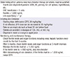
After initiating iron chelation therapy, serum ferritin levels and organ functions including cardiac, hepatic and renal functions should be checked monthly during the first 3 months and then at least once every 3 months. It is recommended that serum ferritin levels be maintained below 1,000 ng/mL and that, when ferritin levels are <500 ng/mL at two successive tests, iron chelation should be discontinued. If ferritin levels continue to increase after initiating therapy, increase the DFX dose up to 30 mg/kg/day. After discontinuing iron chelation, if serum ferritin levels increase above 1,000 ng/mL, restart DFX at the same dose. This guideline is summarized in Table 1.
ADVERSE EVENTS
Gastrointestinal disturbances
Safety data for DFX have been reported in many studies for patients with various anemias (53-56). In the EPIC study, DFX was well tolerated with a clinically manageable side effect profile (5). According to previous studies, the most common drug-related adverse events (AE) were gastrointestinal (GI) disturbances such as abdominal pain (6%), nausea (22%), vomiting (8%), and diarrhea (15%). These events were seen to be generally mild-to-moderate in severity and dose dependent, tended to occur early in the course of treatment and usually lasted less than 1 week, and in general, resolved spontaneously without the need for dose adjustment or discontinuation of treatment (5).
Expert panel recommendations for GI disturbances
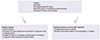
Management of diarrhea requires an exact diagnosis to exclude other etiologies. The expert panel recommends hydration and the use of loperamide for a maximum of 2 days, with evening pre-prandial DFX dosing. The panel does not recommend administration before bedtime because of the risk of esophageal irritation and bleeding. The panel does not recommend DFX dose reduction in cases of mild diarrhea. In cases of persisting moderate-to-severe diarrhea, dose reduction to 10 mg/kg/day should be considered for moderate diarrhea but cease DFX administration for severe diarrhea. When moderate diarrhea has resolved, the DFX dose should be increased in 5 mg/kg/day steps to the target dose. In severe cases, the panel recommends re-initiating the DFX dose at 10 mg/kg/day and adjusting the dose in increments of 5 mg/kg each week to the target dose. These recommendations are summarized in Fig. 1.
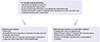
In cases of abdominal pain the panel recommends evening pre-prandial dosing and consumption of soft food for several hours after taking DFX. The panel does not recommend the use of narcotic analgesic drugs or non-steroidal anti-inflammatory drugs because of side effects. If patients have persistent mild-to-moderate pain, dose reductions should be considered before discontinuation. Reduce the dose in steps of 5 mg/kg/day and increment the dose in steps of 5 mg/kg/day after the pain has resolved. If persistent severe abdominal pain occurs, temporarily discontinue DFX treatment and re-initiate and escalate in 5 mg/kg/day steps after the abdominal pain has resolved. These recommendations are summarized in Fig. 2.
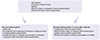
For the management of nausea and vomiting the expert panel recommends hydration, and preprandial administration of DFX in the evening should be considered. In cases of severe vomiting reduce the DFX dose in steps of 5 mg/kg/day. When the symptoms have resolved, re-initiate DFX and increase the dose in increments up to the target dose. These recommendations are summarized in Fig. 3.
Renal adverse events
Some mild increases in serum creatinine levels have been observed and these appear to be dose dependent. In patients with β-thalassemia, 3% overall had serum creatinine values that increased more than 33% above baseline but were still within the normal range (57). In patients with AA, 25% experienced an increase in serum creatinine to more than 33% above baseline and higher than the upper limit of normal (ULN) on two consecutive visits, but there were no progressive increases. The only factor identified as having a significant impact on serum creatinine was concomitant use of the immunosuppressive agent cyclosporine, a drug with a well-recognized potential to impair renal function. Careful monitoring of renal function is necessary in patients with AA who are receiving concomitant cyclosporine and DFX (4).
Expert panel recommendations for renal adverse events
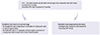
The management of serum creatinine increases should be individualized. It is recommended that serum creatinine levels be assessed in duplicate before initiating therapy and monitored monthly thereafter. Patients who have additional renal risk factors, such as preexisting renal or co-morbid conditions, are elderly, or are receiving medicine that depresses renal function should be monitored weekly during the first month after initiation or modification of the DFX dose and monitored monthly thereafter. In patients who exhibit serum creatinine elevations to more than 33% above baseline but still within the normal range at two consecutive visits, the daily dose should be reduced by 10 mg/kg. If there is a progressive increase in serum creatinine beyond ULN, DFX should be discontinued (58). Once the creatinine level has returned to within the normal range, and if the clinical benefit is expected to exceed the potential risks, DFX should be re-initiated at 10 mg/kg/day and increased in 5 mg/kg/day steps to the target dose. These recommendations are summarized in Fig. 4.
Hepatic and skin adverse events
In the 1-yr trials, up to 2% of patients developed elevations in serum transaminases. These elevations were not related to dose (5). In previous studies, up to 11% of patients developed dose-dependent skin eruptions and most symptoms were mild to moderate (5).
Expert panel recommendations for hepatic and skin adverse events
As a precaution, liver function should be monitored monthly. Furthermore, following any severe or persistent elevations in serum transaminase levels, dose modification should be considered and a hepatologist should be consulted. Therapy may be restarted in patients once serum transaminase levels have returned to within normal limits and levels should be monitored monthly.
In cases of mild-to-moderate skin adverse events, the expert panel does not recommend dose reduction because most skin eruptions resolved spontaneously without treatment. But if the skin eruptions persist or are worse after 1 week the panel advises a temporary discontinuation of DFX treatment until the skin eruptions have disappeared. Short-term use of a steroid should be considered. DFX should then be reinitiated and the dose should be escalated in 5 mg/kg/day steps. These recommendations are summarized in Fig. 5.
Other general considerations
Although the increases in adverse events described above have been reported as dose dependent, recent data showed that the safety profile of DFX in patients who received more than 30 mg/kg/day was consistent with that of patients who received less than 30 mg/kg/day (59). Interestingly, this study also demonstrated that no adverse events were observed following escalation to over 30 mg/kg/day that were not present at lower doses.
There was a higher incidence of drug-related AEs and higher treatment discontinuation rates in patients with MDS than in patients with other chronic anemias (5). This may be related to the risk of disease progression, preexisting co-morbidities, use of concomitant medication, and the advanced ages of patients with MDS.
Most AEs resolved spontaneously and no patients with renal or hepatic failure or drug-related cytopenia have been reported in any of the studies to date. The only contraindication to DFX is prior hypersensitivity to the drug.
DFX has not been investigated in pregnant or breast feeding women, or in pediatric patients younger than 2 yr. Iron chelation should be ceased as soon as pregnancy is confirmed. Caution should be considered in patients older than 65 yr due to a greater frequency of preexisting suppressed hepatic, renal, or cardiac function.
CONCLUSION
Iron overload is a major concern in patients with chronic anemia for whom regular transfusions are necessary. Evidence from many clinical trials has demonstrated clear reduction of the iron burden and improvement in organ function after administration of DFX to patients with transfusion-induced iron overload. This guideline could help many Korean physicians to anticipate potential effects, alert patients to the likelihood of key events, and readily provide effective management according to the recommendations outlined.
 XML Download