

 PDF
PDF ePub
ePub Citation
 Print
Print


INTRODUCTION
Recent advances in hematopoietic stem cell transplantation (HSCT) and supportive therapies have resulted in an increase in the number of long-term survivors. However, these long-term survivors are prone to later complications as a result of their exposure to intensive chemotherapy and radiotherapy. In particular, growth disorders after HSCT are one of the most common problems, and they may influence final adult height (FAH) (1-7). Furthermore, hypogonadism and hypothyroidism after HSCT may result in a growth disorder or accelerate growth impairment (3, 8, 9). Chronic graft-versus-host disease (cGVHD), which develops after HSCT, may also result in growth disorders because it affects multiple organs and requires treatment with steroids (9-11). Radiotherapy (RT) during induction chemotherapy and total body irradiation (TBI) are known to result in growth impairment. Radiation causes growth disorders by causing growth hormone (GH) deficiencies or secretory dysfunction secondary to radiation exposure of the hypothalamic/pituitary region or damage to the growth plates of the long bones and vertebrae. On the other hand, some studies reported that growth impairment might also occur in the absence of an RT history (1, 3).
Some large studies on growth after HSCT (2, 4, 12), have been conducted, but these have been in patients with various primary diseases that require diverse modes of treatment and treatment duration before HSCT. In addition, height outcome after HSCT differs across the primary diseases (2, 12). Moreover, height outcome was different between acute myeloid leukemia (AML) and acute lymphoblastic leukemia (ALL) (2, 13). Therefore, a study on growth after HSCT for a single primary disease is necessary.
AML patients receive HSCT during their first remission and are not treated with steroids during their relatively short induction period. By selecting only AML patients, we could minimize confounding factors affecting growth after HSCT. Previous studies of only AML patients have included various age groups (14, 15), which may have influenced the evaluation of growth after HSCT and of FAH.
We divided the patients into two groups to evaluate the growth patterns shortly after HSCT and to determine factors that influence FAH. To evaluate growth during the 5 yr after HSCT, we selected patients whose puberty began 5 yr after HSCT to minimize the effects of pubertal growth spurts. We evaluated FAH and factors influencing FAH in patients who reached FAH without relapse.
MATERIALS AND METHODS
Patients selection
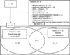
Between January 1996 and June 2010, 97 AML patients received HSCT (Fig. 1).
Exclusion: Twenty-two patients relapsed within 2 yr of HSCT and died, seven received HSCT more than twice, five were already at FAH at the time of HSCT, and two had Down syndrome. Two patients had received part of their induction chemotherapy at other hospitals, 10 had insufficient medical data, one had osteosarcoma before the leukemia diagnosis, one was receiving induction chemotherapy for relapse after HSCT, and one was receiving maintenance therapy for relapse. Nine patients had not reached FAH and had entered puberty 1.3 to 2 yr after HSCT. These 60 patients were excluded from the study, leaving 37 patients.
Inclusion: Among the 37 eligible patients, we evaluated growth over 5 yr after HSCT in 24 patients who entered puberty 5 yr after HSCT (Group 1). Of these, one patient who was diagnosed with hypogonadism showed pubertal signs about 3.2 yr after transplantation with low-dose estrogen replacement therapy. During estrogen replacement therapy, the patient did not undergo a growth spurt and her maximal growth velocity was 5 cm/year. In two patients, puberty commenced 4.8 yr following transplantation. In nine of 24 patients, puberty began 5.3-9.3 yr after transplantation. In the remaining 12 patients, puberty had not commenced at time of follow-up.
We also evaluated FAH and factors influencing FAH in 19 patients who reached FAH without relapse (Group 2). Six of the patients were in both groups.
Chemotherapy, conditioning regimen, radiotherapy and other therapies
Thirty-six of the 37 patients received the same induction and 2-4 cycles of consolidation chemotherapy in accordance with the standard protocol (16). If cycles of consolidation therapy were more than four sessions, patients received maintenance therapies before HSCT. Only one patient in Group 2 received multiple different induction and maintenance therapies.
Carmustine (BCNU)-based conditioning (BCVAC) consisted of BCNU (250 then 200 mg/m2, total dose 450 mg/m2), etoposide (200 mg/m2, twice daily, 4 consecutive days, total 8 doses), cytarabine (2,000 mg/m2, twice daily, 4 consecutive days, total 8 doses) and cyclophosphamide (50 mg/kg, 2 consecutive days, total 2 doses). Busulfan with cyclophosphamide-based conditioning (BuCy) consisted of busulfan (intravenously, 0.8 mg/kg, four times a day, 4 consecutive days, total 16 doses; or orally, 4 mg/kg/day, 4 consecutive days, total 4 doses) and cyclophosphamide (60 mg/kg/day, 2 consecutive days, total 2 doses). Busulfan with fludarabine-based conditioning (BuFlu) consisted of busulfan (intravenously, 0.8 mg/kg, four times a day, 4 consecutive days, total 16 doses; or 120 mg/m2/day for 4 consecutive days, total 4 doses) and fludarabine (40 mg/m2, 6 consecutive days, total 6 doses). The TBI-based conditioning (TBIAcCy) consisted of TBI (300 cGy, 4 consecutive days, total 1200 cGy), cytarabine (3,000 mg/m2, twice daily, 2 consecutive days, total 4 doses) and cyclophosphamide (60 mg/kg, 2 consecutive days, total 2 doses).
Seven of the 37 patients received RT (six in Group 1 and five in Group 2, four in both group). Three patients received cranial irradiation (CI) as prophylaxis for central nervous system involvement (< 18 Gy in two patients, 18 Gy in one patient). Three patients received craniospinal irradiation (CSRT) because of central nervous system involvement (16 and 24 Gy for CI, 12 and 6 Gy for spine). One patient received TBI as described above.
Seventeen of the 37 patients received steroid therapy after HSCT. All patients had taken prednisolone. Fourteen patients received steroid therapy for GVHD prophylaxis for a total of 20-78 days, including the tapering period. Of the remaining three of the 17 patients, one patient had taken prednisolone for the treatment of cGVHD for 174 days, and two patients had taken it for more than a year.
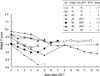
Four patients received GH treatment after HSCT. All of these patients were included in Group 1. One of the four patients was a non-RT patient. The non-RT patient received GH treatment at 7 yr after HSCT and was excluded from Group 2 because of an absence of FAH. The other three patients were RT patients (Fig. 2, S5, S6, S7). Two of the four patients (Fig. 2, S5 and S7) reached FAH, and were included in both Groups 1 and 2.
Height measurement, evaluation of pubertal status and endocrine function
Auxological data and hormone status were collected. Height was measured using a Harpenden stadiometer (Holtain Ltd., Crymych, UK) at the time of diagnosis, at HSCT, and every 6-12 months thereafter. In Group 1, height data were collected for five years. Measurements were taken once a year and within a scheduled two month period. Only data collected during the two month period were used. Because some measurements could not be collected during the annual collection period, there were some missing data. Height was expressed as a Z-score standardized for sex and age on the basis of Korean references (17). A GH stimulation test was done in patients with growth velocity was below 4cm/yr. FAH was defined as a height velocity of less than 1 cm per year or a bone age ≥ 17 yr for males and ≥ 15 yr for females by the Greulich-Pyle method (18). Target height (TH) was calculated from the average parental height plus 6.5 cm for males and minus 6.5 cm for females.
Pubertal stage was assessed by method of Marshall and Tanner (19, 20), every 3-6 months. Onset of puberty was defined as a testicular volume ≥ 4 mL or Tanner breast stage ≥ B2. Hypogonadism was defined as a lack of pubertal signs at 14 yr of age in males and 13 yr in females, with abnormally elevated basal FSH (> 15 IU/mL) and LH levels (> 10 IU/mL) and decreased level of sex hormone (6, 12). In our study, the minimum values of LH and FSH levels of the hypogonadism patients were 14 and 40.7 IU/mL, respectively. A thyroid function test was performed at regular intervals of 6-12 months. Hypothyroidism was defined as a free T4 level below 0.8 ng/dL and an elevated TSH level above the normal range. Acute and chronic GVHD were diagnosed and classified using the Seattle criteria (21).
Statistical analysis
The mean height Z-score was expressed as mean ± standard deviation (SD). The statistical analyses were performed using SPSS statistical software, version 19 (SPSS Inc., Chicago, IL, USA), and R package (R Development Core Team, 2005, http://www.R-project.org). A P value < 0.05 was considered to be statistically significant.
To evaluate growth during the first 5 yr after HSCT, summary measure analysis was used to find statistical differences between factors. Summary measure analysis of serial measurements is a useful and simple tool in medical research (22, 23). In addition, summary measure analysis can be used when there are missing values (22, 23). The overall mean 5-yr height Z-score and the area under curve (AUC) of 5-yr height Z-score were chosen for summary measures. To compare these summary measures, independent t-tests and Mann-Whitney tests were used. To calculate AUC, height both at HSCT and at 5 yr after HSCT was needed. Therefore the AUC calculation was available in only 14 patients. P values from the Mann-Whitney tests were used to evaluate statistical significance. Scatterplots with linear regression lines and smooth curved fit lines with standard error intervals were used to analyze the patterns of growth after HSCT. Smoothed curved fit lines were built by locally estimated scatterplot smoothing (LOESS) method.
To evaluate factors influencing FAH, multivariate regression analysis was performed to determine factors that influence FAH. Independent t-test and Mann-Whitney tests were performed to compare mean FAH Z-scores between patients by gender, RT, cGVHD and steroid treatment. The TH Z-scores were compared to FAH height Z-scores using the Wilcoxon signed rank test and paired t-test.
RESULTS
Five years growth after HSCT (Group 1)
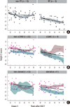
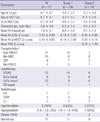
Patient characteristics are shown in Table 1. Fig. 3 shows scatterplots of height Z-scores at yearly intervals with linear regression lines and smooth curved fit lines with standard error intervals. In Fig. 3B and 3C, pink-color indicates non-RT patients and blue-color indicates RT patients. To evaluate effect of age at HSCT, we divided patients in two subgroups with most equal proportions (age < 5 yr, n = 14; age > 5 yr, n = 10). There were no statistical differences by age at HSCT (< 5 and > 5 yr) or by gender (Table 2). The mean 5 yr height Z-score were smaller in RT patients than non-RT patients (-1.82 ± 1.00 vs -0.27 ± 0.99), and this difference was statistically significant (P = 0.009). Further, in the AUC analysis (n = 14), RT patients were significantly shorter than non-RT patients (P = 0.020). The mean 5-yr height Z-score was smaller in cGVHD patients than in non-cGVHD patients, but the significance from mean and AUC analyses was inconsistent (Table 2). However, when differentiated by a history of RT, as in Fig. 3B, only cGVHD patients that had received RT (blue) were shorter. Also, among steroid-treated patients, only steroid-treated patients that had received RT (blue) were shorter than their counterparts (Fig. 3C). The non-RT patients had lower height Z-scores during the first 2-3 yr after HSCT, but they showed catch-up growth 3 yr after HSCT (non-RT in Fig. 3A, pink colored line in 3B and 3C). Hypogonadism and hypothyroidism were not significantly associated with growth impairment during the 5 yr after HSCT.
FAH after HSCT (Group 2)
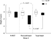
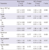
Patient characteristics are shown in Table 1. Nineteen patients reached FAH without relapse after HSCT. Mean FAH height Z-scores of the RT patients were significantly smaller than those of the non-RT patients (Fig. 4; -0.29 ± 0.88 vs -2.07 ± 1.35; P = 0.010). FAH Z-scores of RT patients were smaller than their calculated TH Z-scores, and differences were statistically significant (P = 0.043).
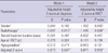
We performed a multivariate regression analysis to evaluate factors influencing FAH. Age at HSCT, gender, history of RT, steroid treatment duration and history of hypogonadism were entered into the multivariate regression model (Table 3). Histories of cGVHD and hypothyroidism were not entered into the model because there were only two patients in each category. Since the steroid doses were almost the same because of similar treatment protocols, the steroid treatment duration was instead entered into the models to adjust the cumulative steroid dose. Because CI and CSRT are performed during induction chemotherapy before HSCT, the regression analysis was also performed after adjusting for the height Z-score at diagnosis (Table 3; Model 1). History of RT was significantly correlated with FAH Z-score after adjusting for the height Z-score at diagnosis (P = 0.047) or at HSCT (P = 0.033). Height Z-scores at diagnosis or at HSCT were also significantly correlated with the FAH Z-scores. Female gender was significantly and positively correlated with FAH in model 2. However, in model 1, the correlation was not significant. Age at HSCT, steroid treatment duration and hypogonadism were not significantly associated with FAH.
Growth of the RT patients
Three RT patients received GH treatment (Fig. 2; S5, S6, S7) after 3.5-4.2 yr from HSCT. After starting the treatment, the height Z-scores for these patients increased or stopped decreasing. No adverse effects were found during the follow-up. GH stimulation test was done in 3 patients. The most growth-impaired patient, whose Z-score dropped to approximately -4 (S7), was the only patient found to have GH deficiency in our study. In another two patients who received CSRT (S5, S6), a GH stimulation test did not reveal a GH deficiency. The three other RT patients (S1, S2 and S3) did not show catch-up growth, although S2 required further follow-up. One RT patient (S4) was initially thought to show catch-up growth after 5 yr, but this growth was found to be pubertal growth. After the pubertal period, the Z-score of this patient decreased to the prepubertal level.
DISCUSSION
HSCT may complicate growth impairment. Growth after HSCT may be influenced by disease type, various therapeutic agents, pre-HSCT treatment duration and many other factors (2, 12, 13). However, most previous studies on growth after HSCT included patients with various diseases, such as leukemia, myelodysplastic syndrome, aplastic anemia and genetic and metabolic diseases (2, 4).
The uniqueness of this study is that all of the included patients had the same primary disease. Second, we adjusted for pubertal growth spurts. Finally, by including only AML patients, we could minimize confounding factors affecting growth after HSCT.
In this study, we found that RT patients did not show catch-up growth after 3 yr from HSCT. History of RT was associated with reduced FAH. Younger age at HSCT, cGVHD, steroid treatment and hypogonadism were not significantly associated with growth impairment after HSCT.
Afify et al. (14) reported that 23 AML patients who received HSCT with the BuCy conditioning regimen showed no significant differences in height Z-score change for 5 yr, and their age at HSCT was not associated with growth impairment. Cohen et al. (4) reported that there was no significant growth impairment in patients without history of RT, and neither GVHD nor steroids influenced FAH. However, cGVHD was a significant factor associated with growth impairment during the 2 yr following HSCT in the studies by Adan et al. (11) and Sanders et al. (10). Bernard et al. reported that conditioning with TBI significantly decreased FAH (1), and Sanders et al. showed that cranial irradiation before HSCT significantly decreased FAH (2). In contrast, other studies have indicated that growth impairment also occurs after HSCT without radiotherapy. Bernard et al. (1) reported that a Busulfan-based conditioning regimen without RT also had adverse effects on growth after HSCT. Bakker et al. (3) also showed that BuCy conditioning for HSCT often resulted in growth impairment.
In this study, there was a group of patients who had lower height Z-scores during the first 2-3 yr after HSCT, but they showed catch-up growth 3 yr after HSCT. Although this finding was not significant, the smooth curved fit line in Fig. 3 indicates that the non-RT patients exhibited a similar pattern. Also, in Fig. 3B and 3C, non-RT patients (pink color) showed similar patterns. Other studies have also reported a nonsignificant decrease in height Z-scores 2.5-5 yr after HSCT (1, 3, 14). This pattern of growth impairment, in the first 3 yr after HSCT seems to be caused by induction chemotherapy and chemotherapy during HSCT. Chemotherapy may result in growth impairment by mechanisms such as damage to cartilage growth plates (24), an increase in nutritional requirements, malnutrition during treatment and reduced peripheral tissue response to growth factors (25). However, the RT patients did not show catch-up growth 3 yr after HSCT. Although three patients received GH treatment during 5 yr after HSCT in Group 1, they were all RT patients. Therefore, there may have been more height loss in RT patients without GH treatment.
Four of the 24 patients were diagnosed with cGVHD (Group 1). These four patients showed prominent growth impairment in the first 2 yr after HSCT. A similar result was found when RT patients were excluded from the analysis. In cGVHD patients, the overall linear regression line showed decreasing height Z-scores during the following 5 yr (Fig. 3B). But the smooth curved fit line showed that only cGVHD patients with RT history (colored blue in Fig. 3B) continued to decrease in the 5 yr after HSCT. Sanders et al. (10) reported that cGVHD patients showed significant growth impairment in the first 2 yr after HSCT compared with non-cGVHD patients. In their study, cGVHD remained a cause of growth impairment 3 yr after HSCT. This result may have been observed because all the patients in their study received TBI. In addition, in their study, cGVHD patients with and without steroid therapy both showed growth impairment. This result indicates that cGVHD alone can cause growth impairment. cGVHD is thought to cause growth impairment by causing the liver to decrease IGF-1 production (9). Adan et al. (11) reported that patients with post-HSCT complications, including cGVHD, had significantly lower serum IGF-1 levels and lower height Z-scores during the first 2 yr after HSCT. Although there were only small numbers of cGVHD patients in our study, cGVHD seems to affect the first 2 yr of growth after HSCT, but cGVHD patients without a history of RT seemed to show catch-up growth as the disease improved.
History of cGVHD, steroid treatment, age at HSCT, and hypogonadism did not significantly affect FAH. A history of RT was significantly and negatively correlated with FAH. Some studies have reported that younger age at TBI correlated significantly with a greater loss of FAH (2, 5, 6). This loss of FAH was thought to occur because a younger age at TBI results in earlier damage to growth plates, eventually resulting in a greater decrease of FAH.
In this study, there was no significant correlation between age at HSCT and FAH. Cohen et al. (4) reported that a younger age at HSCT resulted in shorter FAH. However, in their study, 144 of the 181 subjects received TBI as a part of the conditioning regimen. Other studies have reported that a younger age at HSCT is associated with greater height loss as well (2, 5, 6, 8). In these studies, every subject, or a significant proportion of subjects, received TBI. On the other hand, De Simone et al. reported that thalassemia patients who received HSCT before 7 yr of age reached significantly greater FAH than those who received HSCT after 7 yr of age (7). Therefore, we think a greater loss of FAH is associated with younger age at TBI or RT, not younger age at HSCT.
CI and CSRT also may result in growth impairment after HSCT. CI can affect growth by damaging hypothalamic/pituitary region and causing GH deficiency or secretory dysfunction (8, 13, 26). Sanders et al. (2) showed that GH deficiency was more frequent in subjects who received CI than those without CI. Although CI can affect GH secretion, prevalence of GH deficiency after 18 Gy of radiation dose has been inconsistent among studies (27). Hence, GH neurosecretory dysfunction might have been existed in those without GH deficiency in provocation tests. CSRT may result in not only GH insufficiency, but also direct damages to growth plate of vertebrae. Therefore, we think CSRT result in more severe growth impairment with altered body proportions, than CI.
We attempted to evaluate the effect of cGVHD on FAH, but there were only two patients reached their FAH. However, in Group 1, cGVHD patients without a history of RT showed catch-up growth starting 3 yr after HSCT, suggesting that cGVHD does not significantly affect FAH. The use of steroids did not significantly affect FAH. Cohen et al. (4) and Shalitin et al. (12) also reported that neither cGVHD nor steroid treatment affected FAH.
In our study, hypothyroidism and hypogonadism did not result in significant growth impairment. This might be due to quick thyroid hormone replacement after detection of hypothyroidism in regular thyroid function test follow-up. In the study by Bakker et al. (26), patients with hypogonadism achieved a better height outcome than patients with spontaneous pubertal onset. The explanation for this is that the time of growth is prolonged by delayed bone age progression in hypogonadism patients. Another study also suggested that patients with hypogonadism might have superior height outcome through medical adjustment of pubertal onset (28).
The use of GH has risks in patients with a history of primary malignancies and a history of RT (29). Isfan et al. (30) recently reported a study of GH therapy in 25 acute leukemia patients treated with HSCT, CI and TBI. The mean height Z-score gain was 0.59, and no secondary neoplasms were observed. However, we believe that GH treatment has risks, and only limited numbers of patients, such as those with predicted FAH Z-scores less than -2, should consider treatment. We suggest that a prospective study with large numbers of subjects is needed to examine GH treatment risk factors.
Unexpectedly, there was a significant positive correlation between female gender and FAH in model 2, but not in model 1. Although previous studies also showed that girls had better height outcomes than boys (2, 4, 26), the reason for this difference is not clear. Therefore, additional specific studies are needed.
This study has some limitations. The study was retrospective and therefore could not prove causal relationships between the studied factors. Patients who received HSCT with stem cells of allogenic origin might show more prominent growth impairment (12). However, in our study, the proportion of patients with autologous peripheral HSCT was relatively large. FAH in patients without relapse might stand for a selected group of patients with better prognosis. There were fewer cGVHD patients in our study. The effect of RT on growth impairment might be less prominent because of the GH treatment received by RT patients.
In summary, growth impairment may occur in AML patients after HSCT. In RT patients, catch-up growth does not occur and FAH will be reduced. In non-RT patients, growth impairment seems to be temporary and mitigated by catch-up growth. Chronic GVHD seems to impair growth after HSCT only temporarily. Younger age at HSCT, steroid treatment and hypogonadism are not significantly associated with growth impairment. Therefore, growth hormone treatment is not recommened in non-RT patients and careful monitoring of catch-up growth is needed.
 XML Download